RIOK3 / Serine/threonine-protein kinase RIO3 · IHC design guide

Design Immunohistochemistry for RIOK3

Plan paraffin-section RIOK3 IHC around cytoplasmic tissue staining (HPA tissue IHC). Duodenal glandular cells show high staining and can serve as a positive reference (HPA tissue IHC); keep fixation consistent across sections (standard IHC practice).

Evidence assembled Oct 2026 · For research use; verify linked source records and product datasheet before use
Immunohistochemistry protocol sheet for RIOK3 (IHC for RIOK3): expected localisation Cytoplasmic tissue staining (HPA tissue IHC), antibody A09262-1, validated IHC image, and IHC protocol steps
Printable RIOK3 IHC protocol sheet — expected localisation Cytoplasmic tissue staining (HPA tissue IHC), antibody A09262-1, controls and protocol steps. Open the full RIOK3 IHC guide →

RIOK3 Immunohistochemistry Experimental Design Guide

Expected localisation, validated protocols, controls and antibodies — the at-a-glance facts below, then the full design guide.

Must know before staining
Expected localisation Cytoplasmic tissue staining (HPA tissue IHC)
Staining pattern Cytoplasmic signal across tissues; adipocytes undetected (HPA tissue IHC)
Antigen retrieval Tris-EDTA pH 9.0 HIER, heat-mediated (datasheet A09262-1)
Positive control ⓘ Duodenum+4 more · see all
Negative control ⓘ Adipose tissue
Important caveats
Reasons your staining may differ from the expected pattern.
Fixation Matched tissue-IHC evidence does not establish the fixation claim. Validate the specimen-specific method before use. (selected-SKU IHC image A09262-1)
Caveat Medium staining–RNA consistency; verify specificity (HPA tissue IHC)
Regulation Expression regulation not established (UniProt)
Isoform / epitope Two isoforms; epitope coverage unspecified (UniProt)
Section 1

Recommended RIOK3 IHC & IF Protocols

Compare the catalog antibody’s IHC-P protocol (datasheet A09262-1) with two published RIOK3 IHC protocols (PMC10008507; PMC10920805).

Recommended immunohistochemistry (IHC-P) protocol parameters
SampleParaffin-embedded human Gastric adenocarcinoma tissue; fixative not specified (datasheet A09262-1)
FixationImage fixative and duration unreported (datasheet A09262-1); verify before use.
Sectioning4–5 µm sections on charged slides (standard)
DeparaffinisationXylene, graded ethanol series to water (standard)
Antigen retrievalHeat retrieval: Tris-EDTA pH 9.0 (datasheet A09262-1); 20 min, 95–100 °C (standard)
Peroxidase block3% H2O2, 10 min, room temperature (standard)
Blocking10% normal serum of the secondary host, 30 min, room temperature (standard)
Primary antibodyRabbit anti-RIOK3, 1:50 recommended; image 1:200 (datasheet A09262-1)
Primary incubationOvernight at 4 °C (standard)
DetectionHRP-polymer secondary, DAB chromogen 5–10 min (standard)
CounterstainHematoxylin, blue, dehydrate and mount (standard)
Expected resultRIOK3-positive staining in glandular cells of duodenum (HPA tissue IHC: High). HPA tissue profile: Cytoplasmic expression in all tissue types. No signal in the no-primary control.
💡Decision noteStart with heat-mediated Tris-EDTA, pH 9.0, for the catalog antibody (datasheet A09262-1); the pancreatic study does not specify which retrieval solution was used for RIOK3 (PMC10008507).
Section 2

What Is the Expected RIOK3 Staining Pattern?

RIOK3 is cytosolic and has no transmembrane segment (UniProt O14730 topology). In paraffin-section IHC, expect predominantly cytoplasmic staining across tissue types, including high staining in duodenal glandular cells and esophageal squamous epithelial cells (HPA tissue IHC). Use cell identity as well as compartment to judge the slide: HPA rates its tissue staining Approved, with medium consistency against RNA data and external verification pending (HPA tissue IHC).

What am I looking at on my slide?
Cytoplasmic staining in duodenal glands, esophageal squamous epithelium, or lymph-node germinal centers.This fits HPA's reported high staining in those cell types (HPA tissue IHC). Judge staining within the named cells; a dark region alone does not identify which cells produced the signal (general IHC practice).
Strong nuclear-only staining, with little cytoplasmic signal in the same cells.This departs from the reported cytosolic location and cytoplasmic tissue pattern (UniProt O14730; HPA tissue IHC). Treat it as suspect, then check morphology, controls, and the antibody's validated IHC conditions before assigning RIOK3 localization (general IHC practice).
Prominent staining in adipocytes, especially when expected positive cells stain weakly.HPA reports RIOK3 as not detected in adipocytes (HPA tissue IHC). Investigate antibody cross-reactivity or endogenous detection activity with appropriate controls; this pattern alone cannot distinguish those causes (general IHC practice).
Diffuse chromogen covers cells and extracellular areas, obscuring cell borders.The distribution cannot establish cytoplasmic RIOK3 staining (HPA tissue IHC). Background may arise from nonspecific antibody binding or the detection system; compare a no-primary control and review blocking, washes, and chromogen development (general IHC practice).
No signal in well-preserved duodenal glands or esophageal squamous epithelium.These are high-staining cell populations in HPA tissue IHC, so absence warrants a technical review (HPA tissue IHC). Check the IHC-validated antibody's instructions, controls, retrieval, and detection; a negative slide alone does not establish absent RIOK3 (general IHC practice).
💡Expected RIOK3 appearanceCall a result positive when identifiable high-staining cell populations show predominantly cytoplasmic chromogen (HPA tissue IHC); isolated nuclear staining or strong adipocyte staining is discordant and needs control-based review (UniProt O14730; HPA tissue IHC).
How each factor affects the staining
Tissue and cell selectionHPA reports high staining in duodenal glandular cells, esophageal squamous cells, lymph-node germinal-center cells, ovarian follicle cells, placental trophoblasts, and rectal glands; adipocytes are not detected (HPA tissue IHC). Select and score specific cell populations rather than whole-section darkness (general IHC practice).
Compartment and assayUniProt places RIOK3 in the cytosol, and HPA tissue IHC describes cytoplasmic expression (UniProt O14730; HPA tissue IHC). For the separate IF/ICC guide: HPA also approves plasma-membrane and cytosolic localization, with additional Golgi and centriolar-satellite localization (HPA ICC-IF).
Strength of tissue evidenceHPA rates the tissue pattern Approved, with medium consistency between antibody staining and RNA expression and external verification pending (HPA tissue IHC). Treat the listed cell patterns as expectations, then confirm a new specimen or unusual pattern with controls (general IHC practice).
Isoforms and antibody coverageUniProt lists two RIOK3 isoforms (UniProt O14730). The supplied evidence gives no epitope or isoform coverage for the IHC-validated antibody, so it cannot predict whether both isoforms contribute to staining; follow the antibody's documented validation (general IHC practice).
Why is my staining missing, weak or wrong?
SituationLikely causeNext action
Known high-staining cells are blank, including the positive control.A failed staining run is plausible; HPA reports high staining in duodenal glandular and esophageal squamous cells (HPA tissue IHC). The slide alone does not locate the failed step (general IHC practice).Review the IHC-validated antibody's paraffin-section instructions, antigen retrieval, antibody dilution, detection reagents, and control performance; repeat with an appropriate positive section (general IHC practice).
Signal is weak in a chosen comparison tissue.Expected levels differ by cell type: HPA reports medium staining in endometrial glands and keratinocytes, and low staining in seminal-vesicle glands and smooth muscle cells (HPA tissue IHC).Compare the same identified cell type across sections, and include a reported high-staining population to assess assay performance before interpreting a weak field (HPA tissue IHC; general IHC practice).
Adipocytes stain as strongly as the intended positive cells.This conflicts with HPA's not-detected adipocyte result; cross-reactivity or endogenous detection activity are possible explanations, not established diagnoses (HPA tissue IHC; general IHC practice).Inspect a no-primary control, review endogenous enzyme blocking if using enzyme-based detection, and compare the IHC-validated antibody with an independent validated reagent where available (general IHC practice).
Nuclei dominate while cytoplasm is faint.That distribution differs from UniProt's cytosolic assignment and HPA's cytoplasmic tissue profile (UniProt O14730; HPA tissue IHC). Counterstain or nonspecific signal may complicate compartment reading (general IHC practice).Check cell boundaries and counterstain, compare a no-primary control, and reassess localization under the antibody's validated staining conditions (general IHC practice).
Brown signal is widespread and cell boundaries are hard to see.Diffuse background can reflect nonspecific binding, inadequate washing, or overdevelopment; appearance alone does not identify which step caused it (general IHC practice).Use a no-primary control to assess detection background, then review blocking, washing, antibody concentration, and chromogen development while retaining a positive control (general IHC practice).
Membrane-associated signal appears alongside cytoplasmic staining.HPA ICC-IF approves plasma-membrane and cytosolic localization, while HPA tissue IHC summarizes the tissue pattern as cytoplasmic (HPA ICC-IF; HPA tissue IHC). The assays provide different localization evidence.Record both compartments and the affected cell type; assess the IHC signal against tissue controls, and consult the separate IF/ICC guide for fluorescence localization (HPA tissue IHC; HPA ICC-IF; general IHC practice).

Sample controls for RIOK3 IHC & IF

🧪Run duodenum first: glandular cells should show RIOK3 staining (HPA: High in duodenal glandular cells). Use adipose tissue as the negative tissue (HPA: Not detected in adipocytes); on the duodenal slide, neighboring cells without specific staining should remain near background, but their RIOK3-negative status is unverified (HPA: High in duodenal glandular cells).
Positive control tissue: Duodenum (Glandular cells, HPA High)
Negative control tissue: Adipose tissue (HPA Not detected)
ICC-IF cell lines (HPA subcellular resource): HPA ICC-IF images show RIOK3 in A-431, U-251MG, U2OS, with annotated localisation: Plasma membrane (approved), Cytosol (approved) (HPA subcellular).
Technical controls: Include a no-primary, secondary-only control and a host-species-matched isotype control for a monoclonal antibody or matched normal IgG for a polyclonal antibody (standard IHC practice). If available, use RIOK3 knockout material as a biological negative; quench endogenous peroxidase and check for nonspecific glandular background in duodenum (standard chromogenic IHC practice).
⚠️Feasibility: No target-specific fixation window or fixation effect is reported in the supplied evidence, and the selected A09262-1 tissue-IHC caption does not state a fixative (A09262-1 caption: fixative unreported). The caption uses Tris-EDTA at pH 9.0 for retrieval and a 1:200 primary dilution, but does not establish that retrieval is required (A09262-1 caption). The supplied evidence does not establish whether frozen sections or IF/ICC would be easier; for duodenal glandular staining, check that luminal or diffuse background does not obscure cell-associated signal (HPA: High in duodenal glandular cells; standard IHC practice).

HPA tissue IHC evidence for RIOK3

Comprehensive Human Protein Atlas IHC scoring per tissue (reliability: Approved — Medium consistency between antibody staining and RNA expression data. Pending external verification.). Rows are taken directly from the HPA tissue chart — click any row's HPA link to view the source.

Positive expression · recommended positive controls

TissueCell typeLevelEvidenceSource
Duodenum Glandular cells High Protein (IHC) HPA →
Endometrium Glandular cells Medium Protein (IHC) HPA →
Esophagus Squamous epithelial cells High Protein (IHC) HPA →
Lymph node Germinal center cells High Protein (IHC) HPA →
Ovary Follicle cells High Protein (IHC) HPA →

Undetected expression · recommended negative controls

TissueCell typeLevelEvidenceSource
Adipose tissue Adipocytes Not detected Protein (IHC) HPA →
Section 3

Advanced RIOK3 IHC Tips

Troubleshoot RIOK3 staining in paraffin sections by checking retrieval, cytoplasmic localisation, antibody specificity, and tissue matched controls (datasheet A09262-1; UniProt O14730; HPA tissue IHC).

What retrieval should I try first when RIOK3 staining is weak?
Start with heat-mediated Tris-EDTA at pH 9.0 for paraffin-section IHC (datasheet A09262-1). The selected antibody caption pairs that retrieval with a 1:200 primary dilution incubated at 4°C overnight in human gastric adenocarcinoma sections (datasheet A09262-1). Keep section thickness, heating conditions, and detection chemistry consistent while comparing retrieval runs (standard IHC practice). If staining remains weak, test an alternative retrieval condition on adjacent sections as a fallback, with a positive tissue control in every run (standard IHC practice; HPA: high staining in duodenal glandular cells). Judge improvement by clearer cellular staining without a matching increase in stromal or tissue-edge background (standard IHC practice).
Could fixation explain weak RIOK3 staining in my paraffin sections?
Target-specific fixation sensitivity is unknown from the supplied evidence; the selected paraffin-section caption does not state a fixative (datasheet A09262-1). Record the fixative, time before fixation, fixation duration, and processing schedule for each specimen, then compare sections processed together (standard IHC practice). Keep Tris-EDTA retrieval at pH 9.0 and the 1:200 primary dilution constant during that comparison (datasheet A09262-1). Include a control with expected staining, such as duodenal glandular cells, to distinguish a weak specimen from a weak run (HPA: high in duodenal glandular cells; standard IHC practice). Interpret differences as processing-associated until replicated under matched conditions (standard IHC practice).
Should I accept membrane or nuclear staining as RIOK3 signal?
Use cytoplasmic staining as the main tissue-IHC expectation because the tissue profile reports cytoplasmic expression across tissue types (HPA tissue IHC). RIOK3 is annotated in the cytosol and has no transmembrane segment (UniProt O14730 localisation and topology). Plasma membrane and cytosol are both approved locations in cell imaging, with additional Golgi and centriolar-satellite localisation, so a membrane-associated pattern deserves careful comparison with controls (HPA subcellular). Score nuclear-only staining separately and check whether it persists in a primary-antibody omission control (UniProt O14730 localisation; standard IHC practice). Compare the same cell population across adjacent sections before assigning a compartment-specific result (standard IHC practice).
How can isoforms or epitope placement affect my RIOK3 IHC result?
RIOK3 has 2 annotated isoforms, and its protein kinase domain spans residues 251–519 (UniProt O14730). The supplied antibody caption gives no immunogen or mapped epitope, so isoform coverage cannot be assigned from that caption (datasheet A09262-1). Several modified residues are annotated, including phosphoserines at 8 and 112, but their effects on this antibody's staining are unknown (UniProt O14730; datasheet A09262-1). If a mapped epitope becomes available, compare it with isoform sequences and modified sites before interpreting selective staining (standard IHC practice). Use an independent antibody with a disclosed epitope or an appropriate loss-of-target control to investigate unexpected staining (standard IHC practice).
How should I assess RIOK3 in multiplex immunofluorescence?
Treat IF conditions as a separate optimisation from this antibody's paraffin-section IHC caption (datasheet A09262-1). For tissue multiplexing, pair RIOK3 with a marker for the expected cell population, such as an epithelial marker when examining duodenal glandular cells (HPA: high in duodenal glandular cells; standard IF practice). Choose spectrally separated fluorophores and consider a far-red channel when tissue autofluorescence obscures shorter wavelengths (standard IF practice). Because RIOK3 is cytosolic and has no transmembrane segment, assess mild permeabilisation for intracellular access; the antibody's actual epitope location remains unspecified (UniProt O14730; datasheet A09262-1). Include single-stain and omission controls before interpreting colocalisation (standard IF practice).
What should I change when RIOK3 chromogenic staining looks diffuse?
First compare the stained section with primary-antibody omission and tissue controls to locate background from detection reagents or tissue (standard IHC practice). The selected caption reports a 1:200 primary incubation at 4°C overnight and a 1:200 secondary incubation for 45 minutes at room temperature (datasheet A09262-1). If background rises with primary antibody, titrate its concentration and review wash and blocking conditions while keeping retrieval at pH 9.0 (datasheet A09262-1; standard IHC practice). For peroxidase-based DAB detection, check the peroxidase block and DAB development time as general workflow controls (standard IHC practice). Preserve interpretable cytoplasmic staining while reducing diffuse deposit (HPA tissue IHC; standard IHC practice).
How should I quantify RIOK3 staining across tissue samples? ⚠ ANSWER MARKED FOR VERIFICATION
Define the cell population and tissue region before scoring, then apply the same rules to every section (standard IHC practice). For chromogenic staining, report the percentage of positive cells and an H-score from 0–300, calculated from the percentages at each intensity level (standard IHC practice). If counting discrete positive cells, report density per mm² of viable tissue and normalise to the measured tissue area (standard IHC practice). Score comparable cell types separately because reported staining varies across tissues and cell populations (HPA tissue IHC). Keep retrieval, detection, exposure of the chromogen, and positivity thresholds consistent, and exclude folds or necrotic areas from the denominator (standard IHC practice).
How do I distinguish convincing RIOK3 staining from artefact?
Look for reproducible cellular staining with a substantial cytoplasmic component, consistent with the reported tissue pattern and cytosolic annotation (HPA tissue IHC; UniProt O14730 localisation). High staining in duodenal glandular cells or esophageal squamous epithelial cells can provide a reference, while adipocytes are reported as undetected (HPA tissue IHC). Treat staining confined to tissue edges, folds, or necrotic areas as suspect and compare it with intact regions (standard IHC practice). If a peroxidase-based reaction appears in an omission control, investigate endogenous enzyme activity and the blocking step (standard IHC practice). Interpret isolated nuclear-only staining cautiously because it does not match the supplied localisation evidence (HPA tissue IHC; UniProt O14730 localisation).
Boster reagents

Best RIOK3 / Serine/threonine-protein kinase RIO3 IHC Antibodies

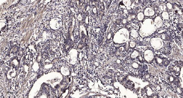
The catalog antibody has an IHC image from a paraffin-embedded human gastric adenocarcinoma section (A09262-1 image caption); its listed reactivity covers human and mouse (A09262-1 catalog).

Real IHC data Immunohistochemical analysis of paraffin-embedded human Gastric adenocarcinoma. 1, Antibody was diluted at 1:200(4° overnight). 2, Tris-EDTA,pH9.0 was used for antigen retrieval. 3,Secondary antibody was diluted at 1:200(room temperature, 45min).
Anti-RIOK3/Sudd Antibody
Cat # A09262-1

A09262-1 is listed for IHC and reacts with human and mouse samples (A09262-1 catalog). Its image shows IHC on paraffin-embedded human gastric adenocarcinoma (A09262-1 image caption); the fixative is unreported (A09262-1 image caption).

Which to pick: Choose A09262-1 for tissue IHC when its demonstrated paraffin-section example is relevant (A09262-1 image caption); the fixative is unreported (A09262-1 image caption). No IF/ICC application or image is listed for A09262-1, so its suitability for IF/ICC is unverified (A09262-1 catalog). A09262-1 is a rabbit polyclonal antibody listed as reactive with human and mouse (A09262-1 catalog), but the supplied IHC image demonstrates human tissue only (A09262-1 image caption).

Each figure is that product's own IHC / IF validation image from its datasheet.

References

  1. UniProt Consortium. UniProt entry O14730 (RIOK3_HUMAN, Serine/threonine-protein kinase RIO3).
  2. Human Protein Atlas. RIOK3 tissue IHC expression (reliability: Approved).
  3. Human Protein Atlas. RIOK3 subcellular location (ICC-IF): Mainly localized to the plasma membrane and cytosol. In addition localized to the Golgi apparatus and centriolar satellites..
  4. Human Protein Atlas. RIOK3 antibody validation summary (2 antibodies).
  5. RIOK3 promotes mTORC1 activation by facilitating SLC7A2-mediated arginine uptake in pancreatic ductal adenocarcinoma. Aging 2023 — PMC10008507.
  6. RIOK3 sustains colorectal cancer cell survival under glucose deprivation via an HSP90α-dependent pathway. Oncogenesis 2024 — PMC10920805.
  7. RIOK3 promotes pancreatic ductal adenocarcinoma cell invasion and metastasis by stabilizing FAK. Heliyon 2022 — PMC9379581.
  8. The Atypical Kinase RIOK1 Promotes Tumor Growth and Invasive Behavior. EBioMedicine 2017 — PMC5478185.
  9. PubMed PMID:9602165 — UniProt-cited evidence.
  10. PubMed PMID:16177791 — UniProt-cited evidence.
  11. PubMed PMID:15489334 — UniProt-cited evidence.