RPL39 / Large ribosomal subunit protein eL39 · IHC design guide

Design Immunohistochemistry for RPL39

Plan RPL39 paraffin-section IHC around reported cytoplasmic staining in hepatocytes, myocytes and late spermatids (HPA tissue IHC). Compare positive and undetected tissue controls, then optimize the catalog antibody within its stated IHC dilution range (HPA tissue IHC; datasheets).

Evidence assembled Oct 2026 · For research use; verify linked source records and product datasheet before use
Immunohistochemistry protocol sheet for RPL39 (IHC for RPL39): expected localisation Cytoplasmic staining in several tissues (HPA tissue IHC), antibody A10219-1, validated IHC image, and IHC protocol steps
Printable RPL39 IHC protocol sheet — expected localisation Cytoplasmic staining in several tissues (HPA tissue IHC), antibody A10219-1, controls and protocol steps. Open the full RPL39 IHC guide →

RPL39 Immunohistochemistry Experimental Design Guide

Expected localisation, validated protocols, controls and antibodies — the at-a-glance facts below, then the full design guide.

Must know before staining
Expected localisation Cytoplasmic staining in several tissues (HPA tissue IHC)
Staining pattern Cytoplasmic in hepatocytes, myocytes and late spermatids (HPA tissue IHC)
Antigen retrieval Citrate pH 6.0 HIER, 95–98 °C, 20 min (rule: cytoplasmic / membrane antigen)
Positive control ⓘ Liver+2 more · see all
Negative control ⓘ Adipose tissue+4 more · see all
Important caveats
Reasons your staining may differ from the expected pattern.
Fixation Keep paraffin-section fixation consistent (standard IHC practice; not target-specific); target response is uncertain.
Caveat Staining may reflect proteins from multiple genes (HPA tissue IHC)
Regulation Low tissue specificity at the RNA level (HPA tissue RNA)
Isoform / epitope No annotated isoforms or processing to alter the epitope map (UniProt)
Section 1

Recommended RPL39 IHC & IF Protocols

Compare the catalog antibody’s IHC-P protocol with the published testicular-slide protocol (PMC11872710).

Recommended immunohistochemistry (IHC-P) protocol parameters
SampleParaffin-embedded Human brain tissue; fixative not specified (datasheet A10219-1)
FixationImage fixative and duration unreported (datasheet A10219-1); verify before use.
Sectioning4–5 µm sections on charged slides (standard)
DeparaffinisationXylene, graded ethanol series to water (standard)
Antigen retrievalHeat-induced epitope retrieval in citrate buffer, pH 6.0, 20 min at 95–98 °C (standard rule: cytoplasmic / membrane antigen)
Peroxidase block3% H2O2, 10 min, room temperature (standard)
Blocking10% normal serum of the secondary host, 30 min, room temperature (standard)
Primary antibodyRabbit anti-RPL39, 1:100-1:300 (datasheet A10219-1)
Primary incubationOvernight at 4 °C (standard)
DetectionHRP-polymer secondary, DAB chromogen 5–10 min (standard)
CounterstainHematoxylin, blue, dehydrate and mount (standard)
Expected resultRPL39-positive staining in hepatocytes of liver (HPA tissue IHC: Medium). HPA tissue profile: Cytoplasmic expression in several different tissue types. No signal in the no-primary control.
💡Decision noteStart with citrate pH 6.0 at 95–98 °C for 20 min (page retrieval rule); try boiling EDTA for 10 min if needed (PMC11872710).
Section 2

What Is the Expected RPL39 Staining Pattern?

RPL39 is a cytoplasmic component of the large ribosomal subunit and has no annotated transmembrane segment (UniProt P62891). On IHC slides, HPA reports cytoplasmic staining across several tissues, including hepatocytes, skeletal myocytes, and elongated or late spermatids (HPA tissue IHC). Treat this as a tentative pattern: HPA rates the tissue profile and its listed IHC antibody as Uncertain (HPA tissue IHC; HPA antibody HPA047105).

What am I looking at on my slide?
Cytoplasmic signal in hepatocytes, skeletal myocytes, or elongated or late spermatids.This matches HPA's reported Medium staining in these cells and UniProt's cytoplasmic location. It is a useful comparison pattern, but HPA rates the IHC evidence Uncertain; appearance alone does not confirm antibody specificity (HPA tissue IHC; UniProt P62891).
Predominantly nuclear or cell-surface signal, with little cytoplasmic staining.This conflicts with the annotated cytoplasmic location and lack of a transmembrane segment (UniProt P62891). Check the counterstain and staining controls before interpreting the signal as RPL39; a compartment mismatch raises concern for artefact or off-target detection (general IHC practice).
Strong staining in a cell population HPA lists as Not detected.Compare the exact cell type, not just the tissue name: HPA lists adipocytes and adrenal glandular cells as Not detected, for example (HPA tissue IHC). Unexpected signal may reflect cross-reactivity or endogenous detection activity (general IHC practice). HPA's Uncertain rating means its negative calls are tentative (HPA tissue IHC).
Diffuse color covers tissue structures and obscures cell boundaries.A slide-wide haze cannot establish cytoplasmic localisation (general IHC practice). Review the no-primary control, blocking, washes, and detection background before scoring cells; the expected compartment is cytoplasm (general IHC practice; UniProt P62891).
No signal in hepatocytes, skeletal myocytes, or elongated or late spermatids.These are HPA-reported Medium IHC examples, so absence warrants a workflow check (HPA tissue IHC; general IHC practice). They are not independently established positive controls: HPA rates the tissue profile Uncertain and notes low agreement with RNA data (HPA tissue IHC).
💡Expected RPL39 appearanceA plausible positive slide shows Medium cytoplasmic staining in hepatocytes, skeletal myocytes, or elongated or late spermatids; dominant nuclear, surface, or slide-wide signal is suspect, and the pattern remains tentative because HPA rates its IHC evidence Uncertain (HPA tissue IHC; UniProt P62891; general IHC practice).
How each factor affects the staining
Compartment and topologyRPL39 is annotated in the cytoplasm, as part of the large ribosomal subunit, with no transmembrane segment (UniProt P62891). Use cytoplasmic staining as the localisation comparison; investigate a dominant surface or nuclear pattern (general IHC practice).
Cell-type comparisonHPA reports Medium staining in hepatocytes, skeletal myocytes, and elongated or late spermatids; selected glandular cells and kidney tubule cells are Low (HPA tissue IHC). Compare like cell types because staining levels differ within the reported profile (HPA tissue IHC).
Strength of IHC evidenceHPA calls the tissue profile Uncertain, citing low consistency between antibody staining and RNA expression and caution that the target may represent protein from more than one gene (HPA tissue IHC). Its listed antibody, HPA047105, is also IHC Uncertain (HPA antibodies).
Antigen retrieval and fixationNo RPL39-specific retrieval condition or fixation sensitivity is supplied in the UniProt and HPA records. Record the conditions used and assess them with slide controls; avoid attributing a weak result to RPL39-specific epitope masking without evidence (general IHC practice).
IF/ICC Q&AWhat should IF/ICC show? Cytoplasmic localisation is the UniProt-based expectation (UniProt P62891). HPA supplies no main ICC-IF location or cell-line images, so it does not establish an observed IF pattern; assess IF on its own guide page (HPA subcellular).
Why is my staining missing, weak or wrong?
SituationLikely causeNext action
No staining in an HPA-reported Medium cell type.A missed staining step or inadequate detection is possible (general IHC practice); the HPA positive call itself is Uncertain (HPA tissue IHC).Check section integrity, reagent sequence, retrieval record, and detection controls; repeat with the IHC-validated antibody's documented conditions where available (general IHC practice).
Predominantly nuclear staining.The pattern conflicts with UniProt's cytoplasmic annotation; counterstain interpretation or nonspecific signal may be involved (UniProt P62891; general IHC practice).Inspect the counterstain and no-primary control, then reassess cytoplasmic signal separately from nuclear color (general IHC practice).
Strong staining in an HPA Not detected cell type.Cross-reactivity or endogenous detection activity is possible; HPA's negative calls also have Uncertain reliability (general IHC practice; HPA tissue IHC).Verify the cell identity and compare no-primary and detection controls before calling the staining RPL39-positive (general IHC practice).
Diffuse color obscures cell boundaries.Background from detection reagents, blocking, or washing can prevent compartment scoring (general IHC practice).Review the no-primary control, blocking and wash steps, and detection exposure; score only signal that resolves to cells (general IHC practice).
Only weak signal in an HPA Low cell type.Low staining is reported for several glandular cell populations and kidney tubule cells (HPA tissue IHC).Compare a reported Medium cell type in the same run before interpreting weak staining as assay failure; retain HPA's Uncertain reliability caveat (HPA tissue IHC; general IHC practice).
The slide looks plausible, but specificity remains unclear.HPA rates both the tissue profile and listed antibody as Uncertain and flags possible detection of protein from more than one gene (HPA tissue IHC; HPA antibodies).Report the compartment, cell type, controls, and uncertainty explicitly; seek an independent specificity check before making a definitive RPL39 claim (general IHC practice).

Sample controls for RPL39 IHC & IF

🧪Run liver first; hepatocytes should stain (HPA: Medium in hepatocytes). Use adipose tissue as the negative comparison; adipocytes should show no specific staining (HPA: Not detected in adipocytes). The supplied HPA rows do not identify validated negative cells within liver, so cells lacking signal there should show only counterstain or background and should not be scored as confirmed internal negatives (HPA: liver row).
Positive control tissue: Liver (Hepatocytes, HPA Medium)
Negative control tissue: Adipose tissue (HPA Not detected)
ICC-IF cell lines (HPA subcellular resource): HPA carries no ICC-IF cell line for RPL39; derive a cell-line control from the positive tissue's cell type (Hepatocytes) and confirm it by RNA or western blot first.
Technical controls: Include a no-primary, secondary-only control and an isotype control matched to the primary antibody’s host species and clonality. Use RPL39 knockout material or, if the immunizing peptide is available, a peptide-block control to assess specificity. For liver chromogenic IHC, block endogenous peroxidase and check for tissue pigment before interpreting signal.
⚠️Feasibility: A target-specific fixation window and retrieval requirement are unreported in the supplied evidence; optimize retrieval empirically for paraffin sections. The selected A10219-1 paraffin-section caption does not report a fixative, so it does not establish fixation (selected-SKU tissue-IHC caption). The evidence does not establish whether frozen sections or IF/ICC are easier; liver pigment and endogenous peroxidase can complicate chromogenic interpretation (HPA: liver positive row). The selected A10219-1 tissue-IHC caption documents paraffin sections, but does not specify the fixative (selected-SKU IHC image A10219-1).

HPA tissue IHC evidence for RPL39

Comprehensive Human Protein Atlas IHC scoring per tissue (reliability: Uncertain — Low consistency between antibody staining and RNA expression data. Pending external verification. Caution, targets protein from more than one gene.). Rows are taken directly from the HPA tissue chart — click any row's HPA link to view the source.

Positive expression · recommended positive controls

TissueCell typeLevelEvidenceSource
Liver Hepatocytes Medium Protein (IHC) HPA →
Skeletal muscle Myocytes Medium Protein (IHC) HPA →
Testis Elongated or late spermatids Medium Protein (IHC) HPA →

Undetected expression · recommended negative controls

TissueCell typeLevelEvidenceSource
Adipose tissue Adipocytes Not detected Protein (IHC) HPA →
Adrenal gland Glandular cells Not detected Protein (IHC) HPA →
Appendix Glandular cells Not detected Protein (IHC) HPA →
Bone marrow Hematopoietic cells Not detected Protein (IHC) HPA →
Bronchus Respiratory epithelial cells Not detected Protein (IHC) HPA →
Section 3

Advanced RPL39 IHC Tips

Troubleshoot RPL39 chromogenic IHC by checking retrieval, cytoplasmic localisation, controls and scoring before interpreting differences in staining.

What retrieval conditions should I try first for RPL39 in paraffin sections?
Start with citrate buffer at pH 6.0 for heat-induced epitope retrieval at 95–98 °C for 20 min (page retrieval setting). After cooling, compare staining in matched sections processed with the same antibody dilution, detection reagents and development time (standard IHC practice). If staining is weak, test a modest change in heating time on adjacent sections while watching for tissue damage; the supplied evidence does not establish a second RPL39 retrieval condition. Use the expected cytoplasmic pattern to judge improvement (UniProt P62891 localisation), and include a no-primary control to reveal detection background (standard IHC practice).
Could fixation explain weak or patchy RPL39 staining?
RPL39-specific sensitivity to fixation is unknown from the supplied evidence, so a fixation effect must be tested rather than assumed. The selected antibody caption documents paraffin-embedded human brain and a 1:100 dilution, but does not state the fixative or fixation duration (caption A10219-1). Compare sections with documented processing histories using the same pH 6.0 retrieval, antibody dilution and chromogen development conditions (page retrieval setting; standard IHC practice). Assess morphology alongside cytoplasmic staining (UniProt P62891 localisation); uneven signal at folds or section edges should prompt a processing check before any biological interpretation (standard IHC practice).
Where should convincing RPL39 staining appear in chromogenic IHC?
Expect predominantly cytoplasmic signal because RPL39 is annotated in the cytoplasm and is a component of the large ribosomal subunit (UniProt P62891 localisation and subunit). It has no annotated transmembrane segment, so a crisp membrane-only outline requires independent validation before assignment to RPL39 (UniProt P62891 topology). HPA describes cytoplasmic staining across several tissues, while rating its tissue IHC evidence uncertain because staining and RNA expression have low consistency (HPA tissue IHC). Compare intact cells with a no-primary section under identical pH 6.0 retrieval and chromogen development (page retrieval setting; standard IHC practice), then record compartment and cell type separately.
How should I assess an unexpected staining pattern without an epitope map?
Do not assign an unexpected pattern to an RPL39 isoform: the supplied record annotates 0 isoforms and a 51-residue chain (UniProt P62891 isoforms and processing). It also annotates no glycosylation sites or modified residues, but those annotations do not identify the antibody's epitope or prove that processing cannot affect staining (UniProt P62891 modifications). Ask for the catalog antibody's immunogen or epitope information before making an epitope-specific explanation (standard IHC practice). Meanwhile, compare the pattern with expected cytoplasmic localisation and a no-primary control (UniProt P62891 localisation; standard IHC practice), holding retrieval at pH 6.0 (page retrieval setting).
How can IF help assess a doubtful RPL39 IHC pattern?
Use IF as a separate localisation check, since the supplied HPA subcellular record lists no ICC/IF images for RPL39 (HPA subcellular). Multiplex RPL39 with a validated marker for the cell type being assessed, and inspect merged and single-channel images to avoid mistaking overlap for shared localisation (standard IF practice). Choose a far-red fluorophore when tissue autofluorescence compromises shorter wavelengths, and include single-label and no-primary controls (standard IF practice). Because RPL39 is cytoplasmic with no annotated transmembrane segment, test gentle permeabilisation, such as 0.1% Triton X-100 for 5 min, for access to intracellular epitopes; that treatment is a starting condition, not RPL39 validation (UniProt P62891 localisation and topology; standard IF practice).
How do I distinguish diffuse RPL39 staining from chromogenic background?
Run a no-primary control through the same peroxidase block and DAB development used for test sections; both are general chromogenic IHC steps (standard IHC practice). If control sections stain, check endogenous enzyme activity, incomplete blocking and nonspecific detection reagents before changing the RPL39 interpretation (standard IHC practice). The selected paraffin-brain caption reports 1:100 antibody dilution but provides no complete background-optimisation method (caption A10219-1). Titrate antibody concentration and development time on matched sections while retaining citrate pH 6.0 retrieval as the starting condition (page retrieval setting; standard IHC practice). Genuine target assignment also requires a plausible cytoplasmic pattern (UniProt P62891 localisation).
What should I measure when comparing RPL39 IHC across sections? ⚠ ANSWER MARKED FOR VERIFICATION
Score cytoplasmic staining in a defined cell population using the percentage of positive cells and an intensity-weighted H-score from 0–300 (UniProt P62891 localisation; standard IHC scoring). Record the number of evaluable cells and normalise counts to that denominator, or report positive-cell density per mm² of viable tissue when cell counting is impractical (standard IHC practice). Keep section thickness, pH 6.0 retrieval, antibody dilution and DAB development consistent across the comparison (page retrieval setting; standard IHC practice). Exclude folds, damaged edges and necrotic areas by a rule set before scoring, and report cell type and compartment rather than a whole-section average alone (standard IHC practice).
When is an RPL39-positive IHC result biologically convincing?
Favor reproducible staining within intact cytoplasm over a membrane-only or extracellular pattern, given RPL39's cytoplasmic annotation and lack of a transmembrane segment (UniProt P62891 localisation and topology). Verify the stained cell type against morphology and appropriate controls; HPA reports medium hepatocyte, myocyte and late-spermatid staining but rates its tissue IHC reliability uncertain (HPA tissue IHC). Staining confined to section edges, necrotic material or the no-primary control warrants investigation of processing or endogenous enzyme activity (standard IHC practice). Use matched sections and the same citrate pH 6.0 retrieval when repeating the result (page retrieval setting), and describe unresolved signal as provisional rather than confirmed RPL39 expression.
Boster reagents

Best RPL39 / Large ribosomal subunit protein eL39 IHC Antibodies

Both anti-RPL39 antibodies have IHC images from human paraffin sections (catalog image captions); both list human, mouse and rat reactivity (catalog applications/reactivity). A10219-1 also lists IF, without an IF image (catalog applications/images).

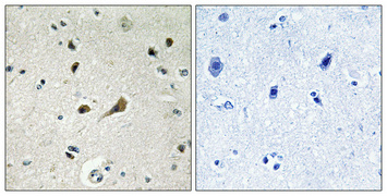
Real IHC data Immunohistochemistry validation of RPL39 using Anti-Ribosomal Protein L39 RPL39 Antibody (A10219-1). Immunohistochemical analysis of paraffin-embedded Human brain. Antibody was diluted at 1:100 (4°C
Anti-Ribosomal Protein L39 RPL39 Antibody
Cat # A10219-1
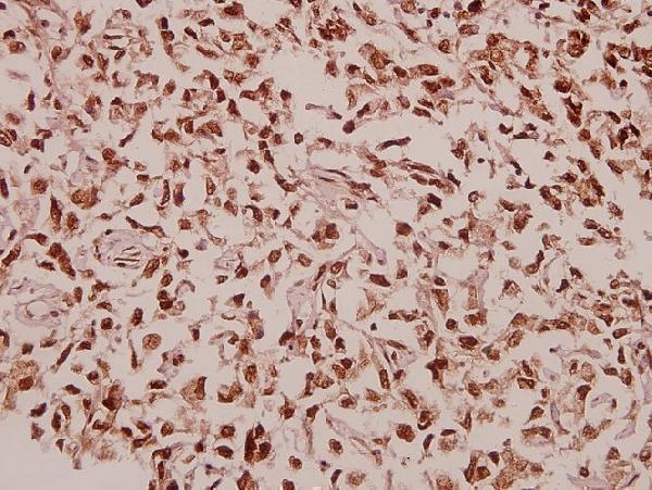
Real IHC data Immunohistochemistry (IHC) analyzes of RPL39 (S39) pAb in paraffin-embedded human colorectal carcinoma tissue at 1:50.
Anti-RPL39 (S39) Antibody
Cat # A10219S39

A10219-1 lists IHC and IF and shows IHC staining of paraffin-embedded human brain at 1:100 (catalog applications; A10219-1 image caption). A10219S39 lists IHC and shows staining of paraffin-embedded human colorectal carcinoma at 1:50 (catalog applications; A10219S39 image caption).

Which to pick: For tissue IHC, choose the SKU whose pictured sample is closest to yours: A10219-1 for paraffin-embedded human brain or A10219S39 for paraffin-embedded human colorectal carcinoma (respective IHC image captions). For IF/ICC, start with A10219-1 because it lists IF at 1:200–1:1000; no IF image or ICC validation is supplied (catalog applications/dilutions/images). Both list human, mouse and rat reactivity, but their IHC images show human tissue only; the fixative and antibody clonality are unreported (catalog reactivity, image captions and clone fields).

Each figure is that product's own IHC / IF validation image from its datasheet.

References

  1. UniProt Consortium. UniProt entry P62891 (RL39_HUMAN, Large ribosomal subunit protein eL39).
  2. Human Protein Atlas. RPL39 tissue IHC expression (reliability: Uncertain).
  3. Human Protein Atlas. RPL39 subcellular location (ICC-IF): Highest expression in HTCEpi: 7723.1 nTPM.
  4. Human Protein Atlas. RPL39 antibody validation summary (1 antibodies).
  5. Identification and validation of a 17-gene signature to improve the survival prediction of gliomas. Frontiers in immunology 2022 — PMC9556650.
  6. An integrated transcriptomic analysis unveils the regulatory roles of RNA binding proteins during human spermatogenesis. Frontiers in endocrinology 2025 — PMC11872710.
  7. Acylglycerol kinase promotes ovarian cancer progression and regulates mitochondria function by interacting with ribosomal protein L39. Journal of experimental & clinical cancer research : CR 2022 — PMC9358817.
  8. PubMed PMID:8908372 — UniProt-cited evidence.
  9. PubMed PMID:8764829 — UniProt-cited evidence.
  10. PubMed PMID:11875025 — UniProt-cited evidence.