This website uses cookies to ensure you get the best experience on our website.


- Table of Contents
Plan chromogenic TM4SF1 IHC in paraffin sections using lung alveolar cells as a staining reference (HPA tissue IHC). Start the catalog antibody at 5 μg/mL (datasheet: A10368), and assess membranous and cytoplasmic staining in light of the uncertain tissue evidence (HPA tissue IHC).

Expected localisation, validated protocols, controls and antibodies — the at-a-glance facts below, then the full design guide.
| Expected localisation | Membranous and cytoplasmic staining (HPA tissue IHC) | |
| Staining pattern | Alveolar and glandular cells show staining in a few tissues (HPA tissue IHC) | |
| Antigen retrieval | Citrate pH 6.0 HIER, 95–98 °C, 20 min (rule: cytoplasmic / membrane antigen) | |
| Positive control | Adrenal gland+4 more · see all | |
| Negative control | Adipose tissue+4 more · see all |
| Fixation | Keep fixation consistent across paraffin sections (standard IHC practice; not target-specific) | |
| Caveat | Staining has low concordance with RNA expression (HPA tissue IHC) | |
| Regulation | Expression regulation is unreported (UniProt) | |
| Isoform / epitope | No annotated isoforms; epitope side still matters (UniProt) |
The catalog antibody’s IHC-P protocol (datasheet) is accompanied by three published TM4SF1 IHC protocols (PMC6392956; PMC6010756; PMC12174346).
| Sample | Tissue sections; selected-image fixative not specified (standard IHC workflow) |
| Fixation | Image fixative and duration unreported (datasheet A10368); verify before use. |
| Sectioning | 4–5 µm sections on charged slides (standard) |
| Deparaffinisation | Xylene, graded ethanol series to water (standard) |
| Antigen retrieval | Heat-induced epitope retrieval in citrate buffer, pH 6.0, 20 min at 95–98 °C (standard rule: cytoplasmic / membrane antigen) |
| Peroxidase block | 3% H2O2, 10 min, room temperature (standard) |
| Blocking | 10% normal serum of the secondary host, 30 min, room temperature (standard) |
| Primary antibody | Rabbit anti-TM4SF1, 5 μg/mL (datasheet A10368) |
| Primary incubation | Overnight at 4 °C (standard) |
| Detection | HRP-polymer secondary, DAB chromogen 5–10 min (standard) |
| Counterstain | Hematoxylin, blue, dehydrate and mount (standard) |
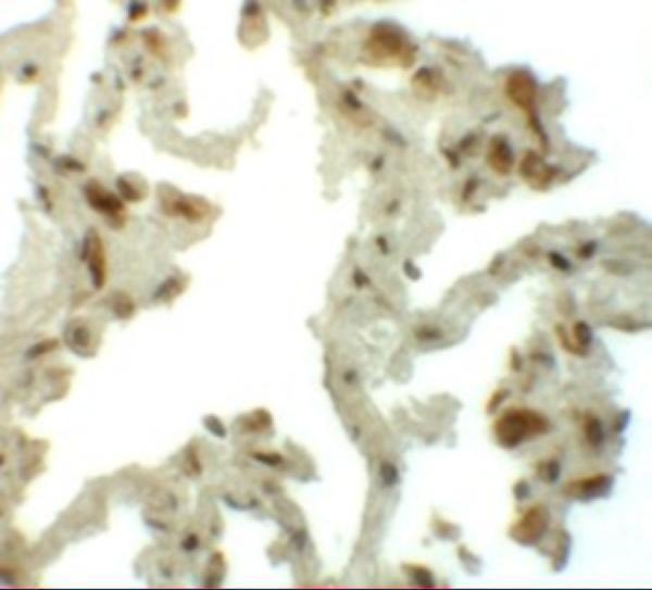
| Expected result | TM4SF1-positive staining in glandular cells of adrenal gland (HPA tissue IHC: Medium). HPA tissue profile: Cytoplasmic and membranous expression in a few tissues. No signal in the no-primary control. |
TM4SF1 is a membrane protein with 4 transmembrane segments (UniProt P30408 topology). In paraffin-section IHC, expect membranous staining, potentially with cytoplasmic staining, in selected glandular or alveolar cells (HPA tissue IHC). HPA reports medium staining in several normal tissues, while UniProt also reports expression in some normal endothelial cells (HPA tissue IHC; UniProt P30408 tissue specificity). Interpret apparent positives cautiously: HPA rates its tissue IHC profile Uncertain because antibody staining and RNA data have low consistency (HPA tissue IHC reliability).
| Membranous staining in breast glandular cells or lung alveolar cells, with possible cytoplasmic staining (HPA tissue IHC). | This fits the reported compartment and cell distribution: both examples have medium staining in HPA, and UniProt places TM4SF1 at the membrane (HPA tissue IHC; UniProt P30408 subcellular location). Record the stained cell type and compartment separately; HPA's Uncertain reliability limits how confidently this pattern alone identifies TM4SF1 (HPA tissue IHC reliability). |
| Predominantly nuclear staining, without a convincing membrane-associated pattern (UniProt P30408 subcellular location). | Nuclear localisation is unsupported by the supplied TM4SF1 record; UniProt assigns the protein to membranes, and HPA describes cytoplasmic and membranous tissue staining (UniProt P30408; HPA tissue IHC). Treat an isolated nuclear signal as suspect and review counterstain alignment and detection controls (general IHC practice). |
| Staining in a cell population reported as not detected, such as adipocytes or marrow hematopoietic cells (HPA tissue IHC). | Check cell identification, cross-reactivity and endogenous detection activity before calling the signal TM4SF1 (general IHC practice). HPA lists those populations as not detected, but its Uncertain IHC reliability means a disagreement needs independent assessment rather than automatic rejection (HPA tissue IHC). |
| Diffuse staining across tissue and background, obscuring cell boundaries (general IHC practice). | A widespread haze cannot establish the compartment-specific pattern expected from UniProt membrane localisation and HPA tissue staining (UniProt P30408; HPA tissue IHC). Compare a no-primary control, wash adequacy and detection background, then judge staining only where cellular outlines remain interpretable (general IHC practice). |
| No detectable staining in breast glandular cells or lung alveolar cells (HPA tissue IHC). | HPA reports medium staining in these populations, so review section quality, antigen retrieval, antibody concentration and detection performance (HPA tissue IHC; general IHC practice). A negative result does not by itself establish biological absence: the reference tissue profile and both listed IHC antibody assessments are Uncertain (HPA tissue IHC; HPA antibodies). |
| Compartment and topology (UniProt P30408 topology). | TM4SF1 has 4 membrane-spanning segments and extracellular as well as cytoplasmic regions (UniProt P30408 topology). Score membrane association first; HPA also reports cytoplasmic tissue staining, so cytoplasmic signal alone needs cautious interpretation (HPA tissue IHC). The supplied record does not locate either antibody's epitope (HPA antibodies). |
| Normal-tissue distribution (HPA tissue IHC). | HPA reports medium staining in adrenal, breast and fallopian-tube glandular cells, lung alveolar cells and pancreatic exocrine glandular cells (HPA tissue IHC). It reports selected other populations as low or not detected; compare like cell types rather than assigning one expected intensity to every tissue (HPA tissue IHC). |
| Antibody evidence (HPA antibodies). | HPA labels tissue IHC reliability Uncertain and lists IHC assessments as Uncertain for HPA002823 and CAB002760 (HPA tissue IHC; HPA antibodies). Reproducing a plausible pattern with one antibody does not resolve that uncertainty; use controls and an independent check when the interpretation is consequential (general IHC practice). |
| Glycosylation and processing (UniProt P30408). | UniProt annotates 2 glycosylation sites, at residues 129 and 159, and a single chain spanning residues 1–202; it annotates no signal peptide or propeptide (UniProt P30408). These features do not identify the antibodies' epitopes or establish a target-specific fixation or retrieval effect (UniProt P30408; HPA antibodies). |
| IF/ICC: what pattern is expected? (HPA subcellular ICC-IF). | For the separate IF/ICC guide, membrane localisation is the supported starting expectation (HPA subcellular ICC-IF; UniProt P30408). HPA supplies a membrane summary but no main-location assignment or cell-line ICC-IF images here; this record does not support an IF protocol choice or a validated cell-line example (HPA subcellular ICC-IF). |
| Situation | Likely cause | Next action |
|---|---|---|
| No signal in an HPA medium-staining reference population (HPA tissue IHC). | The assay may have failed, or this section may differ from HPA's uncertain tissue observation (general IHC practice; HPA tissue IHC reliability). | Verify tissue identity and morphology, then review retrieval, antibody concentration and detection controls using the assay's documented conditions (general IHC practice). Do not infer a TM4SF1-specific retrieval failure from these sources (HPA tissue IHC; UniProt P30408). |
| Only nuclear staining appears (UniProt P30408 subcellular location). | The compartment conflicts with UniProt membrane localisation and HPA's cytoplasmic/membranous description (UniProt P30408; HPA tissue IHC). | Check counterstain alignment and no-primary background; reassess with an independent antibody or method if the finding matters (general IHC practice). Avoid scoring an isolated nuclear signal as the expected pattern (UniProt P30408; HPA tissue IHC). |
| A broad brown haze hides cell outlines (general IHC practice). | Non-specific staining or detection background may be obscuring cellular localisation (general IHC practice). | Compare the no-primary control and review blocking, washing and detection settings; score only interpretable cells (general IHC practice). HPA's reported cytoplasmic staining does not make tissue-wide haze a positive result (HPA tissue IHC). |
| Adipocytes or marrow hematopoietic cells appear strongly positive (HPA tissue IHC). | Cell misidentification, cross-reactivity or endogenous detection activity is possible; HPA reports these cells as not detected (general IHC practice; HPA tissue IHC). | Confirm the cell type and inspect appropriate detection controls before attributing staining to TM4SF1 (general IHC practice). Document persistent disagreement because HPA rates its IHC profile Uncertain (HPA tissue IHC reliability). |
| Cytoplasmic signal dominates, with little membrane definition (HPA tissue IHC). | HPA allows cytoplasmic staining, but a poorly resolved signal may also prevent a confident compartment call (HPA tissue IHC; general IHC practice). | Review morphology, staining concentration and detection background; report membrane and cytoplasmic components separately (general IHC practice). Do not claim cytoplasmic staining alone proves specificity under HPA's Uncertain assessment (HPA tissue IHC reliability). |
| Endothelial staining appears beside epithelial staining (UniProt P30408 tissue specificity). | Some normal endothelial expression is reported by UniProt, so this distribution can have a biological explanation (UniProt P30408 tissue specificity). | Identify endothelial cells morphologically and score them separately from glandular or alveolar cells (general IHC practice; HPA tissue IHC). Check controls before treating every vascular signal as specific, given HPA's Uncertain tissue IHC reliability (HPA tissue IHC). |
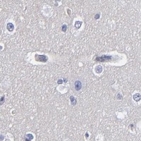


Comprehensive Human Protein Atlas IHC scoring per tissue (reliability: Uncertain — Low consistency between antibody staining and RNA expression data. Pending external verification.). Rows are taken directly from the HPA tissue chart — click any row's HPA link to view the source.
| Tissue | Cell type | Level | Evidence | Source |
|---|---|---|---|---|
| Adrenal gland | Glandular cells | Medium | Protein (IHC) | HPA → |
| Breast | Glandular cells | Medium | Protein (IHC) | HPA → |
| Fallopian tube | Glandular cells | Medium | Protein (IHC) | HPA → |
| Lung | Alveolar cells | Medium | Protein (IHC) | HPA → |
| Pancreas | Exocrine glandular cells | Medium | Protein (IHC) | HPA → |
| Tissue | Cell type | Level | Evidence | Source |
|---|---|---|---|---|
| Adipose tissue | Adipocytes | Not detected | Protein (IHC) | HPA → |
| Appendix | Glandular cells | Not detected | Protein (IHC) | HPA → |
| Bone marrow | Hematopoietic cells | Not detected | Protein (IHC) | HPA → |
| Caudate | Glial cells | Not detected | Protein (IHC) | HPA → |
| Cerebellum | Cells in granular layer | Not detected | Protein (IHC) | HPA → |
Troubleshoot TM4SF1 chromogenic IHC by checking retrieval, tissue context, membrane localisation and staining controls before scoring (UniProt P30408; HPA tissue IHC).
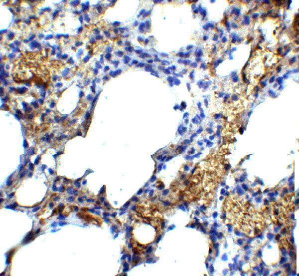
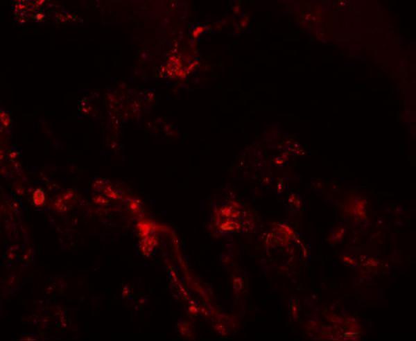
A10368 has IHC images from human and mouse lung and IF images from human and mouse lung (catalog image captions); rat reactivity is listed without an image (catalog reactivity).
A10368 will render with an IHC image from human lung at 5 μg/mL (catalog IHC image caption). Its additional images show IHC in mouse lung and IF in human and mouse lung (catalog image captions).
Which to pick: Choose A10368 for paraffin section IHC because IHC-P is listed and its own IHC images show staining in human and mouse lung (catalog applications; catalog IHC image captions); the fixative is unreported (catalog IHC image captions). For tissue IF, A10368 has human and mouse lung images at 20 μg/mL (catalog IF image captions); ICC validation is unreported (catalog applications). For work across species, A10368 lists human, mouse and rat reactivity, with IHC images for human and mouse only (catalog reactivity; catalog IHC image captions); clonality is unreported (catalog clone field).