This website uses cookies to ensure you get the best experience on our website.


- Table of Contents
Plan TRNT1 staining in paraffin sections using the catalog antibody’s 2–5 μg/ml IHC range (datasheet A06030-1). This guide covers the reported nuclear and cytoplasmic tissue pattern and its uncertain reliability when interpreting staining (HPA tissue IHC).

Expected localisation, validated protocols, controls and antibodies — the at-a-glance facts below, then the full design guide.
| Expected localisation | Nuclear/cytoplasmic staining, uncertain (HPA tissue IHC) | |
| Staining pattern | Nuclear and cytoplasmic staining across cell types (HPA tissue IHC) | |
| Antigen retrieval | EDTA pH 8.0 HIER, heat-mediated (datasheet A06030-1) | |
| Positive control | Adipose tissue+4 more · see all | |
| Negative control | Skeletal muscle |
| Fixation | Keep fixation conditions consistent across sections. (standard IHC practice; not target-specific) | |
| Caveat | Tissue staining has uncertain reliability (HPA tissue IHC) | |
| Regulation | Staining varies by cell type (HPA tissue IHC) | |
| Isoform / epitope | 3 isoforms; epitope coverage is unreported (UniProt) |
Compare the catalog antibody’s IHC-P protocol with a published TRNT1 tissue microarray protocol (PMC12464730).
| Sample | Paraffin-embedded human liver cancer tissue; fixative not specified (datasheet A06030-1) |
| Fixation | Image fixative and duration unreported (datasheet A06030-1); verify before use. |
| Sectioning | 4–5 µm sections on charged slides (standard) |
| Deparaffinisation | Xylene, graded ethanol series to water (standard) |
| Antigen retrieval | Heat retrieval: EDTA pH 8.0 (datasheet A06030-1); 20 min, 95–100 °C (standard) |
| Peroxidase block | 3% H2O2, 10 min, room temperature (standard) |
| Blocking | 10% goat serum (datasheet A06030-1) |
| Primary antibody | Rabbit anti-TRNT1, 2-5 μg/ml (datasheet A06030-1) |
| Primary incubation | Overnight at 4 °C (datasheet A06030-1) |
| Detection | HRP-conjugated secondary, DAB chromogen (datasheet A06030-1) |
| Counterstain | Hematoxylin, blue, dehydrate and mount (standard) |
| Expected result | TRNT1-positive staining in adipocytes of adipose tissue (HPA tissue IHC: Medium). HPA tissue profile: General nuclear and cytoplasmic expression. No signal in the no-primary control. |
TRNT1 is reported in mitochondria, cytoplasm and nucleus and has no transmembrane segment (UniProt Q96Q11). In paraffin sections, expect nuclear and cytoplasmic staining in several cell types, including adipocytes and glandular cells (HPA tissue IHC: general nuclear and cytoplasmic expression; Medium in these cells). Treat compartment calls cautiously: tissue IHC reliability is Uncertain, with subcellular location contradicted by external data (HPA tissue IHC).
| Moderate nuclear and cytoplasmic staining in adipocytes or glandular cells. | This matches the reported tissue pattern and Medium staining in these cells (HPA tissue IHC). Score the identified cells and compartments separately; the Uncertain reliability rating limits how confidently this establishes TRNT1 localisation (HPA tissue IHC). |
| Crisp staining confined to cell borders, with little intracellular signal. | An exclusively border-like pattern conflicts with the reported nuclear and cytoplasmic tissue pattern and with TRNT1 lacking a transmembrane segment (HPA tissue IHC; UniProt Q96Q11 topology). Check morphology and detection controls before treating it as specific. |
| Strong staining in skeletal muscle myocytes. | This differs from the HPA observation of Not detected in these cells (HPA tissue IHC). Consider cross-reactivity or endogenous detection activity, but do not call the tissue universally negative: TRNT1 RNA has low tissue specificity (HPA tissue IHC). |
| Uniform chromogen across cells, stroma and empty spaces. | That distribution is difficult to reconcile with cell-associated nuclear and cytoplasmic staining (HPA tissue IHC). Assess background with an appropriate negative detection control, then review blocking, washing and chromogen development as general IHC workflow checks. |
| No cellular signal in adipocytes or glandular cells expected to stain. | HPA reports Medium staining in adipocytes and glandular cells, so an entirely blank result warrants a technical check (HPA tissue IHC). Confirm tissue identity and run conditions with a positive control before interpreting the absence biologically. |
| Tissue and cell context | HPA reports Medium staining in adipocytes, glandular cells, respiratory epithelial cells, glial cells and endothelial cells, but Not detected in skeletal muscle myocytes (HPA tissue IHC). Use the identified cell population when judging a section. |
| Compartment evidence | Tissue IHC reports general nuclear and cytoplasmic expression with Uncertain reliability; ICC-IF supports mitochondria (HPA tissue IHC; HPA subcellular). Do not require a visibly mitochondrial pattern in chromogenic paraffin IHC. |
| Antibody validation | The listed antibody HPA036938 is rated Uncertain for IHC and Supported for ICC (HPA antibodies). A positive IHC pattern therefore needs appropriate tissue and detection controls before it is assigned to TRNT1. |
| Isoforms and processing | UniProt lists three isoforms and a chain spanning residues 42–434 (UniProt Q96Q11). The supplied evidence gives no antibody epitope, so it cannot predict which forms contribute to staining or justify an epitope-specific retrieval choice. |
| IF/ICC Q&A: Should IF show the same pattern? | HPA supports mitochondrial localisation in ICC-IF images from A-431 and U2OS, while tissue IHC reports nuclear and cytoplasmic expression with Uncertain reliability (HPA subcellular; HPA tissue IHC). Evaluate each application against its own evidence. |
| Fixation sensitivity | Target-specific fixation effects are unreported in the supplied UniProt and HPA records. If signal is weak, review fixation and antigen retrieval as general IHC workflow variables; do not infer a TRNT1-specific masking effect. |
| Situation | Likely cause | Next action |
|---|---|---|
| No staining in an expected positive cell population | The run may have failed, or the sampled population may differ from the HPA example (HPA tissue IHC: Medium in adipocytes and glandular cells). | Verify cell identity, positive-control performance and detection reagents; review the validated IHC procedure, including retrieval and antibody dilution, without assuming a TRNT1-specific fixation effect. |
| Strong myocyte staining | This disagrees with the HPA skeletal-muscle observation and may reflect nonspecific antibody binding or endogenous detection activity (HPA tissue IHC: myocytes Not detected). | Compare with a negative detection control and inspect the cell type and signal distribution; repeat with controlled detection conditions before reporting a new positive pattern. |
| Staining appears only along cell borders | Border-only signal does not fit the reported nuclear and cytoplasmic tissue pattern or the absence of a transmembrane segment (HPA tissue IHC; UniProt Q96Q11 topology). | Check section morphology, counterstain and negative controls; score intracellular signal separately from edge staining. |
| Diffuse colour obscures nuclei and cytoplasm | Background may arise from general staining or detection conditions; uniform colour is unlike the reported cell-associated pattern (HPA tissue IHC). | Use a negative detection control, then review blocking, washing and chromogen development; reassess whether individual cells and compartments remain distinguishable. |
| Nuclear IHC signal seems inconsistent with mitochondrial IF | The applications have different reported patterns: tissue IHC is nuclear and cytoplasmic with Uncertain reliability, while ICC-IF supports mitochondria (HPA tissue IHC; HPA subcellular). | Record the application and compartment for each observation; evaluate IHC with tissue controls and avoid treating the ICC-IF result as proof of an IHC artefact. |
| Signal varies among cells in the same section | HPA reports different staining levels across cell populations, including Low and Medium examples (HPA tissue IHC). Cell mixture can change the apparent intensity of a field. | Identify and score each cell population separately, compare like cells across sections, and document any result that departs from the HPA examples. |


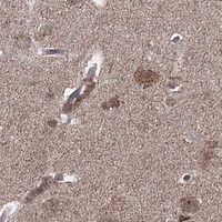
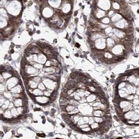
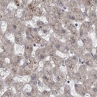
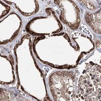


Comprehensive Human Protein Atlas IHC scoring per tissue (reliability: Uncertain — Medium consistency between antibody staining and RNA expression data. Subcellular location is contradicted by external data. Pending external verification.). Rows are taken directly from the HPA tissue chart — click any row's HPA link to view the source.
| Tissue | Cell type | Level | Evidence | Source |
|---|---|---|---|---|
| Adipose tissue | Adipocytes | Medium | Protein (IHC) | HPA → |
| Adrenal gland | Glandular cells | Medium | Protein (IHC) | HPA → |
| Appendix | Glandular cells | Medium | Protein (IHC) | HPA → |
| Breast | Adipocytes | Medium | Protein (IHC) | HPA → |
| Bronchus | Respiratory epithelial cells | Medium | Protein (IHC) | HPA → |
| Tissue | Cell type | Level | Evidence | Source |
|---|---|---|---|---|
| Skeletal muscle | Myocytes | Not detected | Protein (IHC) | HPA → |
Troubleshoot TRNT1 chromogenic IHC in paraffin sections using the selected antibody’s tissue protocol and compartment-specific interpretation.
The IHC-validated antibody has real staining data from human paraffin-embedded tissue sections (catalog IHC image captions). No IF/ICC data are supplied (catalog applications and image captions).
A06030-1 is a human-reactive rabbit polyclonal antibody listed for IHC (catalog: reactivity, host, clonality, applications). Its IHC captions show paraffin-embedded human liver cancer, lung cancer, rectum adenocarcinoma, and invasive bladder urothelial carcinoma sections (catalog IHC image captions).
Which to pick: Choose A06030-1 for human paraffin-section IHC; its figure shows staining in human liver cancer tissue, with the fixative unreported (A06030-1 IHC figure caption). No SKU in the payload is validated for IF/ICC or cross-species use (catalog: applications, reactivity, IF image captions).