This website uses cookies to ensure you get the best experience on our website.


- Table of Contents
Plan paraffin IHC for TXNDC5 using the cytoplasmic staining reported in exocrine pancreatic cells and leukocyte subsets (HPA tissue IHC). Start with the catalog antibody at 2–5 μg/ml (datasheet A05772-1) and include cardiomyocytes as a comparison where staining was not detected (HPA tissue IHC).

Expected localisation, validated protocols, controls and antibodies — the at-a-glance facts below, then the full design guide.
| Expected localisation | Cytoplasmic staining in tissue (HPA tissue IHC) | |
| Staining pattern | Cytoplasm of leukocyte subsets and exocrine pancreatic cells (HPA tissue IHC) | |
| Antigen retrieval | EDTA pH 8.0 HIER, heat-mediated (datasheet A05772-1) | |
| Positive control | Duodenum+4 more · see all | |
| Negative control | Caudate+4 more · see all |
| Fixation | Keep fixation consistent across sections (standard IHC practice; not target-specific) | |
| Caveat | Positive cell subsets may be unevenly distributed (HPA tissue IHC) | |
| Regulation | No expression regulation annotated (UniProt) | |
| Isoform / epitope | 2 isoforms; verify epitope coverage for both (UniProt) |
The catalog antibody’s IHC-P protocol uses EDTA pH 8.0 retrieval (datasheet: A05772-1). The published protocols below cover gastric mucosa, colon biopsies, and mouse ankle joints (PMC4315038; PMC11786489; PMC3541148).
| Sample | Paraffin-embedded human breast cancer tissue; fixative not specified (datasheet A05772-1) |
| Fixation | Image fixative and duration unreported (datasheet A05772-1); verify before use. |
| Sectioning | 4–5 µm sections on charged slides (standard) |
| Deparaffinisation | Xylene, graded ethanol series to water (standard) |
| Antigen retrieval | Heat retrieval: EDTA pH 8.0 (datasheet A05772-1); 20 min, 95–100 °C (standard) |
| Peroxidase block | 3% H2O2, 10 min, room temperature (standard) |
| Blocking | 10% goat serum (datasheet A05772-1) |
| Primary antibody | Rabbit anti-TXNDC5, 2-5 μg/ml (datasheet A05772-1) |
| Primary incubation | Overnight at 4 °C (datasheet A05772-1) |
| Detection | HRP-conjugated secondary, DAB chromogen (datasheet A05772-1) |
| Counterstain | Hematoxylin, blue, dehydrate and mount (standard) |
| Expected result | TXNDC5-positive staining in paneth cells of duodenum (HPA tissue IHC: High). HPA tissue profile: cytoplasmic expression in several tissue types, most abundant in a subset of leukocytes, exocrine pancreas and a subset of decidual cells. No signal in the no-primary control. |
TXNDC5 is an endoplasmic reticulum (ER) lumen protein with no transmembrane segment (UniProt Q8NBS9 topology). In paraffin IHC, expect cytoplasmic staining, especially in pancreatic exocrine glandular cells, intestinal Paneth and goblet cells, and tonsillar non-germinal center cells (HPA: High in each). HPA rates tissue staining Enhanced, while reporting medium consistency with RNA expression; interpret the pattern alongside cell identity (HPA: tissue IHC reliability).
| Granular or reticular cytoplasmic staining in pancreatic exocrine cells or intestinal Paneth cells. | This fits an ER-associated pattern in cells reported High by tissue IHC (UniProt Q8NBS9 localization; HPA: High in both). Compare the signal with neighboring cell types on the same section before calling the tissue uniformly positive. |
| Predominantly nuclear, sharply membranous, or extracellular staining. | Treat this as an unexpected compartment pattern: TXNDC5 is ER-luminal and has no transmembrane segment (UniProt Q8NBS9 topology). Review morphology and detection controls before assigning it to TXNDC5; HPA reports cytoplasmic tissue expression (HPA: tissue IHC profile). |
| Strong signal in cells reported as not detected, such as cardiomyocytes. | This conflicts with the listed cell-level reference (HPA: Not detected in cardiomyocytes). Check for cross-reactivity or endogenous chromogenic activity using appropriate controls (general IHC practice). A tissue name alone does not identify which cells produced the signal. |
| Broad, uniform color over cells and surrounding tissue, obscuring cell boundaries. | This is difficult to score as cellular TXNDC5 staining. Assess background with a primary-omission control and inspect blocking, washing, and detection steps (general IHC practice). The reference pattern is cytoplasmic and cell selective (HPA: tissue IHC profile). |
| No visible staining in pancreatic exocrine cells on an otherwise interpretable section. | This fails a listed high-expression reference (HPA: High in pancreatic exocrine glandular cells). Confirm that the expected cells are present, then review the IHC detection workflow and antibody validation before concluding that TXNDC5 is absent (general IHC practice). |
| Compartment and processing | TXNDC5 enters the secretory pathway through a signal peptide at residues 1–32; the annotated chain spans 33–432 (UniProt Q8NBS9 processing). Its ER-lumen location supports a cytoplasmic IHC pattern, but the payload gives no antibody epitope, so processing cannot predict antibody-specific staining. |
| Cell-level reference strength | Tissue IHC is rated Enhanced, with medium consistency between antibody staining and RNA expression (HPA: tissue IHC reliability). HPA lists High staining in selected cells, not every cell within those tissues (HPA: tissue IHC profile). Use cell identity when comparing sections. |
| Isoforms and target scope | Two isoforms are annotated (UniProt Q8NBS9 isoforms). Their epitopes and tissue-specific staining are not supplied, so the isoform count alone cannot explain a positive, weak, or absent IHC result. Keep an isoform-based explanation provisional until antibody-specific evidence is available. |
| IF/ICC Q&A: What pattern should I expect? | An ER-associated intracellular pattern is the supported expectation: HPA approves the ER as the main ICC-IF location, and UniProt places TXNDC5 in the ER lumen (HPA: subcellular; UniProt Q8NBS9 localization). This informs interpretation only; IF/ICC procedures belong in its separate guide. |
| Situation | Likely cause | Next action |
|---|---|---|
| Pancreatic exocrine cells show no signal. | The expected High cell population may be absent from the field, or the IHC workflow may have failed (HPA: High in pancreatic exocrine glandular cells; general IHC practice). | Verify cell identity and section quality; review the antibody's IHC validation, detection reagents, and control results before interpreting the negative (general IHC practice). |
| Color is mainly nuclear or outlines cell membranes. | The dominant compartment conflicts with an ER-luminal protein lacking a transmembrane segment (UniProt Q8NBS9 topology). | Recheck counterstain and cell boundaries, then compare with a primary-omission control and a listed High cell population (general IHC practice; HPA: High in pancreatic exocrine cells). |
| Cardiomyocytes stain strongly. | That cell-level result conflicts with the HPA reference and may reflect cross-reactivity or endogenous detection activity (HPA: Not detected in cardiomyocytes; general IHC practice). | Inspect a primary-omission control and the detection chemistry; confirm that the colored cells are cardiomyocytes before scoring TXNDC5 (general IHC practice). |
| Diffuse chromogen masks cellular detail. | Background may arise from endogenous detection activity or incomplete blocking or washing (general IHC practice); the diffuse appearance does not establish TXNDC5 localization. | Compare a primary-omission control, review blocking and washes, and score only clearly cellular staining against the cytoplasmic reference (general IHC practice; HPA: tissue IHC profile). |
| Only some cells within a positive tissue stain. | HPA reports expression in specific populations, including intestinal Paneth and goblet cells, rather than a tissue-wide uniform pattern (HPA: High in Paneth and goblet cells). | Identify the stained cell types and compare them with the appropriate HPA cell-level entry before calling the section inconsistent (general IHC practice; HPA: tissue IHC profile). |
| A weak section raises concern about fixation or retrieval. | The supplied sources give no TXNDC5-specific fixation sensitivity or retrieval condition; HPA staining levels and UniProt topology do not establish one (HPA: tissue IHC; UniProt Q8NBS9 topology). | Review the catalog antibody's IHC-P instructions and run suitable positive and detection controls when optimizing the general paraffin IHC workflow (general IHC practice). |


Comprehensive Human Protein Atlas IHC scoring per tissue (reliability: Enhanced — Medium consistency between antibody staining and RNA expression data.). Rows are taken directly from the HPA tissue chart — click any row's HPA link to view the source.
| Tissue | Cell type | Level | Evidence | Source |
|---|---|---|---|---|
| Duodenum | Paneth cells | High | Protein (IHC) | HPA → |
| Endometrium | Cells in endometrial stroma | Medium | Protein (IHC) | HPA → |
| Pancreas | Exocrine glandular cells | High | Protein (IHC) | HPA → |
| Small intestine | Goblet cells | High | Protein (IHC) | HPA → |
| Stomach | Glandular cells | Medium | Protein (IHC) | HPA → |
| Tissue | Cell type | Level | Evidence | Source |
|---|---|---|---|---|
| Caudate | Glial cells | Not detected | Protein (IHC) | HPA → |
| Heart muscle | Cardiomyocytes | Not detected | Protein (IHC) | HPA → |
| Hippocampus | Glial cells | Not detected | Protein (IHC) | HPA → |
| Liver | Cholangiocytes | Not detected | Protein (IHC) | HPA → |
| Oral mucosa | Squamous epithelial cells | Not detected | Protein (IHC) | HPA → |
Troubleshoot TXNDC5 staining in paraffin sections by checking retrieval, cellular distribution and controls before interpreting chromogenic signal.
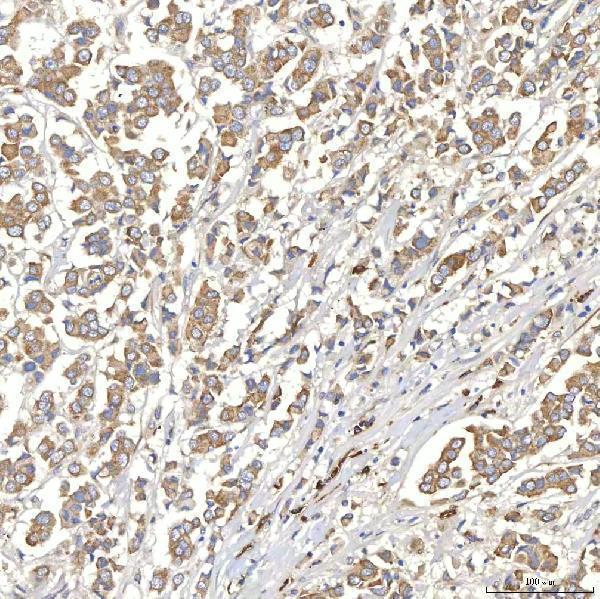
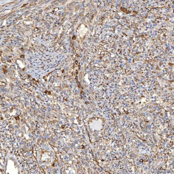
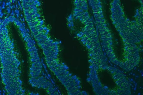
The IHC-validated A05772-1 has paraffin-section IHC images from human breast, larynx, prostate, and rectum cancers, plus a paraffin-section IF image from human colon cancer (catalog image captions).
A05772-1 is listed for IHC in human, mouse, and rat; its IHC images show paraffin-embedded human breast, larynx, prostate, and rectum cancer sections (catalog: applications, reactivity, IHC image captions). Its IF image shows a paraffin-embedded human colon cancer section (catalog: IF image caption).
Which to pick: For tissue IHC, choose A05772-1: its images document paraffin-section staining at 2 μg/ml, while the catalog recommends 2–5 μg/ml (IHC image captions; catalog: IHC dilution). For fluorescence work, the same SKU has paraffin-section IF data at 5 μg/ml; ICC is not listed, and clonality is unreported (IF image caption; catalog: applications, clone). For cross-species IHC, the catalog lists human, mouse, and rat reactivity, but the supplied IHC images show human tissue only; the fixative is unreported (catalog: reactivity; IHC image captions).