This website uses cookies to ensure you get the best experience on our website.


- Table of Contents
Plan paraffin-section UBASH3B IHC around the reported cytoplasmic staining, highest in lymphoid tissue (HPA tissue IHC). Use matched fixation and tissue controls to assess staining, bearing in mind the reported low consistency between staining and RNA expression (HPA tissue IHC).

Expected localisation, validated protocols, controls and antibodies — the at-a-glance facts below, then the full design guide.
| Expected localisation | Cytoplasmic tissue staining; nucleus also annotated (HPA tissue IHC; UniProt) | |
| Staining pattern | Cytoplasmic staining, highest in lymphoid tissue (HPA tissue IHC) | |
| Antigen retrieval | Citrate pH 6.0 HIER, heat-mediated (datasheet A09570-1) | |
| Positive control | Adrenal gland+4 more · see all | |
| Negative control | Adipose tissue+4 more · see all |
| Fixation | Keep fixation consistent across sections. (standard IHC practice; not target-specific) | |
| Caveat | Staining has low consistency with RNA expression (HPA tissue IHC) | |
| Regulation | Expression regulation not annotated (UniProt) | |
| Isoform / epitope | No annotated isoforms or processing; chain spans 1–649 (UniProt) |
Compare the catalog antibody’s IHC-P protocol (datasheet A09570-1) with a published prostate cancer tissue protocol (PMC6974685).
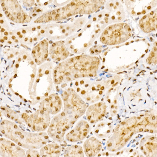
| Sample | Paraffin-embedded human lung cancer tissue; fixative not specified (datasheet A09570-1) |
| Fixation | Image fixative and duration unreported (datasheet A09570-1); verify before use. |
| Sectioning | 4–5 µm sections on charged slides (standard) |
| Deparaffinisation | Xylene, graded ethanol series to water (standard) |
| Antigen retrieval | Heat retrieval: Citrate pH 6.0 (datasheet A09570-1); 20 min, 95–100 °C (standard) |
| Peroxidase block | 3% H2O2, 10 min, room temperature (standard) |
| Blocking | 10% normal serum of the secondary host, 30 min, room temperature (standard) |
| Primary antibody | Rabbit anti-UBASH3B, 1:50-1:200 (datasheet A09570-1) |
| Primary incubation | Overnight at 4 °C (standard) |
| Detection | HRP-polymer secondary, DAB chromogen 5–10 min (standard) |
| Counterstain | Hematoxylin, blue, dehydrate and mount (standard) |
| Expected result | UBASH3B-positive staining in glandular cells of adrenal gland (HPA tissue IHC: High). HPA tissue profile: Cytoplasmic expression in several tissues with highest expression levels in lymphoid tissues. No signal in the no-primary control. |
UBASH3B is a cytoplasmic protein with a reported nuclear location and no transmembrane segment (UniProt Q8TF42). In tissue IHC, expect cytoplasmic staining in selected cells, including lymph node germinal center cells and lung macrophages (HPA: High in both). Treat the pattern as a guide rather than a definitive identity test: HPA rates its tissue IHC Approved but reports low consistency with RNA expression and pending external verification (HPA: tissue IHC reliability).
| Cytoplasmic chromogen in lymph node germinal center cells or appendix lymphoid tissue. | This matches reported high staining in those cell populations (HPA: High in lymph node germinal center cells and appendix lymphoid tissue). Judge the signal within the named cells; the HPA reliability caveat still applies (HPA: tissue IHC reliability). |
| Predominantly surface rim or extracellular staining, with little cytoplasmic signal. | Investigate the compartment assignment: UBASH3B has no transmembrane segment and is annotated in cytoplasm and nucleus (UniProt Q8TF42). This pattern alone cannot establish an artefact; compare cell morphology and controls (general IHC practice). |
| Strong staining in adipocytes or soft tissue fibroblasts while expected positive cells are also stained. | Those cell types are reported as not detected (HPA: adipocytes and soft tissue fibroblasts). Consider cross-reactivity or endogenous detection activity, then assess a reagent control and tissue morphology (general IHC practice). |
| Diffuse chromogen across cells, stroma and blank spaces, with unclear cellular boundaries. | This is difficult to score as cellular UBASH3B staining (general IHC practice). Check a no-primary control for detection background and review blocking, washes and chromogen development as general IHC steps (general IHC practice). |
| No signal in lymph node germinal center cells or lung macrophages. | These are reported high-staining populations (HPA: lymph node germinal center cells and lung macrophages). Confirm the expected cells are present, then review the antibody's IHC validation and routine assay controls before interpreting the sample as negative (general IHC practice). |
| How strong is the tissue-pattern evidence? | HPA labels the tissue IHC profile Approved, yet notes low consistency between staining and RNA expression and says external verification is pending (HPA: tissue IHC reliability). Use its high and not-detected calls as comparators, with that limitation stated. |
| Which cells provide useful comparisons? | Lymph node germinal center cells, lung macrophages and pancreatic exocrine glandular cells are reported High (HPA: tissue IHC). Adipocytes and smooth muscle cells are reported Not detected (HPA: tissue IHC). Compare named cells rather than an entire tissue. |
| What compartment should guide scoring? | HPA describes cytoplasmic expression across several tissues (HPA: tissue IHC profile). UniProt also lists the nucleus (UniProt Q8TF42). Score the cytoplasmic pattern first; interpret isolated nuclear IHC cautiously in light of the sample and controls (general IHC practice). |
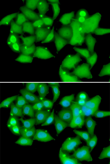
| Does IF/ICC add a localisation check? | Yes: HPA reports approved cytosolic localisation with additional nucleoplasm and nuclear body localisation in ICC-IF (HPA: subcellular). This supports possible nuclear signal, but an ICC-IF image does not establish the intensity or distribution expected in a paraffin tissue section (general IHC practice). |
| What does antibody validation establish? | Two listed antibodies have Approved IHC status, HPA038605 and HPA038607; HPA038606 has Approved ICC status (HPA: antibody validation). These are application-specific labels. The supplied record does not report an Enhanced IHC designation (HPA: antibody validation). |
| Situation | Likely cause | Next action |
|---|---|---|
| Known-positive lymphoid cells are unstained. | The expected population may be absent from the section, or the assay may have failed; HPA reports high staining in lymph node germinal center cells (HPA: tissue IHC; general IHC practice). | Check the section's cell morphology and an appropriate positive control, then review the IHC-validated antibody and detection run (general IHC practice). |
| All compartments show a faint, even brown haze. | Nonspecific reagent or detection background can obscure cell boundaries (general IHC practice). HPA's cell-specific levels do not explain uniform haze (HPA: tissue IHC). | Inspect a no-primary control; review blocking, washing and chromogen development using general IHC workflow checks (general IHC practice). |
| Macrophages stain, but nearby adipocytes also stain strongly. | HPA reports High in lung macrophages and Not detected in adipocytes, so the adipocyte signal conflicts with its reference pattern (HPA: tissue IHC). | Verify cell identification and examine a no-primary control; if adipocyte staining persists, investigate antibody specificity (general IHC practice). |
| Only nuclei stain in an otherwise suitable positive tissue. | A nuclear location is possible (UniProt Q8TF42; HPA: ICC-IF), but tissue IHC is described chiefly as cytoplasmic (HPA: tissue IHC profile). | Check nuclear boundaries and controls, then report the nuclear-only pattern separately rather than scoring it as the expected tissue IHC pattern (general IHC practice). |
| A surface rim is the dominant staining pattern. | The rim differs from the reported cytoplasmic tissue pattern and from the absence of a transmembrane segment (HPA: tissue IHC profile; UniProt Q8TF42 topology). | Review morphology and background controls; interpret the rim cautiously unless an independent specificity check supports it (general IHC practice). |
| Different tissues give inconsistent positives and negatives. | Expression varies by named cell population, and HPA reports low staining-to-RNA consistency with external verification pending (HPA: tissue IHC). | Record cell-level localisation and intensity alongside positive and negative comparators; avoid treating disagreement with one tissue as definitive assay failure (general IHC practice). |
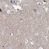
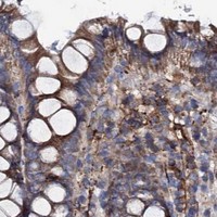
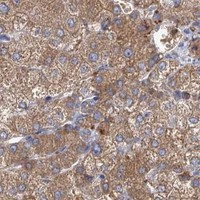
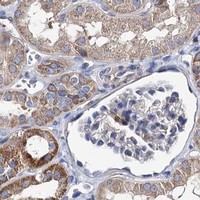


Comprehensive Human Protein Atlas IHC scoring per tissue (reliability: Approved — Low consistency between antibody staining and RNA expression data. Pending external verification.). Rows are taken directly from the HPA tissue chart — click any row's HPA link to view the source.
| Tissue | Cell type | Level | Evidence | Source |
|---|---|---|---|---|
| Adrenal gland | Glandular cells | High | Protein (IHC) | HPA → |
| Appendix | Lymphoid tissue | High | Protein (IHC) | HPA → |
| Cerebellum | Cells in granular layer | High | Protein (IHC) | HPA → |
| Epididymis | Glandular cells | High | Protein (IHC) | HPA → |
| Heart muscle | Cardiomyocytes | High | Protein (IHC) | HPA → |
| Tissue | Cell type | Level | Evidence | Source |
|---|---|---|---|---|
| Adipose tissue | Adipocytes | Not detected | Protein (IHC) | HPA → |
| Endometrium | Cells in endometrial stroma | Not detected | Protein (IHC) | HPA → |
| Ovary | Ovarian stroma cells | Not detected | Protein (IHC) | HPA → |
| Smooth muscle | Smooth muscle cells | Not detected | Protein (IHC) | HPA → |
| Soft tissue | Fibroblasts | Not detected | Protein (IHC) | HPA → |
Troubleshooting chromogenic IHC for UBASH3B in paraffin sections, with one IF/ICC question for comparing localization.
Anti-UBASH3B antibodies cover human, mouse and rat samples (catalog: reactivity). Paraffin-section IHC images are available for all three species; IF/ICC is listed without an image (A09570-1: image captions and applications).
A09570-1 has paraffin-section IHC images from human lung cancer, mouse kidney and rat lung, and is listed for IF/ICC (A09570-1: image captions and applications). A09570-3 is listed for IHC in human, mouse and rat, but has no IHC image in the payload (A09570-3: applications, reactivity and image captions).
Which to pick: For tissue IHC, choose A09570-1 when an illustrated paraffin-section example matters: its captions show staining in all three listed species (A09570-1: IHC image captions). For IF/ICC, choose A09570-1 because IF/ICC is listed for it, whereas A09570-3 lists IHC but no IF/ICC; both are rabbit polyclonals with human, mouse and rat reactivity (catalog: applications, dilution_raw and reactivity). The A09570-1 captions specify paraffin sections and citrate retrieval, but do not report the fixative (A09570-1: IHC image captions).