UBE2M / NEDD8-conjugating enzyme Ubc12 · IHC design guide

Design Immunohistochemistry for UBE2M

Plan paraffin-section IHC for UBE2M using the catalog antibody at 1–2 μg/ml (datasheet A07478-1). Compare cytoplasmic staining in glandular or neuronal cells with low or undetected cell populations, while accounting for the medium consistency between staining and RNA data (HPA tissue IHC).

Evidence assembled Oct 2026 · For research use; verify linked source records and product datasheet before use
Immunohistochemistry protocol sheet for UBE2M (IHC for UBE2M): expected localisation General cytoplasmic staining (HPA tissue IHC), antibody A07478-1, validated IHC image, and IHC protocol steps
Printable UBE2M IHC protocol sheet — expected localisation General cytoplasmic staining (HPA tissue IHC), antibody A07478-1, controls and protocol steps. Open the full UBE2M IHC guide →

UBE2M Immunohistochemistry Experimental Design Guide

Expected localisation, validated protocols, controls and antibodies — the at-a-glance facts below, then the full design guide.

Must know before staining
Expected localisation General cytoplasmic staining (HPA tissue IHC)
Staining pattern Cytoplasmic staining in glandular and neuronal cells (HPA tissue IHC)
Antigen retrieval EDTA pH 8.0 HIER, heat-mediated (datasheet A07478-1)
Positive control ⓘ Adrenal gland+4 more · see all
Negative control ⓘ Adipose tissue+2 more · see all
Important caveats
Reasons your staining may differ from the expected pattern.
Fixation Keep fixation consistent across sections. (standard IHC practice; not target-specific)
Caveat Staining and RNA show medium consistency (HPA tissue IHC)
Regulation Expression regulation not annotated (UniProt)
Isoform / epitope Two isoforms; epitope coverage is unspecified (UniProt)
Section 1

Recommended UBE2M IHC & IF Protocols

The catalog antibody’s IHC-P protocol uses heat-mediated EDTA pH 8.0 retrieval (datasheet A07478-1). The published UBE2M IHC protocols below provide tissue-specific alternatives (PMC13156996; PMC13123242; PMC8797438).

Recommended immunohistochemistry (IHC-P) protocol parameters
SampleParaffin-embedded human spleen tissue; fixative not specified (datasheet A07478-1)
FixationImage fixative and duration unreported (datasheet A07478-1); verify before use.
Sectioning4–5 µm sections on charged slides (standard)
DeparaffinisationXylene, graded ethanol series to water (standard)
Antigen retrievalHeat retrieval: EDTA pH 8.0 (datasheet A07478-1); 20 min, 95–100 °C (standard)
Peroxidase block3% H2O2, 10 min, room temperature (standard)
Blocking10% goat serum (datasheet A07478-1)
Primary antibodyRabbit anti-UBE2M, 1-2 μg/ml (datasheet A07478-1)
Primary incubationOvernight at 4 °C (datasheet A07478-1)
DetectionHRP-conjugated secondary, DAB chromogen (datasheet A07478-1)
CounterstainHematoxylin, blue, dehydrate and mount (standard)
Expected resultUBE2M-positive staining in glandular cells of adrenal gland (HPA tissue IHC: Medium). HPA tissue profile: General cytoplasmic expression. No signal in the no-primary control.
💡Decision noteStart with EDTA pH 8.0 for the catalog antibody (datasheet A07478-1); try citrate pH 6.0 when adapting the two published protocols that specify retrieval (PMC13156996; PMC13123242).
Section 2

What Is the Expected UBE2M Staining Pattern?

In paraffin-section IHC, expect generally cytoplasmic UBE2M staining in cells such as adrenal and appendix glandular cells, bronchial respiratory epithelial cells, and neurons in the caudate or cerebral cortex (HPA: tissue IHC). HPA rates its tissue staining Approved, with medium agreement with RNA data and external verification pending (HPA: tissue IHC). UBE2M has no transmembrane segment, but UniProt does not assign it a subcellular location (UniProt P61081: topology; subcellular annotation).

What am I looking at on my slide?
Cytoplasmic chromogen is visible in adrenal or appendix glandular cells, with moderate staining relative to nearby tissue.This matches the reported cell type, intensity, and general tissue pattern (HPA: Medium in adrenal and appendix glandular cells; general cytoplasmic expression). Judge the signal within identified cells and against controls; HPA's Approved rating has medium RNA agreement and awaits external verification (HPA: tissue IHC reliability).
A paraffin-section IHC slide shows predominantly nuclear staining, with little cytoplasmic signal in expected positive cells.That differs from HPA's general cytoplasmic tissue IHC pattern and warrants a localisation and detection check (HPA: tissue IHC). Nuclear signal alone is not proof of an artefact: HPA reports mainly nucleoplasmic UBE2M in ICC-IF, and UniProt gives no subcellular annotation (HPA: ICC-IF; UniProt P61081: subcellular annotation).
Adipocytes, skeletal myocytes, or vaginal squamous cells stain strongly while the expected positive cells do not.Those cell types were reported as Not detected in HPA tissue IHC, so the distribution raises concern for antibody cross-reactivity or endogenous chromogenic detection activity (HPA: tissue IHC; general IHC practice). Check cell identity and controls before assigning the signal to UBE2M; Not detected is an observed result, not a universal biological absence (HPA: tissue IHC).
Colour spreads across extracellular space, tissue edges, or many unrelated cells without clear cell boundaries.That appearance is less interpretable than the cell-associated cytoplasmic pattern reported in tissue IHC (HPA: general cytoplasmic expression; general IHC practice). Background can arise from nonspecific binding, endogenous detection activity, or chromogen deposition (general IHC practice). Its cause cannot be identified from distribution alone; compare detection and antibody controls.
There is no cellular signal in an adrenal or appendix section expected to contain glandular cells.HPA reports Medium staining in those glandular cells, so their absence of signal calls for a run check (HPA: adrenal and appendix tissue IHC). Confirm the relevant cells are present, then review antibody and detection steps with a concurrent positive control (general IHC practice). A failed stain alone does not establish lack of UBE2M.
💡Expected UBE2M appearanceCall the IHC result positive when identifiable adrenal or appendix glandular cells show predominantly cytoplasmic, approximately medium staining (HPA: tissue IHC); cell-free deposits or strong staining confined to HPA Not detected cell types should trigger a false-positive check (HPA: tissue IHC; general IHC practice).
How each factor affects the staining
Tissue and cell choiceAdrenal and appendix glandular cells and bronchial respiratory epithelial cells are Medium examples; adipocytes and skeletal myocytes are Not detected examples (HPA: tissue IHC). Use the stated cell types when comparing sections, since an organ-wide score can hide which cells carry the signal (general IHC practice).
IHC versus IF/ICC: should their compartments match?No direct match is established: tissue IHC is described as generally cytoplasmic, whereas ICC-IF is mainly nucleoplasmic, with nucleoli, nuclear bodies, and cytosol also reported (HPA: tissue IHC; HPA: ICC-IF). Interpret each assay against its own HPA observation; this section gives no IF/ICC protocol.
Antibody validationHPA054551, HPA057800, and CAB004993 each have Approved IHC status; the first two also have Approved ICC status (HPA: antibody list). HPA describes tissue IHC reliability as Approved with medium RNA agreement and external verification pending; no IHC Enhanced status is supplied (HPA: tissue IHC reliability; antibody list).
Isoforms and epitopeUniProt lists two UBE2M isoforms and a UBC core at residues 29–173 (UniProt P61081: isoforms; domain). The supplied record gives no antibody epitope or isoform reactivity, so a compartment difference or absent stain cannot be assigned to isoform recognition from these data.
Processing and retrieval evidenceUniProt reports no signal peptide, propeptide, or transmembrane segment (UniProt P61081: processing; topology). No target-specific retrieval or fixation sensitivity is supplied. Retrieval is a general paraffin-IHC workflow variable; changes in UBE2M signal after retrieval would need empirical controls (general IHC practice).
Why is my staining missing, weak or wrong?
SituationLikely causeNext action
Expected glandular cells are unstained.Possible assay failure or missing target cells; HPA reports Medium staining in adrenal and appendix glandular cells (HPA: tissue IHC).Verify cell identity and tissue preservation, then compare a concurrent expected-positive section and the run's detection control (general IHC practice). Record any retrieval or dilution adjustment as an empirical test, since UBE2M-specific effects are unreported.
Staining is mainly nuclear in paraffin IHC.The distribution differs from HPA's general cytoplasmic tissue IHC profile; ICC-IF separately reports a mainly nucleoplasmic location (HPA: tissue IHC; ICC-IF).Check morphology, antibody specificity controls, and whether the observation came from IHC or ICC-IF (general IHC practice). Report the compartment and assay explicitly; do not infer an IHC artefact solely from a nuclear signal.
Adipocytes or skeletal myocytes stain as strongly as expected positive cells.These cells are Not detected in the supplied HPA tissue IHC observations; cross-reactivity or endogenous detection activity is possible (HPA: tissue IHC; general IHC practice).Confirm cell identity, examine a no-primary control, and assess the detection system's endogenous activity controls (general IHC practice). Treat the unexpected staining as unresolved until its source is checked.
Background covers the section or obscures cell borders.Nonspecific reagent binding, endogenous detection activity, or excess chromogen can produce diffuse colour (general IHC practice).Compare no-primary and detection controls; review blocking, washes, antibody concentration, and chromogen development against the established run conditions (general IHC practice). Reassess whether cytoplasmic signal remains cell associated.
Staining appears only at section edges or as scattered deposits.Edge-associated or particulate chromogen can mimic positive cells without reproducing the expected cell-associated cytoplasmic pattern (HPA: tissue IHC; general IHC practice).Inspect unaffected tissue regions and the counterstain for intact cellular detail; compare a no-primary control and repeat the stain if deposits prevent scoring (general IHC practice).
A negative-looking cell type is used to reject an otherwise plausible positive result.HPA's Not detected and Low entries describe observations in specified cells, while its overall tissue profile is general cytoplasmic expression (HPA: tissue IHC).Score the named cells separately: compare Medium reference cells with Not detected or Low reference cells under the same run (HPA: tissue IHC; general IHC practice). Do not treat one unstained cell population as a failed assay.

Sample controls for UBE2M IHC & IF

🧪Run breast first: glandular cells should show medium staining (HPA: breast glandular cells, Medium). Use skeletal muscle myocytes as the negative tissue (HPA: skeletal muscle myocytes, Not detected); if adipocytes are present on the breast slide, they should lack specific staining and can provide an internal reference (HPA: adipose tissue adipocytes, Not detected).
Positive control tissue: Adrenal gland (Glandular cells, HPA Medium)
Negative control tissue: Adipose tissue (HPA Not detected)
ICC-IF cell lines (HPA subcellular resource): HPA ICC-IF images show UBE2M in HEK293, MCF-7, U2OS, NIH 3T3, with annotated localisation: Nucleoplasm (enhanced) (HPA subcellular).
Technical controls: Include a no-primary, secondary-only control and rabbit IgG isotype control matched to the primary antibody’s clonality and concentration (hero caption: rabbit primary antibody). Use UBE2M knockout tissue or a validated peptide block as a biological specificity control; quench endogenous peroxidase before DAB detection, especially in blood-rich areas (standard IHC practice).
⚠️Feasibility: Paraffin-section IHC is demonstrated with heat retrieval in EDTA at pH 8.0 (selected-SKU tissue-IHC caption: A07478-1); whether retrieval is essential has not been established. The caption does not report a fixative, and the supplied evidence reports no target-specific fixation window or fixation effect. Frozen-section performance and its relative ease are unreported; ICC-IF images exist for this target (HPA: UBE2M subcellular), but they do not establish an IF tissue protocol. Breast adipose areas may provide little target signal, so interpret weak staining by cell type (HPA: adipose tissue adipocytes, Not detected).

HPA tissue IHC evidence for UBE2M

Comprehensive Human Protein Atlas IHC scoring per tissue (reliability: Approved — Medium consistency between antibody staining and RNA expression data. Pending external verification.). Rows are taken directly from the HPA tissue chart — click any row's HPA link to view the source.

Positive expression · recommended positive controls

TissueCell typeLevelEvidenceSource
Adrenal gland Glandular cells Medium Protein (IHC) HPA →
Appendix Glandular cells Medium Protein (IHC) HPA →
Breast Glandular cells Medium Protein (IHC) HPA →
Bronchus Respiratory epithelial cells Medium Protein (IHC) HPA →
Caudate Neuronal cells Medium Protein (IHC) HPA →

Undetected expression · recommended negative controls

TissueCell typeLevelEvidenceSource
Adipose tissue Adipocytes Not detected Protein (IHC) HPA →
Skeletal muscle Myocytes Not detected Protein (IHC) HPA →
Vagina Squamous epithelial cells Not detected Protein (IHC) HPA →
Section 3

Advanced UBE2M IHC Tips

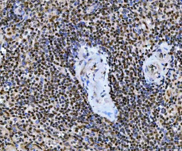
Use the catalog antibody’s paraffin-section spleen image as a workflow reference, and interpret staining alongside tissue morphology and controls (caption A07478-1; standard IHC practice).

Which retrieval conditions should I start with for UBE2M paraffin sections?
Start with heat-mediated retrieval in EDTA at pH 8.0 for this catalog antibody (datasheet A07478-1). Apply the same retrieval to reference and test sections in matched runs; the spleen image caption does not report fixation or heating duration (caption A07478-1). If staining is weak, vary heating duration and cooling systematically before trying another buffer, while checking tissue morphology after each change (standard IHC practice). Hold primary incubation at the demonstrated 2 μg/ml overnight at 4°C during that comparison, and include a no-primary slide to identify detection background (caption A07478-1; standard IHC practice).
How should I investigate weak UBE2M staining after fixation?
Target-specific fixation sensitivity is unknown: the catalog spleen caption identifies a paraffin section but does not name its fixative (caption A07478-1). Record the fixative and fixation interval for each specimen, then compare matched sections using EDTA at pH 8.0 and the demonstrated 2 μg/ml primary concentration (datasheet A07478-1; standard IHC practice). Examine morphology and staining together, with a no-primary control for background; changes in processing can affect both epitope access and tissue preservation (standard IHC practice). HPA tissue staining and UBE2M’s lack of a transmembrane segment do not establish which fixative best preserves this antigen (HPA: tissue IHC; UniProt P61081 topology).
How should I interpret cytoplasmic versus nuclear UBE2M staining?
Begin by scoring compartments separately: HPA describes general cytoplasmic tissue-IHC staining, while its ICC/IF data show enhanced nucleoplasmic staining and additional nucleolar, nuclear-body and cytosolic signal (HPA: tissue IHC; HPA: subcellular). UniProt supplies no subcellular annotation for UBE2M, so it cannot resolve that difference between methods (UniProt P61081 subcellular record). Compare candidate staining with a no-primary control, nuclear counterstain and preserved cell morphology before assigning a compartment (standard IHC practice). Record the proportion of stained nuclei and cytoplasm separately under identical DAB development conditions, rather than calling either pattern definitive from this evidence alone (standard IHC practice; HPA: tissue IHC and subcellular).
Could an isoform or modified epitope explain variable UBE2M staining?
UBE2M has 2 recorded isoforms and a UBC core spanning residues 29–173 (UniProt P61081 isoforms and domains). Recorded modifications include N-terminal acetylation, phosphorylation at residue 50 and alternative methylation annotations at residue 169 (UniProt P61081 modified residues). The supplied catalog caption gives no antibody epitope, so these features cannot establish which isoform or modification its tissue stain detects (caption A07478-1; UniProt P61081). When sections differ, first standardise EDTA pH 8.0 retrieval, primary incubation and DAB development, then interpret remaining differences using independent antigen-specific evidence if available (datasheet A07478-1; standard IHC practice).
How can IF help assess an ambiguous UBE2M IHC pattern?
Use IF/ICC as a separate localisation check: HPA reports mainly nucleoplasmic UBE2M, with additional nucleolar, nuclear-body and cytosolic signal, whereas its tissue-IHC profile is generally cytoplasmic (HPA: subcellular; HPA: tissue IHC). Multiplex UBE2M with a validated marker for the cell population being assessed, and include single-colour controls before comparing channels (standard IF practice). Select a fluorophore whose emission avoids the specimen’s strongest autofluorescence, checking an unstained section to identify that background (standard IF practice). Because UBE2M has no transmembrane segment and the reported IF compartments are intracellular, optimise permeabilisation for nuclear and cytosolic epitope access while monitoring morphology (UniProt P61081 topology; HPA: subcellular; standard IF practice).
What should I check when DAB background obscures UBE2M staining?
Run a no-primary control through the same secondary antibody and DAB steps to identify detection-system or tissue background (standard IHC practice). The catalog spleen workflow used 10% goat serum blocking, 2 μg/ml primary overnight at 4°C, and peroxidase-conjugated secondary for 30 minutes at 37°C (caption A07478-1). If background persists, check peroxidase blocking, washes and DAB development against the no-primary slide; these are general chromogenic-IHC controls (standard IHC practice). Compare stained cells with morphology and an unstained area before increasing primary concentration, since HPA describes generally cytoplasmic tissue staining with low tissue specificity (HPA: tissue IHC; standard IHC practice).
How should I score UBE2M across paraffin tissue sections? ⚠ ANSWER MARKED FOR VERIFICATION
Define the cell population and compartment before scoring, because HPA reports generally cytoplasmic tissue-IHC staining but a different dominant compartment in ICC/IF (HPA: tissue IHC; HPA: subcellular). For cell-level analysis, report percent positive cells and an H-score based on intensity categories; for spatial analysis, report positive-cell density per mm² of evaluable tissue (standard IHC practice). Normalise counts to the number of evaluable cells or measured tissue area, exclude damaged regions by a prespecified rule, and hold DAB development constant across sections (standard IHC practice). Report nuclear and cytoplasmic scores separately, alongside no-primary background and the antibody concentration used (standard IHC practice; caption A07478-1: 2 μg/ml).
How can I distinguish convincing UBE2M staining from artefact?
Treat staining as more convincing when it follows intact cell boundaries, recurs in comparable sections and exceeds the matched no-primary control (standard IHC practice). HPA describes general cytoplasmic tissue-IHC staining and low tissue specificity, while reporting low staining in spleen white-pulp cells; a strong claim from one cell type needs appropriate controls (HPA: tissue IHC). Investigate signal confined to section edges, necrotic areas or implausible compartments, and check whether endogenous peroxidase produces DAB colour on the no-primary slide (standard IHC practice). UBE2M participates in NEDD8 attachment, but its IHC signal alone does not measure neddylation activity; score staining and function as separate findings (UniProt P61081 function; standard IHC interpretation).
Boster reagents

Best UBE2M / NEDD8-conjugating enzyme Ubc12 IHC Antibodies

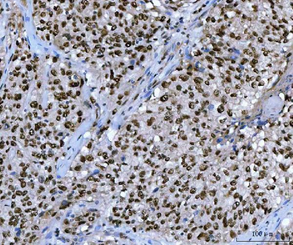
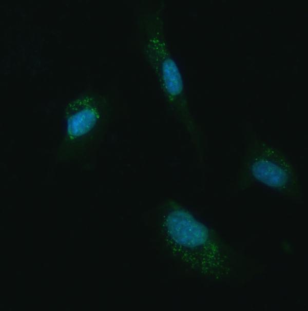
A07478-1 has IHC images from human paraffin sections and IF images from U87 cells and human paraffin sections (catalog image captions); listed reactivity also includes mouse and rat (catalog reactivity).

Real IHC data IHC analysis of UBC12/UBE2M using anti-UBC12/UBE2M antibody (A07478-1). UBC12/UBE2M was detected in a paraffin-embedded section of human spleen tissue. Heat mediated antigen retrieval was performed in EDTA buffer (pH 8.0, epitope retrieval solution). The tissue section was blocked with 10% goat serum. The tissue section was then incubated with 2 μg/ml rabbit anti-UBC12/UBE2M Antibody (A07478-1) overnight at 4°C. Peroxidase Conjugated Goat Anti-rabbit IgG was used as secondary antibody and incubated for 30 minutes at 37°C. The tissue section was developed using HRP Conjugated Rabbit IgG Super Vision Assay Kit (Catalog # SV0002) with DAB as the chromogen.
Anti-UBC12/UBE2M Antibody ®
Cat # A07478-1

A07478-1 is listed for IHC and has images from human spleen, lung squamous cell carcinoma, cervical cancer, and breast cancer paraffin sections (catalog applications; IHC image captions). A07478-1 is listed for IF/ICC and has IF images from U87 cells and human breast and ovarian cancer paraffin sections (catalog applications; IF image captions).

Which to pick: Choose A07478-1 for tissue IHC using paraffin sections, as shown in its own IHC captions; the fixative is unreported (catalog IHC image captions). For IF/ICC, A07478-1 has the listed applications and corresponding IF images (catalog applications; IF image captions). For work involving mouse or rat, A07478-1 lists reactivity with both species, although its supplied IHC and IF images use human material; clonality is unreported (catalog reactivity; image captions; clone field).

Each figure is that product's own IHC / IF validation image from its datasheet.

References

  1. UniProt Consortium. UniProt entry P61081 (UBC12_HUMAN, NEDD8-conjugating enzyme Ubc12).
  2. Human Protein Atlas. UBE2M tissue IHC expression (reliability: Approved).
  3. Human Protein Atlas. UBE2M subcellular location (ICC-IF): Mainly localized to the nucleoplasm. In addition localized to the nucleoli, nuclear bodies and cytosol..
  4. Human Protein Atlas. UBE2M antibody validation summary (3 antibodies).
  5. UBE2M-mediated EGFR neddylation drives keratinocyte proliferation in psoriasis. iScience 2026 — PMC13156996.
  6. UBE2M promotes malignant phenotypes of prostate cancer through mediating NAA10 neddylation. Journal of translational medicine 2026 — PMC13123242.
  7. UBE2M Identified by CRISPR Screening as a Key Regulator of Cisplatin-Induced Acute Kidney Injury via the p53 Pathway. Endocrine, metabolic & immune disorders drug targets 2026 — PMC13284637.
  8. Overexpression of UBE2M through Wnt/β-Catenin signaling is associated with poor prognosis and chemotherapy resistance in colorectal cancer. Translational cancer research 2020 — PMC8797438.
  9. PubMed PMID:9694792 — UniProt-cited evidence.
  10. PubMed PMID:10207026 — UniProt-cited evidence.
  11. PubMed PMID:15489334 — UniProt-cited evidence.