This website uses cookies to ensure you get the best experience on our website.


- Table of Contents
This guide covers cytoplasmic and nuclear UBE2N staining in paraffin sections, with the strongest reported signal in late spermatids (HPA tissue IHC). It includes an IHC starting concentration of 2–5 μg/mL for catalog antibody A02462-2 in human sections (datasheet A02462-2).

Expected localisation, validated protocols, controls and antibodies — the at-a-glance facts below, then the full design guide.
| Expected localisation | Cytoplasmic and nuclear tissue staining (HPA tissue IHC) | |
| Staining pattern | Cytoplasmic and nuclear staining; strongest in testis spermatids (HPA tissue IHC) | |
| Antigen retrieval | EDTA pH 8.0 HIER, heat-mediated (datasheet A02462-2) | |
| Positive control | Testis+4 more · see all | |
| Negative control | Adipose tissue+4 more · see all |
| Fixation | Keep fixation consistent across sections (standard IHC practice; not target-specific); Matched tissue-IHC evidence does not establish the fixation claim. Validate the specimen-specific method before use. (selected-SKU IHC image A02462-2) | |
| Caveat | Assay may detect proteins from more than one gene (HPA tissue IHC) | |
| Regulation | Expression regulation is unreported (UniProt) | |
| Isoform / epitope | No annotated isoforms or processing; one 1–152 chain (UniProt) |
The catalog antibody’s IHC-P protocol uses EDTA pH 8.0 heat retrieval (datasheet: A02462-2). Four published UBE2N IHC methods follow (PMC6639966; PMC9945078; PMC10039148; PMC11075218).
| Sample | Paraffin-embedded human cervical cancer tissue; fixative not specified (datasheet A02462-2) |
| Fixation | Image fixative and duration unreported (datasheet A02462-2); verify before use. |
| Sectioning | 4–5 µm sections on charged slides (standard) |
| Deparaffinisation | Xylene, graded ethanol series to water (standard) |
| Antigen retrieval | Heat retrieval: EDTA pH 8.0 (datasheet A02462-2); 20 min, 95–100 °C (standard) |
| Peroxidase block | 3% H2O2, 10 min, room temperature (standard) |
| Blocking | 10% goat serum (datasheet A02462-2) |
| Primary antibody | Rabbit anti-UBE2N, 2-5 μg/ml (datasheet A02462-2) |
| Primary incubation | Overnight at 4 °C (datasheet A02462-2) |
| Detection | HRP-conjugated secondary, DAB chromogen (datasheet A02462-2) |
| Counterstain | Hematoxylin, blue, dehydrate and mount (standard) |
| Expected result | UBE2N-positive staining in elongated or late spermatids of testis (HPA tissue IHC: High). HPA tissue profile: General cytoplasmic nuclear expression, most abundant in testis. No signal in the no-primary control. |
UBE2N is found in the nucleus and cytoplasm (UniProt P61088: subcellular location), and HPA describes general nuclear and cytoplasmic tissue staining, most abundant in testis (HPA: tissue IHC profile). In paraffin sections, elongated or late spermatids are the clearest expected positive cells, with High staining (HPA: High in testis spermatids). HPA rates the tissue pattern Supported, while cautioning that its antibody targets proteins from more than one gene (HPA: reliability description).
| Nuclear and cytoplasmic staining in elongated or late spermatids, stronger than nearby weakly stained cells (HPA: High in testis spermatids; tissue IHC profile). | This fits the reported positive cell population and both annotated compartments (HPA: testis IHC; UniProt P61088: subcellular location). Judge the signal by identifiable cells and compartments, since HPA's Supported pattern includes a multiple-gene antibody caution (HPA: reliability description). |
| A sharply membrane-restricted pattern dominates otherwise intact cells (UniProt P61088: no transmembrane segment). | Treat this as unexpected and investigate localisation or staining artefact: UBE2N has no annotated transmembrane segment and is reported in the nucleus and cytoplasm (UniProt P61088: topology and subcellular location). A membrane outline alone does not establish UBE2N-positive cells. |
| Strong signal appears in adipocytes or lung alveolar type I cells (HPA: Not detected in these cell types). | These are cell-specific discrepancies, not proof that the whole tissue is negative (HPA: tissue IHC). Check cross-reactivity and, with chromogenic detection, endogenous enzyme activity as possible causes (general IHC practice). HPA's antibody caution makes an independent specificity check useful (HPA: reliability description). |
| Diffuse chromogen covers several compartments and extracellular areas without clear cell boundaries. | This cannot be scored as the reported nuclear and cytoplasmic pattern (HPA: tissue IHC profile; UniProt P61088: subcellular location). Background from detection or nonspecific binding can obscure the expected cell contrast (general IHC practice); compare a matched negative control before interpreting intensity. |
| Elongated or late spermatids lack convincing staining in an otherwise interpretable testis section (HPA: High in testis spermatids). | The result conflicts with HPA's clearest positive reference (HPA: testis IHC). First confirm that the relevant spermatids are present, then assess the staining run with a known-positive section and controls (general IHC practice). An absent signal alone cannot establish absent UBE2N. |
| Cell and tissue context (HPA: tissue IHC). | HPA reports Medium staining in adrenal, appendix, breast and duodenal glandular cells, bronchial respiratory epithelium, cerebral cortex neuropil and colon endothelium (HPA: tissue IHC). Its Not detected entries name particular cell populations; they do not define an entire organ as a negative control (HPA: tissue IHC). |
| Antibody evidence (HPA: reliability description; antibody validation). | The tissue profile is Supported and antibody HPA044976 is IHC Supported, with a warning that the antibody targets proteins from more than one gene (HPA: tissue reliability; antibody validation). Interpret unexpected staining with that specificity limit in mind; these labels do not establish that every positive cell contains UBE2N alone. |
| Topology and processing (UniProt P61088: topology and processing). | UBE2N is a 152-residue chain with no annotated transmembrane segment, signal peptide or propeptide (UniProt P61088: topology and processing). Those annotations support checking intracellular localisation; they do not specify antigen retrieval conditions or predict a fixation effect. |
| IF/ICC Q: What localisation should a separate IF/ICC guide assess? | A: HPA reports mainly nucleoplasmic staining and additional nucleoli fibrillar-center staining in ICC-IF, both Supported, with a multiple-gene antibody caution (HPA: subcellular ICC-IF). That cell-imaging observation can inform localisation review; it is not a paraffin IHC intensity standard (HPA: ICC-IF; tissue IHC). |
| Situation | Likely cause | Next action |
|---|---|---|
| No signal in identifiable elongated or late spermatids (HPA: High in testis spermatids). | The expected positive population may be absent from the viewed area, or the staining run may have failed (HPA: testis IHC; general IHC practice). HPA supplies no target-specific fixation-sensitivity claim here. | Verify cell identity on the counterstain and examine a known-positive testis section in the same run (HPA: High in testis spermatids; general IHC practice). Review retrieval and detection using the validated IHC procedure, without inferring a UBE2N-specific fixation effect. |
| All compartments and empty spaces show diffuse chromogen. | Nonspecific binding, incomplete blocking, or detection background can hide cellular staining (general IHC practice). This appearance cannot be matched to HPA's cellular nuclear and cytoplasmic profile (HPA: tissue IHC profile). | Compare a matched negative control, inspect blocking and washing, and adjust the staining run according to the validated IHC procedure (general IHC practice). Score only resolvable cells and compartments against the HPA reference (HPA: tissue IHC). |
| Strong colour appears in adipocytes, skin fibroblasts, or lung alveolar type I cells (HPA: Not detected in those cell types). | The result could reflect cross-reactivity or endogenous chromogenic detection activity (general IHC practice). HPA also cautions that its antibody targets proteins from more than one gene (HPA: reliability description). | Confirm the stained cell identity, compare a no-primary control, and check the endogenous enzyme blocking step for the detection system (general IHC practice). Treat these HPA entries as cell-specific references, not whole-tissue exclusions (HPA: tissue IHC). |
| Staining forms a crisp cell-surface rim with little intracellular signal. | A membrane-dominant result conflicts with the annotated nuclear and cytoplasmic location and lack of a transmembrane segment (UniProt P61088: subcellular location and topology). It may reflect artefact or off-target staining. | Recheck morphology, compare the no-primary control, and review specificity with an independent control if available (general IHC practice). Do not count a membrane rim alone as the expected UBE2N pattern (UniProt P61088: subcellular location and topology). |
| Nuclear-only staining is seen where the paraffin-section pattern was expected to include cytoplasmic signal (HPA: tissue IHC profile). | HPA's ICC-IF summary is mainly nucleoplasmic, whereas its tissue IHC summary describes general nuclear and cytoplasmic expression (HPA: subcellular ICC-IF; tissue IHC). The methods provide different observations; this discrepancy alone does not identify a cause. | Record nuclear and cytoplasmic scores separately, check the relevant positive cell type, and compare like-for-like paraffin sections before calling the pattern discordant (HPA: tissue IHC; general IHC practice). Keep the ICC-IF localisation observation as context (HPA: subcellular ICC-IF). |
| Medium staining in a sampled tissue is mistaken for a failed positive result (HPA: Medium in several listed cell types). | The strongest supplied reference is testis elongated or late spermatids; HPA assigns Medium to several other specified populations (HPA: tissue IHC). Intensity expectations depend on the sampled cell population. | Identify and score the actual cell type, then compare it with its HPA entry: for example, bronchial respiratory epithelial cells or colon endothelial cells are Medium (HPA: tissue IHC). Use testis spermatids when a High reference is needed (HPA: testis IHC). |
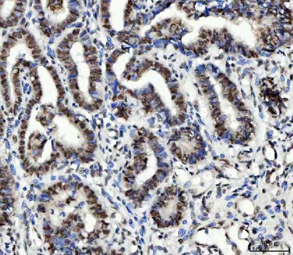
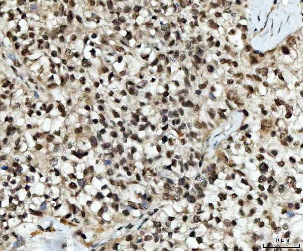
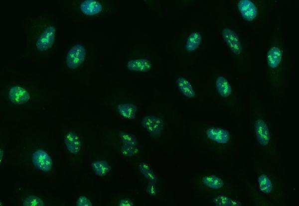
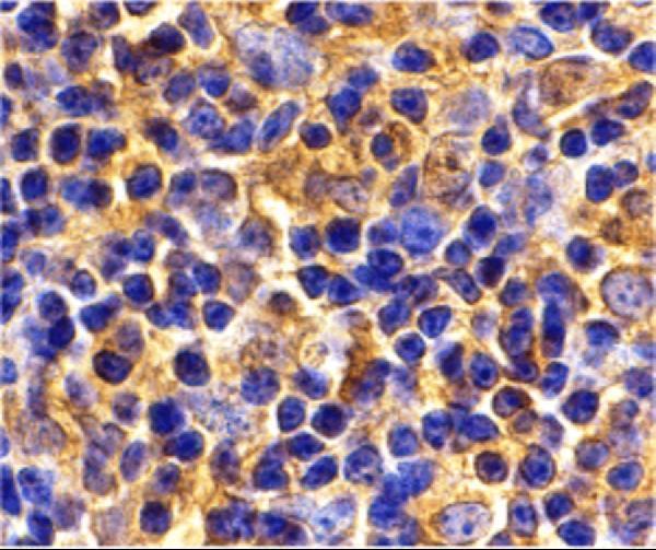
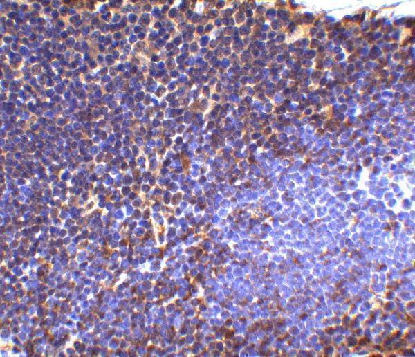
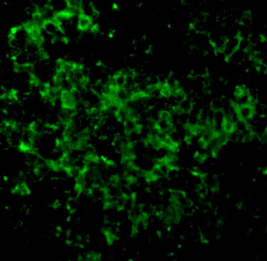
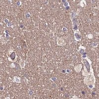


Comprehensive Human Protein Atlas IHC scoring per tissue (reliability: Supported — High consistency between antibody staining and RNA expression data. Caution, targets protein from more than one gene.). Rows are taken directly from the HPA tissue chart — click any row's HPA link to view the source.
| Tissue | Cell type | Level | Evidence | Source |
|---|---|---|---|---|
| Testis | Elongated or late spermatids | High | Protein (IHC) | HPA → |
| Adrenal gland | Glandular cells | Medium | Protein (IHC) | HPA → |
| Appendix | Glandular cells | Medium | Protein (IHC) | HPA → |
| Breast | Glandular cells | Medium | Protein (IHC) | HPA → |
| Bronchus | Respiratory epithelial cells | Medium | Protein (IHC) | HPA → |
| Tissue | Cell type | Level | Evidence | Source |
|---|---|---|---|---|
| Adipose tissue | Adipocytes | Not detected | Protein (IHC) | HPA → |
| Lung | Alveolar cells type I | Not detected | Protein (IHC) | HPA → |
| Ovary | Ovarian stroma cells | Not detected | Protein (IHC) | HPA → |
| Parathyroid gland | Glandular cells | Not detected | Protein (IHC) | HPA → |
| Skin | Fibroblasts | Not detected | Protein (IHC) | HPA → |
Troubleshoot UBE2N staining in paraffin section IHC by checking retrieval, compartment, controls and cell specific scoring before interpreting DAB signal.
Two catalog antibodies have IHC images from human paraffin sections and mouse thymus tissue, plus IF images from A549 and mouse thymus cells (catalog image captions).
A02462-2 will render with human cervical cancer paraffin-section IHC; its catalog also shows IF in A549 cells (A02462-2 image captions). A02462-1 will render with mouse thymus IHC; its catalog also shows IF in mouse thymus cells (A02462-1 image captions).
Which to pick: Choose A02462-2 for human paraffin-section IHC because its own caption documents EDTA retrieval at pH 8.0 and primary antibody at 2 μg/ml; the fixative is unreported (A02462-2 IHC caption). For IF/ICC, choose A02462-2 for A549 cells or A02462-1 for mouse thymus cells, according to their respective IF images (catalog IF captions). For work across species, both list human, mouse and rat reactivity, while the pictured IHC evidence is human tissue for A02462-2 and mouse thymus for A02462-1; the mouse tissue fixative is unreported (catalog reactivity; respective IHC captions).