This website uses cookies to ensure you get the best experience on our website.


- Table of Contents
Plan chromogenic UBR2 IHC on paraffin sections with the catalog antibody at 0.5–1 µg/mL (datasheet A05812-2). Compare its annotated nuclear location (UniProt) with the cytoplasmic and nuclear staining reported in tissues (HPA tissue IHC), and score each compartment separately.

Expected localisation, validated protocols, controls and antibodies — the at-a-glance facts below, then the full design guide.
| Expected localisation | Cytoplasmic and nuclear staining (HPA tissue IHC) | |
| Staining pattern | Broad cytoplasmic and nuclear staining across tissues (HPA tissue IHC) | |
| Antigen retrieval | EDTA pH 8.0 HIER, heat-mediated (datasheet A05812-2) | |
| Positive control | Adipose tissue+4 more · see all | |
| Negative control | None in HPA (detected in all 45 tissues); use no-primary + isotype controls |
| Fixation | Keep fixation consistent across sections. (standard IHC practice; not target-specific) | |
| Caveat | Cytoplasmic staining conflicts with nuclear annotation (HPA tissue IHC; UniProt) | |
| Regulation | DUSP22 promotes UBR2 degradation (UniProt) | |
| Isoform / epitope | 4 isoforms; verify epitope coverage (UniProt) |
Compare the catalog antibody’s IHC-P protocol (datasheet A05812-2) with the published mouse TNBC protocol (PMC12271019).
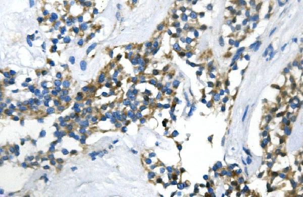
| Sample | Paraffin-embedded human pancreatic cancer tissue; fixative not specified (datasheet A05812-2) |
| Fixation | Image fixative and duration unreported (datasheet A05812-2); verify before use. |
| Sectioning | 4–5 µm sections on charged slides (standard) |
| Deparaffinisation | Xylene, graded ethanol series to water (standard) |
| Antigen retrieval | Heat retrieval: EDTA pH 8.0 (datasheet A05812-2); 20 min, 95–100 °C (standard) |
| Peroxidase block | 3% H2O2, 10 min, room temperature (standard) |
| Blocking | 10% goat serum (datasheet A05812-2) |
| Primary antibody | Rabbit anti-UBR2, 0.5-1μg/ml (datasheet A05812-2) |
| Primary incubation | Overnight at 4 °C (datasheet A05812-2) |
| Detection | Streptavidin-biotin complex (SABC), DAB chromogen (datasheet A05812-2) |
| Counterstain | Hematoxylin, blue, dehydrate and mount (standard) |
| Expected result | UBR2-positive staining in adipocytes of adipose tissue (HPA tissue IHC: High). HPA tissue profile: Ubiquitous cytoplasmic and nuclear expression. No signal in the no-primary control. |
UBR2 is annotated in the nucleus and on meiotic chromosomes; it has no transmembrane segment (UniProt Q8IWV8). Tissue IHC reports widespread cytoplasmic and nuclear staining, including high staining in adipocytes and thyroid glandular cells (HPA tissue IHC). HPA rates its tissue staining Approved, with medium agreement with RNA data and a subcellular pattern contradicted by external data (HPA tissue IHC).
| Cell-associated nuclear staining, with some cytoplasmic staining, across several tissue compartments. | This fits the broad HPA tissue IHC profile. Nuclear signal agrees with UniProt localization; cytoplasmic signal is reported by HPA but has a subcellular evidence caveat. Assess staining within recognizable cells and compare compartments on the same section (HPA tissue IHC; UniProt Q8IWV8). |
| Strong staining in adipocytes or thyroid glandular cells, alongside weaker staining elsewhere. | High staining in these cell types is reported by HPA. Contrast can help orient interpretation, but HPA also reports widespread expression; a weaker neighboring population is not automatically a negative control (HPA tissue IHC). |
| Color is concentrated in acellular spaces or surface deposits, without a convincing intracellular pattern. | That distribution does not match the cell-associated tissue profile or nuclear localization. Treat it as a possible staining artefact and inspect a matched negative control; membrane signal alone needs caution because HPA ICC-IF also reports an additional plasma-membrane location (HPA tissue IHC; HPA ICC-IF; UniProt Q8IWV8; general IHC practice). |
| A single unexpected cell population stains strongly while expected high-staining cells do not. | Consider antibody cross-reactivity or endogenous detection activity before assigning that population as UBR2-positive. HPA lists high staining in defined cell types, but its low-staining entries are not negatives; compare morphology and controls before interpreting the difference (HPA tissue IHC; general IHC practice). |
| Weak, diffuse color covers cells and surrounding tissue, or a known high-staining tissue has no signal. | Diffuse color limits compartment scoring and suggests background. Absent signal in an expected high-staining population calls for a run-level check of controls and detection steps; neither observation alone establishes UBR2 absence (HPA tissue IHC; general IHC practice). |
| Compartment evidence | UniProt places UBR2 in the nucleus and associates it with meiotic chromatin; HPA tissue IHC reports nuclear and cytoplasmic staining and flags external disagreement about subcellular location. Score compartments separately rather than treating all cytoplasmic color as confirmed localization (UniProt Q8IWV8; HPA tissue IHC). |
| Tissue comparison | HPA reports high staining in adipocytes, adrenal and thyroid glandular cells, and smooth muscle cells; pancreatic exocrine glandular cells are listed as low. These are observed staining levels, not guaranteed outcomes for every section or an absolute positive/negative tissue pair (HPA tissue IHC). |
| Antibody evidence | HPA027869 and HPA027880 each have Approved IHC status. HPA's overall tissue rating is Approved with medium RNA–staining consistency; these labels support cautious comparison with its reported pattern, not perfect specificity in a new preparation (HPA antibodies; HPA tissue IHC). |
| Protein forms and topology | UniProt lists 4 isoforms, no signal peptide or propeptide, and no transmembrane segment. The payload gives no antibody epitope or isoform coverage, so a compartment difference cannot be assigned to a particular isoform or shedding event (UniProt Q8IWV8). |
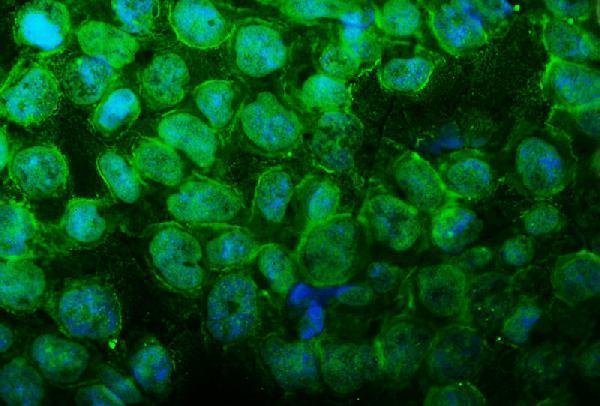
| What should IF/ICC show? | HPA ICC-IF reports mainly nucleoplasmic signal and an additional plasma-membrane location, both Approved, with images in A-431, U-251MG and U2OS cells. Use that as a localization cross-check; its cell-image pattern does not resolve the tissue IHC subcellular disagreement (HPA ICC-IF; HPA tissue IHC). |
| Situation | Likely cause | Next action |
|---|---|---|
| Expected high-staining cells are blank. | The section or detection run may have failed; absence in one slide cannot establish absent UBR2 (HPA tissue IHC; general IHC practice). | Check a same-run positive tissue and detection controls, then review primary-antibody incubation and chromogenic development. If the run still fails, optimize retrieval as a general IHC step; UBR2-specific fixation or retrieval sensitivity is unreported here (general IHC practice). |
| The entire section has diffuse brown color. | Non-specific background or endogenous detection activity may obscure cellular staining (general IHC practice). | Compare a no-primary control and inspect blocking, washes and chromogen exposure. Interpret UBR2 only where cell boundaries and compartments remain distinguishable (general IHC practice). |
| Strong color appears only in an unexpected cell population. | Cross-reactivity or endogenous activity is possible, especially if HPA high-staining cell types are blank; HPA low-staining cells are not defined negatives (HPA tissue IHC; general IHC practice). | Compare the no-primary control, cell morphology and a high-staining reference tissue in the same run. Record the unexpected distribution rather than calling it specific from intensity alone (HPA tissue IHC; general IHC practice). |
| Staining is mainly cytoplasmic, with little nuclear color. | HPA tissue IHC reports cytoplasmic and nuclear expression, while UniProt annotates nuclear localization; HPA flags external subcellular disagreement (HPA tissue IHC; UniProt Q8IWV8). | Score nuclear and cytoplasmic signals separately and compare a control section and the reported HPA pattern. Avoid assigning the cytoplasmic component to a UBR2 isoform without epitope evidence (HPA tissue IHC; UniProt Q8IWV8; general IHC practice). |
| Color pools in lumina or outside intact cells. | Acellular deposition does not match the reported cellular tissue pattern (HPA tissue IHC; general IHC practice). | Inspect the section and no-primary control for deposits or residual chromogen. Exclude acellular color from cell-level scoring (general IHC practice). |
| Pancreatic exocrine cells stain weakly while another tissue stains strongly. | HPA lists pancreatic exocrine glandular cells as low despite UniProt describing high overall pancreatic expression and protein in acinar cells; the sources measure or describe different aspects of expression (HPA tissue IHC; UniProt Q8IWV8). | Do not use pancreatic exocrine cells as a required high-intensity IHC control. Compare with an HPA high-staining cell type and document the tissue and cellular compartment scored (HPA tissue IHC; general IHC practice). |


Comprehensive Human Protein Atlas IHC scoring per tissue (reliability: Approved — Medium consistency between antibody staining and RNA expression data. Subcellular location is contradicted by external data.). Rows are taken directly from the HPA tissue chart — click any row's HPA link to view the source.
| Tissue | Cell type | Level | Evidence | Source |
|---|---|---|---|---|
| Adipose tissue | Adipocytes | High | Protein (IHC) | HPA → |
| Adrenal gland | Glandular cells | High | Protein (IHC) | HPA → |
| Smooth muscle | Smooth muscle cells | High | Protein (IHC) | HPA → |
| Thyroid gland | Glandular cells | High | Protein (IHC) | HPA → |
| Appendix | Glandular cells | Medium | Protein (IHC) | HPA → |
| Tissue | Cell type | Level | Evidence | Source |
|---|---|---|---|---|
| None in HPA: UBR2 is detected in all 45 scored tissues. Use a no-primary (secondary-only) and an isotype control instead. | ||||
Troubleshoot UBR2 staining in paraffin sections using the catalog antibody’s IHC evidence, with IF/ICC considerations kept to a single entry.
A05812-2 has paraffin-section IHC images from human pancreatic and renal cancer tissue and an IF image from HEPG2 cells (A05812-2 IHC/IF image captions). Human and mouse reactivity is listed (A05812-2 catalog reactivity).
A05812-2 will render with its human pancreatic cancer paraffin-section IHC figure; its caption documents EDTA retrieval and DAB detection (A05812-2 IHC image caption). The catalog also lists IF/ICC, and its IF caption documents staining in HEPG2 cells (A05812-2 application list; A05812-2 IF image caption).
Which to pick: Choose A05812-2 for chromogenic IHC on paraffin sections; its own figure documents that preparation, but does not report the fixative (A05812-2 IHC image caption). For IF/ICC, the same SKU has a cell-staining example (A05812-2 IF image caption). For human or mouse samples, A05812-2 lists both species as reactive, while the supplied images document human samples only (A05812-2 catalog reactivity; A05812-2 IHC/IF image captions).