UPK3A / Uroplakin-3a · IHC design guide

Design Immunohistochemistry for UPK3A

Use urinary bladder urothelium as a high-staining reference and adipose tissue as a not-detected comparator (HPA tissue IHC). This guide covers fixation consistency, selective cytoplasmic and membranous staining, and UPK1B-dependent ER exit (standard IHC practice; HPA tissue IHC; UniProt).

Evidence assembled Oct 2026 · For research use; verify linked source records and product datasheet before use
Immunohistochemistry protocol sheet for UPK3A (IHC for UPK3A): expected localisation Urothelial cytoplasm and membrane (HPA tissue IHC), antibody M06534-1, validated IHC image, and IHC protocol steps
Printable UPK3A IHC protocol sheet — expected localisation Urothelial cytoplasm and membrane (HPA tissue IHC), antibody M06534-1, controls and protocol steps. Open the full UPK3A IHC guide →

UPK3A Immunohistochemistry Experimental Design Guide

Expected localisation, validated protocols, controls and antibodies — the at-a-glance facts below, then the full design guide.

Must know before staining
Expected localisation Urothelial cytoplasm and membrane (HPA tissue IHC)
Staining pattern Selective cytoplasmic and membranous urothelial staining (HPA tissue IHC)
Antigen retrieval Citrate pH 6.0 HIER, 95–98 °C, 20 min (rule: cytoplasmic / membrane antigen)
Positive control ⓘ Urinary bladder
Negative control ⓘ Adipose tissue+4 more · see all
Important caveats
Reasons your staining may differ from the expected pattern.
Fixation Keep fixation consistent across paraffin sections (standard IHC practice; not target-specific)
Caveat UPK1B-dependent ER exit may affect membrane staining (UniProt)
Regulation Associated with terminal urothelial differentiation (UniProt)
Isoform / epitope 2 isoforms; lumenal vs cytoplasmic epitope may matter (UniProt)
Section 1

Recommended UPK3A IHC & IF Protocols

Compare the catalog antibody’s IHC-P protocol (datasheet) with the published kidney IHC protocol (PMC3762787).

Recommended immunohistochemistry (IHC-P) protocol parameters
SampleParaffin-embedded human bladder tissue; fixative not specified (datasheet M06534-1)
FixationImage fixative and duration unreported (datasheet M06534-1); verify before use.
Sectioning4–5 µm sections on charged slides (standard)
DeparaffinisationXylene, graded ethanol series to water (standard)
Antigen retrievalHeat-induced epitope retrieval in citrate buffer, pH 6.0, 20 min at 95–98 °C (standard rule: cytoplasmic / membrane antigen)
Peroxidase block3% H2O2, 10 min, room temperature (standard)
Blocking10% normal serum of the secondary host, 30 min, room temperature (standard)
Primary antibodyRabbit monoclonal (clone AFEH-21) anti-UPK3A, 1:1000-1:500 (datasheet M06534-1)
Primary incubationOvernight at 4 °C (standard)
DetectionHRP-polymer secondary, DAB chromogen 5–10 min (standard)
CounterstainHematoxylin, blue, dehydrate and mount (standard)
Expected resultUPK3A-positive staining in urothelial cells of urinary bladder (HPA tissue IHC: High). HPA tissue profile: Selective cytoplasmic and membranous expression in urothelium. No signal in the no-primary control.
💡Decision noteStart with citrate pH 6.0 retrieval at 95–98 °C for 20 min (page antigen retrieval); the paper does not specify retrieval conditions (PMC3762787).
Section 2

What Is the Expected UPK3A Staining Pattern?

In paraffin-section IHC, expect selective cytoplasmic and membranous UPK3A staining in urothelial cells, with high staining reported in urinary bladder (HPA tissue IHC). UPK3A has one transmembrane segment and contributes to the asymmetric unit membrane of differentiated urothelium (UniProt O75631). HPA rates the tissue IHC pattern Enhanced, while reporting medium consistency between antibody staining and RNA expression (HPA tissue IHC).

What am I looking at on my slide?
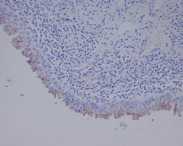
Urothelial cells show strong membrane-associated staining with some cytoplasmic staining.This matches the selective cytoplasmic and membranous pattern and high bladder urothelial signal (HPA tissue IHC). Record which cells and compartments stain when scoring a section (general IHC practice).
Nuclei dominate the chromogenic signal, with little urothelial membrane staining.This is discordant with the reported tissue IHC pattern (HPA tissue IHC). Review counterstain, detection background, and antibody specificity before calling it UPK3A; the uncertain nuclear locations reported by ICC-IF do not validate a nuclear IHC result (HPA subcellular ICC-IF).
Strong staining appears in cells outside the urothelium, including a cell type HPA reports as undetected.Treat the result as suspect, rather than assigning UPK3A expression from stain alone. Cross-reactivity or endogenous detection activity are possible explanations (general IHC practice); HPA reports no detection in adipocytes of adipose tissue and several other sampled cell types (HPA tissue IHC).
Colour spreads across the section without a clear urothelial cell or membrane pattern.Diffuse background does not reproduce the selective HPA pattern (HPA tissue IHC). Compare a reagent control and inspect blocking, washes, and detection chemistry to locate nonspecific signal (general IHC practice).
Bladder urothelium has no detectable signal.This conflicts with the high bladder urothelial staining reported by HPA (HPA tissue IHC). Check that urothelium is present and intact, then review the antibody and IHC detection workflow before interpreting another sample as negative (general IHC practice).
💡Expected UPK3A appearanceCall a result positive when bladder urothelial cells show high, selective cytoplasmic and membranous staining (HPA tissue IHC); nuclear-only or widespread unrelated-cell staining warrants investigation (HPA tissue IHC; general IHC practice).
How each factor affects the staining
Membrane architectureUPK3A has a lumenal region at residues 19–207, a transmembrane segment at 208–235, and a cytoplasmic region at 236–287 (UniProt O75631 topology). This supports compartment-aware scoring, but does not identify the catalog antibody's epitope.
Assembly and traffickingHeterodimer formation with UPK1B is required for UPK3A to leave the endoplasmic reticulum (UniProt O75631). Interpret cytoplasmic staining alongside the observed urothelial membrane pattern; this record does not establish how much IHC signal comes from either pool.
Molecular annotationsUniProt lists glycosylation sites at 74, 139, and 170 and two isoforms (UniProt O75631). Their effects on this antibody's staining, antigen retrieval, or paraffin-section sensitivity are unreported in the supplied sources.
Does the ICC-IF location define the IHC result?No. HPA describes a mainly nuclear-membrane location and additional nuclear bodies in ICC-IF, both marked uncertain (HPA subcellular ICC-IF). For this paraffin-section IHC guide, use the selective urothelial cytoplasmic and membranous tissue pattern as the reference (HPA tissue IHC).
Why is my staining missing, weak or wrong?
SituationLikely causeNext action
Expected bladder urothelium is unstained.The sampled section may lack intact urothelium, or an IHC workflow step may have failed (general IHC practice). HPA reports high staining in bladder urothelial cells (HPA tissue IHC).Confirm urothelial morphology and include a known-positive bladder section in the run; check antibody application and detection steps (general IHC practice). Do not infer UPK3A-specific retrieval or fixation sensitivity from these sources.
Nuclear staining dominates.The result differs from the tissue IHC pattern (HPA tissue IHC). HPA's nuclear-membrane and nuclear-body ICC-IF locations are uncertain and come from a different application (HPA subcellular ICC-IF).Review a reagent control, chromogen distribution, and cell boundaries before scoring; repeat with an independently validated IHC antibody if available (general IHC practice; HPA antibodies: HPA018407 and HPA018415 IHC Enhanced).
Staining is diffuse throughout the section.Background from incomplete blocking, insufficient washing, or the detection system can obscure cell-level localisation (general IHC practice). The expected tissue pattern is selective (HPA tissue IHC).Compare a control omitting the primary antibody, then review blocking, washes, and detection exposure (general IHC practice). Score only signal that resolves to the expected urothelial cells and compartments (HPA tissue IHC).
Unexpected cells stain strongly.Cross-reactivity or endogenous detection activity is possible (general IHC practice). For example, HPA reports adipocytes in adipose tissue as not detected (HPA tissue IHC).Identify the stained cell type, compare a detection control, and seek concordance with an independent IHC-validated antibody before assigning UPK3A positivity (general IHC practice; HPA antibodies: two IHC Enhanced entries).
A low-level signal is difficult to classify.HPA reports low staining in selected cell types, including Purkinje cells and rectal glandular cells, while its tissue IHC reliability note reports medium staining-to-RNA consistency (HPA tissue IHC).Record the cell type, compartment, and intensity; compare with the same run's urothelial positive control and background controls (general IHC practice). Avoid treating every faint deposit as either a confirmed positive or a technical failure.

Sample controls for UPK3A IHC & IF

🧪Run urinary bladder first; its urothelial cells should stain (HPA: High in urinary bladder urothelial cells). Use bronchus respiratory epithelium as a negative tissue (HPA: Not detected in bronchus respiratory epithelial cells); non-urothelial cells on the bladder slide should show only background staining and serve as an internal reference (HPA: High in bladder urothelial cells).
Positive control tissue: Urinary bladder (Urothelial cells, HPA High)
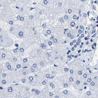
Negative control tissue: Adipose tissue (HPA Not detected)
ICC-IF cell lines (HPA subcellular resource): HPA ICC-IF images show UPK3A in Hep-G2, RT-4, U2OS, with annotated localisation: Nuclear membrane (uncertain) (HPA subcellular).
Technical controls: Include a no-primary, secondary-only control and an isotype-matched control for a monoclonal primary, or normal IgG from the primary host species for a polyclonal primary (standard IHC practice). Confirm target specificity with a matched UPK3A knockout specimen if available, or a peptide-block control when the antibody immunogen is available; quench endogenous peroxidase for chromogenic bladder IHC and assess tissue autofluorescence if using IF (standard IHC/IF practice).
⚠️Feasibility: A target-specific fixation window and fixation effect are unreported in the supplied evidence; the selected catalog antibody’s paraffin-embedded bladder caption does not state a fixative (selected SKU M06534-1 tissue-IHC caption). Antigen-retrieval dependence is also unreported, so evaluate retrieval conditions on paired paraffin sections; the evidence does not establish that frozen sections or IF are easier (selected SKU M06534-1 tissue-IHC caption). Interpret IF localization cautiously: the reported nuclear membrane and nuclear body locations are uncertain (HPA subcellular), while UPK3A is a component of the differentiated urothelial membrane and requires UPK1B for ER exit (UniProt O75631 function and subunit).

HPA tissue IHC evidence for UPK3A

Comprehensive Human Protein Atlas IHC scoring per tissue (reliability: Enhanced — Medium consistency between antibody staining and RNA expression data.). Rows are taken directly from the HPA tissue chart — click any row's HPA link to view the source.

Positive expression · recommended positive controls

TissueCell typeLevelEvidenceSource
Urinary bladder Urothelial cells High Protein (IHC) HPA →

Undetected expression · recommended negative controls

TissueCell typeLevelEvidenceSource
Adipose tissue Adipocytes Not detected Protein (IHC) HPA →
Adrenal gland Glandular cells Not detected Protein (IHC) HPA →
Appendix Glandular cells Not detected Protein (IHC) HPA →
Breast Adipocytes Not detected Protein (IHC) HPA →
Bronchus Respiratory epithelial cells Not detected Protein (IHC) HPA →
Section 3

Advanced UPK3A IHC Tips

Troubleshoot UPK3A chromogenic IHC by checking urothelial staining, membrane localization, processing conditions and matched controls (HPA tissue IHC; UniProt O75631).

Which retrieval conditions should I start with for UPK3A in paraffin sections?
Start with citrate buffer at pH 6.0, heated to 95–98 °C for 20 min (page retrieval setting). Allow sections to cool in the buffer, then compare staining with a matched section processed without primary antibody (standard IHC practice). Assess signal in urothelial cells, where high UPK3A staining is reported, rather than judging retrieval by whole-section color (HPA tissue IHC). If staining remains weak, test an EDTA-based alkaline retrieval condition on adjacent sections as a fallback, keeping antibody concentration and detection constant (standard IHC practice). Reject conditions that raise diffuse background or damage urothelial morphology (standard IHC practice).
How should I assess whether fixation is affecting UPK3A staining?
The selected antibody caption documents staining in paraffin-embedded human bladder but does not report its fixative or fixation duration (selected M06534-1 tissue-IHC caption). Target-specific sensitivity to fixation is therefore unknown; the tissue staining pattern and protein topology do not establish a preferred fixation condition (selected M06534-1 tissue-IHC caption; HPA tissue IHC; UniProt O75631 topology). Record each specimen’s fixative, fixation interval and processing history, then compare sections processed together with the same retrieval and detection settings (standard IHC practice). Include a known positive urothelial section in each run to distinguish a batch-wide staining failure from a specimen-specific loss (HPA tissue IHC; standard IHC practice).
Where should convincing UPK3A staining appear in a bladder IHC section?
Look for selective cytoplasmic and membranous staining in urothelial cells, with high staining reported in urinary bladder urothelium (HPA tissue IHC). UPK3A is a component of the asymmetric unit membrane of terminally differentiated urothelial cells, so assess membrane-associated signal in its epithelial context (UniProt O75631 function). Its transmembrane segment spans residues 208–235, with a lumenal region at 19–207 and a cytoplasmic region at 236–287 (UniProt O75631 topology). ER-associated signal can occur before UPK3A exits with UPK1B, but isolated nuclear staining warrants scrutiny against the urothelial pattern (UniProt O75631 subunit and localization; HPA tissue IHC).
How do epitope location and isoforms affect a negative UPK3A result?
First establish whether the antibody recognizes an epitope in the lumenal 19–207 region or the cytoplasmic 236–287 region; the supplied caption does not identify its epitope (UniProt O75631 topology; selected M06534-1 tissue-IHC caption). UPK3A has 2 annotated isoforms, so a negative result cannot establish that both are absent without isoform coverage information (UniProt O75631 isoforms). The lumenal region includes annotated glycosylation sites at residues 74, 139 and 170; their effect on this antibody’s staining is unreported (UniProt O75631 glycosylation; selected M06534-1 tissue-IHC caption). Compare retrieval conditions and a positive urothelial control before interpreting a negative specimen (standard IHC practice; HPA tissue IHC).
What should an IF follow-up resolve when IHC localization is ambiguous?
Use IF as a follow-up to the chromogenic IHC finding, and multiplex UPK3A with a marker that identifies urothelial cells in the specimen (HPA tissue IHC; standard IF practice). Choose fluorophores after inspecting unstained tissue autofluorescence, and include single-label controls to assess channel bleed-through (standard IF practice). If the antibody epitope is cytoplasmic, assess permeabilisation for access to residues 236–287; if lumenal, determine whether membrane access requires a different permeabilisation condition (UniProt O75631 topology; standard IF practice). HPA reports nuclear membrane and nuclear-body IF localization as uncertain, so reconcile any nuclear signal with the urothelial cytoplasmic and membranous IHC pattern (HPA subcellular; HPA tissue IHC).
How can I reduce background without losing the urothelial UPK3A signal?
Compare the stained section with a no-primary control and the expected urothelial pattern before changing antibody or retrieval conditions (HPA tissue IHC; standard IHC practice). Block endogenous peroxidase for chromogenic detection, use an appropriate protein block, and wash between incubations (standard IHC practice). If background remains diffuse, titrate the primary antibody and detection reagent separately while holding retrieval constant; no working dilution is supplied in the evidence here (standard IHC practice; selected M06534-1 tissue-IHC caption). Check whether color follows tissue folds, cut edges or damaged areas rather than urothelial cell boundaries, and exclude those regions from interpretation (standard IHC practice; HPA tissue IHC).
How should I score UPK3A across sections with different amounts of urothelium? ⚠ ANSWER MARKED FOR VERIFICATION
Define urothelial regions on the counterstained section and score UPK3A within that compartment, since high staining is reported in urinary bladder urothelial cells (HPA tissue IHC; standard IHC practice). Report the percentage of positive urothelial cells and, when intensity matters, an H-score from 0–300 using intensity grades 0–3 (standard IHC practice). Normalize each score to the number of evaluable urothelial cells or measured urothelial area, rather than total section area (standard IHC practice). Apply the same positivity threshold and imaging settings across specimens, excluding folds, necrosis and detached epithelium before comparing scores (standard IHC practice).
Which findings make an apparent UPK3A-positive IHC result suspect?
A convincing result places staining in urothelial cells with cytoplasmic or membranous distribution, consistent with the reported tissue-IHC pattern (HPA tissue IHC). Treat isolated nuclear staining cautiously: nuclear membrane and nuclear-body IF locations are marked uncertain, while UPK3A has a transmembrane segment and a role in the urothelial asymmetric unit membrane (HPA subcellular; UniProt O75631 topology and function). Review signal confined to section edges, folds or necrotic areas, and compare it with the no-primary and peroxidase-block controls (standard IHC practice). Apparent staining in unrelated cell populations requires review against morphology and a positive urothelial control before it is called UPK3A-specific (HPA tissue IHC; standard IHC practice).
Boster reagents

Best UPK3A / Uroplakin-3a IHC Antibodies

The IHC-validated anti-UPK3A antibody has an IHC image from paraffin-embedded human bladder (M06534-1 image caption). Human is the only listed species; no IF data are supplied (catalog: M06534-1).

Real IHC data Immunohistochemical analysis of paraffin-embedded human bladder, using Uroplakin III Antibody.
Anti-Uroplakin3a UPK3A Monoclonal Antibody
Cat # M06534-1

M06534-1 is the only listed SKU and is offered for human IHC and flow cytometry (catalog: M06534-1 applications/reactivity). Its IHC image shows paraffin-embedded human bladder (M06534-1 image caption).

Which to pick: For tissue IHC, choose M06534-1, a rabbit monoclonal (clone AFEH-21) with an image from paraffin-embedded human bladder; the caption does not report a fixative (catalog: M06534-1 host/clone; M06534-1 image caption). No listed SKU has IF/ICC validation (catalog: M06534-1 applications/IF images). No listed SKU has stated reactivity beyond human (catalog: M06534-1 reactivity).

Each figure is that product's own IHC / IF validation image from its datasheet.

References

  1. UniProt Consortium. UniProt entry O75631 (UPK3A_HUMAN, Uroplakin-3a).
  2. Human Protein Atlas. UPK3A tissue IHC expression (reliability: Enhanced).
  3. Human Protein Atlas. UPK3A subcellular location (ICC-IF): Mainly localized to the nuclear membrane. In addition localized to the nuclear bodies..
  4. Human Protein Atlas. UPK3A antibody validation summary (2 antibodies).
  5. Multimodal prognostic models for bladder urothelial carcinoma: uroplakin III combined with serum and demographic data. Frontiers in oncology 2025 — PMC12490981.
  6. Molecular basis of renal adaptation in a murine model of congenital obstructive nephropathy. PloS one 2013 — PMC3762787.
  7. Uroplakin peptide-specific autoimmunity initiates interstitial cystitis/painful bladder syndrome in mice. PloS one 2013 — PMC3745386.
  8. Mesenchymal stromal cell therapy is a long-term regenerative approach for chronic radiation cystitis, involving coordinated modulation of the urothelial, immune, vascular, and fibrotic pathways. Stem cell research & therapy 2026 — PMC13224412.
  9. PubMed PMID:9818021 — UniProt-cited evidence.
  10. PubMed PMID:15461802 — UniProt-cited evidence.
  11. PubMed PMID:10591208 — UniProt-cited evidence.