This website uses cookies to ensure you get the best experience on our website.


- Table of Contents
Plan chromogenic WTAP IHC in paraffin sections with the catalog antibody at 1:50–1:200 (datasheet). Use colon glandular cells with high staining as a positive reference and assess nuclear intensity across cell types (HPA tissue IHC).

Expected localisation, validated protocols, controls and antibodies — the at-a-glance facts below, then the full design guide.
| Expected localisation | Nuclear staining in tissue sections (HPA tissue IHC) | |
| Staining pattern | Widespread nuclear staining at variable intensity (HPA tissue IHC) | |
| Antigen retrieval | Tris-EDTA pH 9.0 HIER, 95–98 °C, 20 min (rule: nuclear antigen) | |
| Positive control | Adrenal gland+4 more · see all | |
| Negative control | Heart muscle |
| Fixation | Keep fixation consistent across paraffin sections. (standard IHC practice; not target-specific) | |
| Caveat | Cardiomyocytes may show no detectable staining (HPA tissue IHC) | |
| Regulation | Ubiquitous expression; variable tissue levels (UniProt; HPA tissue IHC) | |
| Isoform / epitope | 2 isoforms; epitope coverage is unclear (UniProt) |
Compare the catalog antibody's IHC-P protocol (datasheet) with published WTAP IHC protocols for ovarian tissue (PMC6689666) and glioblastoma tissue (PMC11006471).
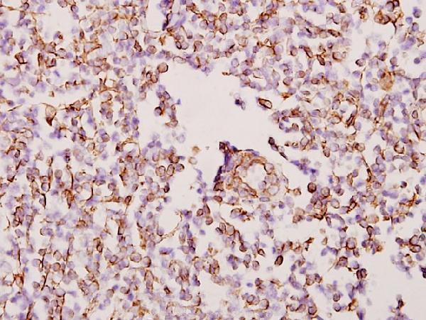
| Sample | Paraffin-embedded human tonsil cancer tissue; fixative not specified (datasheet A04296-1) |
| Fixation | Image fixative and duration unreported (datasheet A04296-1); verify before use. |
| Sectioning | 4–5 µm sections on charged slides (standard) |
| Deparaffinisation | Xylene, graded ethanol series to water (standard) |
| Antigen retrieval | Heat-induced epitope retrieval in Tris-EDTA buffer, pH 9.0, 20 min at 95–98 °C (standard rule: nuclear antigen) |
| Peroxidase block | 3% H2O2, 10 min, room temperature (standard) |
| Blocking | 10% normal serum of the secondary host, 30 min, room temperature (standard) |
| Primary antibody | Rabbit anti-WTAP, 1:50-1:200 (datasheet A04296-1) |
| Primary incubation | Overnight at 4 °C (standard) |
| Detection | HRP-polymer secondary, DAB chromogen 5–10 min (standard) |
| Counterstain | Hematoxylin, blue, dehydrate and mount (standard) |
| Expected result | WTAP-positive staining in glandular cells of adrenal gland (HPA tissue IHC: High). HPA tissue profile: Ubiquitous nuclear expression at variable levels. No signal in the no-primary control. |
WTAP should stain mainly nuclei, with possible weaker cytoplasmic signal (UniProt Q15007: subcellular location). Nuclear staining varies across tissues and cell types; HPA reports high staining in several glandular, hematopoietic, respiratory epithelial, and neuronal populations (HPA: tissue IHC). Treat this as a supported pattern with medium consistency between antibody staining and RNA expression (HPA: reliability). WTAP has no transmembrane segment (UniProt Q15007: topology).
| Nuclear staining predominates in glandular cells of colon, breast, or adrenal gland; intensity varies across cells. | This fits the expected compartment and examples of high staining (UniProt Q15007: mainly nuclear; HPA: High in these glandular cells). Score the nuclei of the named cell population, since HPA describes variable levels across tissues (HPA: ubiquitous nuclear expression). |
| Staining appears concentrated in subnuclear puncta, with little cytoplasmic signal. | Nuclear speckles are consistent with WTAP localisation (UniProt Q15007: nuclear speckle; HPA: enhanced nuclear-speckle location by ICC-IF). Speckles may be easier to distinguish by fluorescence microscopy; a chromogenic IHC result can still be consistent when its dominant signal is nuclear (general IHC interpretation). |
| Strong membrane staining or cytoplasmic staining dominates while nuclei are faint. | This conflicts with the mainly nuclear distribution and lack of a transmembrane segment (UniProt Q15007: subcellular location and topology). Some cytoplasmic WTAP is possible (UniProt Q15007: subcellular location), so assess whether nuclear staining remains predominant before treating the slide as an artefact. |
| Cardiomyocytes stain strongly, or chromogen appears in cells inconsistent with the expected tissue pattern. | HPA reports WTAP as not detected in heart-muscle cardiomyocytes (HPA: tissue IHC). Unexpected cell staining warrants checks for antibody cross-reactivity or endogenous detection activity (general IHC practice); this single HPA observation is a comparison point, not an absolute biological absence claim (HPA: Supported, medium consistency). |
| The entire section has diffuse colour, or a known high-staining population has no nuclear signal. | Diffuse colour limits compartment scoring and may reflect background from antibody, detection reagents, or endogenous activity (general IHC practice). No nuclear signal in a high-staining reference population, such as bone-marrow hematopoietic cells, makes the run inconclusive (HPA: High in hematopoietic cells; general IHC practice). |
| Which compartment should guide scoring? | Use nuclear staining as the primary readout; WTAP also has a reported cytoplasmic fraction (UniProt Q15007: subcellular location). A chromogenic slide need not resolve nuclear speckles to support a nuclear call (general IHC interpretation). |
| How much tissue-to-tissue variation is expected? | Expression is described as ubiquitous, yet HPA reports variable nuclear levels: high in several listed populations, low in salivary-gland glandular cells, liver cholangiocytes, skeletal-muscle myocytes, and splenic white-pulp cells, and not detected in cardiomyocytes (UniProt Q15007: tissue specificity; HPA: tissue IHC). |
| How certain is the antibody-based tissue pattern? | The listed HPA010550 antibody has Supported IHC status, and the tissue profile has medium consistency with RNA expression (HPA: antibody validation and tissue reliability). Use the reported tissue levels as reference patterns while reviewing controls and cellular morphology (general IHC practice). |
| What should IF/ICC show? | Expect WTAP enrichment in nuclear speckles; HPA lists this as an enhanced main location and reports ICC-IF images in A-431, U-251MG, and U2OS cells (HPA: subcellular and antibody validation). This is an IF/ICC localisation reference, not an IHC-P protocol or an IHC tissue-intensity standard. |
| Could isoforms change interpretation? | UniProt lists two WTAP isoforms, but the supplied record gives no antibody epitope or isoform-specific staining evidence (UniProt Q15007: isoforms; HPA: supplied antibody record). Do not assign a different cellular pattern to either isoform from these data. |
| Situation | Likely cause | Next action |
|---|---|---|
| High-staining reference tissue shows no nuclear chromogen. | The run may have a detection or staining-step failure (general IHC practice); HPA reports high WTAP in colon glandular cells (HPA: tissue IHC). | Check the positive control, primary-antibody application, retrieval step, detection reagents, and counterstain; document each condition before changing it (general IHC practice). |
| Colour is diffuse across tissue and obscures nuclei. | Nonspecific antibody binding, endogenous detection activity, or excess chromogen development can raise background (general IHC practice). | Compare a no-primary control; review blocking, primary-antibody concentration, washes, and development time against the validated workflow (general IHC practice). |
| Membranes appear more strongly stained than nuclei. | The distribution conflicts with mainly nuclear WTAP and its absence of a transmembrane segment (UniProt Q15007: location and topology). | Recheck cell boundaries and counterstained nuclei; compare controls and review primary-antibody specificity and detection background before scoring (general IHC practice). |
| Cardiomyocytes appear clearly positive. | This differs from the reported not-detected cardiomyocyte pattern (HPA: tissue IHC); cross-reactivity or endogenous detection activity is possible (general IHC practice). | Inspect a no-primary control and a known high-staining tissue on the same run; describe the discrepancy without treating the HPA result as an absolute absence claim (general IHC practice; HPA: Supported, medium consistency). |
| A low-staining population appears negative. | HPA reports low staining in salivary-gland glandular cells and liver cholangiocytes, so weak signal may be hard to distinguish from background (HPA: tissue IHC; general IHC interpretation). | Use an HPA high-staining cell population as a run control and score only signal distinguishable from the no-primary control (HPA: tissue IHC; general IHC practice). |
| Puncta are apparent in IF/ICC but unclear in chromogenic IHC. | HPA's enhanced nuclear-speckle observation comes from ICC-IF, whereas the IHC tissue profile describes nuclear expression (HPA: subcellular and tissue IHC). | For IHC, assess whether staining is predominantly nuclear in the expected cells; reserve a speckle-level localisation judgment for the separate IF/ICC guide (HPA: tissue IHC and subcellular location; general IHC interpretation). |
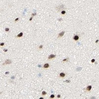


Comprehensive Human Protein Atlas IHC scoring per tissue (reliability: Supported — Medium consistency between antibody staining and RNA expression data.). Rows are taken directly from the HPA tissue chart — click any row's HPA link to view the source.
| Tissue | Cell type | Level | Evidence | Source |
|---|---|---|---|---|
| Adrenal gland | Glandular cells | High | Protein (IHC) | HPA → |
| Appendix | Glandular cells | High | Protein (IHC) | HPA → |
| Bone marrow | Hematopoietic cells | High | Protein (IHC) | HPA → |
| Breast | Glandular cells | High | Protein (IHC) | HPA → |
| Bronchus | Respiratory epithelial cells | High | Protein (IHC) | HPA → |
| Tissue | Cell type | Level | Evidence | Source |
|---|---|---|---|---|
| Heart muscle | Cardiomyocytes | Not detected | Protein (IHC) | HPA → |
Troubleshoot WTAP staining in paraffin sections by checking retrieval, nuclear localisation, controls, and scoring before interpreting biological differences.
A04296-1 has real IHC data from paraffin-embedded human tonsil cancer tissue (IHC image caption). Catalog reactivity lists Human, Mouse, and Rat (catalog: reactivity).
A04296-1 is listed for IHC in Human, Mouse, and Rat (catalog: applications and reactivity). Its IHC image shows paraffin-embedded human tonsil cancer tissue at 1:50 (IHC image caption).
Which to pick: Choose A04296-1 for paraffin-section IHC; its image documents human tonsil cancer tissue, but does not report the fixative (IHC image caption). No IF/ICC application or image is listed for A04296-1, so the payload does not support an IF/ICC recommendation (catalog: applications and IF images). A04296-1 lists Mouse and Rat reactivity alongside Human, but its supplied tissue image documents Human only (catalog: reactivity; IHC image caption).