This website uses cookies to ensure you get the best experience on our website.


- Table of Contents
Plan chromogenic YME1L1 staining in paraffin sections using the cytoplasmic tissue profile (HPA tissue IHC). The catalog antibody has IHC images from mouse and rat heart; compare staining by cell type when assessing results (datasheet A03012-1; HPA tissue IHC).

Expected localisation, validated protocols, controls and antibodies — the at-a-glance facts below, then the full design guide.
| Expected localisation | Cytoplasmic in tissue (HPA tissue IHC); inner mitochondrial membrane (UniProt) | |
| Staining pattern | Cytoplasmic staining in most tissues (HPA tissue IHC) | |
| Antigen retrieval | EDTA pH 8.0 HIER, heat-mediated (datasheet A03012-1) | |
| Positive control | Adrenal gland+4 more · see all | |
| Negative control | Adipose tissue+4 more · see all |
| Fixation | Matched tissue-IHC evidence does not establish the fixation claim. Validate the specimen-specific method before use. (selected-SKU IHC image A03012-1) | |
| Caveat | Staining and RNA have only medium consistency (HPA tissue IHC) | |
| Regulation | Expression regulation unreported (UniProt) | |
| Isoform / epitope | 3 isoforms; epitope differences unreported (UniProt) |
The catalog antibody’s IHC-P protocol (datasheet A03012-1) is accompanied by a published YME1L1 kidney-biopsy IHC protocol (PMC11809329).
| Sample | Paraffin-embedded mouse heart tissue; fixative not specified (datasheet A03012-1) |
| Fixation | Image fixative and duration unreported (datasheet A03012-1); verify before use. |
| Sectioning | 4–5 µm sections on charged slides (standard) |
| Deparaffinisation | Xylene, graded ethanol series to water (standard) |
| Antigen retrieval | Heat retrieval: EDTA pH 8.0 (datasheet A03012-1); 20 min, 95–100 °C (standard) |
| Peroxidase block | 3% H2O2, 10 min, room temperature (standard) |
| Blocking | 10% goat serum (datasheet A03012-1) |
| Primary antibody | Rabbit anti-YME1L1, 1:50 recommended; image 1:100 (datasheet A03012-1) |
| Primary incubation | Overnight at 4 °C (datasheet A03012-1) |
| Detection | HRP-conjugated secondary, DAB chromogen (datasheet A03012-1) |
| Counterstain | Hematoxylin, blue, dehydrate and mount (standard) |
| Expected result | YME1L1-positive staining in glandular cells of adrenal gland (HPA tissue IHC: Medium). HPA tissue profile: Cytoplasmic expression in most tissues. No signal in the no-primary control. |
YME1L1 is an inner mitochondrial membrane protein with one transmembrane segment; its amino terminus faces the matrix and its carboxyl terminus faces the intermembrane space (UniProt Q96TA2 topology). In paraffin section IHC, expect cytoplasmic staining in many tissues, including medium staining in several glandular cell populations and respiratory epithelial cells (HPA: tissue IHC). The HPA tissue profile is Approved, with medium consistency between antibody staining and RNA data (HPA: reliability).
| Cytoplasmic chromogen is visible in glandular cells of appendix, breast, or duodenum, while nuclei remain comparatively clear. | This matches the reported medium staining in those cell populations (HPA: tissue IHC) and the mitochondrial location of YME1L1 (UniProt Q96TA2; HPA: supported mitochondrial ICC-IF). Judge the cell population and compartment together; chromogenic IHC need not resolve individual mitochondria (general IHC practice). |
| Strong, predominantly nuclear or cell surface staining replaces the expected cytoplasmic pattern. | Treat this as a compartment mismatch requiring control review: the established locations are mitochondrial (UniProt Q96TA2; HPA: supported mitochondrial ICC-IF). HPA also reports nuclear bodies in ICC-IF, but labels that additional location uncertain; it does not establish broad nuclear staining as an expected paraffin section result (HPA: subcellular ICC-IF). |
| A cell population listed as not detected stains as strongly as a listed medium population. | Check whether the stained cells were identified correctly and whether staining persists in detection controls (general IHC practice). HPA reports adipocytes and bone marrow hematopoietic cells as not detected (HPA: tissue IHC). Unexpected staining can reflect nonspecific antibody binding or endogenous detection activity; one discrepant slide cannot establish either cause (general IHC practice). |
| Color spreads evenly across tissue, lumina, and areas without recognizable cells. | A uniform deposit lacks the cell and cytoplasmic distribution used to interpret the HPA pattern (HPA: cytoplasmic expression in most tissues). Review reagent controls, blocking, washing, and chromogen development for background (general IHC practice). |
| A glandular population reported as medium shows no convincing cytoplasmic signal. | First verify the sampled cell type and run a known staining reference in the same batch (general IHC practice). HPA reports medium staining in appendix, breast, and duodenal glandular cells (HPA: tissue IHC). An absent signal alone cannot distinguish technical failure from sample variation, especially given medium antibody–RNA consistency (HPA: reliability). |
| Cell type and tissue selection | HPA reports cytoplasmic expression in most tissues but low tissue specificity by RNA (HPA: tissue IHC). Its listed medium and not detected calls apply to the named cell populations, not every cell in those organs (HPA: tissue IHC). Use cell level comparisons when selecting reference sections (general IHC practice). |
| Mitochondrial topology | YME1L1 spans the inner mitochondrial membrane at residues 296–316; residues 1–295 face the matrix and 317–773 face the intermembrane space (UniProt Q96TA2 topology). This supports a mitochondrial expectation, but the record does not identify the antibody epitope or predict its antigen retrieval requirements. |
| Isoforms and antibody interpretation | UniProt lists 3 isoforms (UniProt Q96TA2). The supplied record gives no isoform specific IHC pattern or antibody epitope, so staining cannot be assigned to an individual isoform from location or intensity alone (UniProt Q96TA2; HPA: antibody validation). |
| Strength of antibody evidence | The listed antibody, HPA066953, is Approved for IHC and Supported for ICC (HPA: antibody validation). The tissue profile's medium antibody–RNA consistency calls for cautious interpretation of isolated discrepancies (HPA: reliability). These designations do not establish the cause of unexpected staining in a particular section. |
| Does IF/ICC predict the IHC result? | ICC-IF supports mitochondria as the main location and marks additional nuclear body localisation uncertain (HPA: subcellular ICC-IF). Use that evidence to assess compartment plausibility; score paraffin section staining against the HPA tissue IHC cell populations (HPA: tissue IHC). IF/ICC has a separate guide. |
| Situation | Likely cause | Next action |
|---|---|---|
| No staining in an appendix, breast, or duodenal glandular reference. | The wrong cell population may have been sampled, or the IHC run may have failed; these glandular populations are reported at medium level (HPA: tissue IHC). | Confirm cell identity and section integrity, then review retrieval, primary antibody use, detection reagents, and a same run reference (general IHC practice). Do not infer a YME1L1 specific retrieval requirement from the supplied record. |
| Only adipocytes or bone marrow hematopoietic cells appear positive. | Both named populations are reported as not detected (HPA: tissue IHC); unexpected color could arise from background, detection activity, or cell identification error (general IHC practice). | Recheck morphology and compare with a listed medium population. Inspect a no primary control and the detection chemistry before assigning biological positivity (general IHC practice). |
| Predominantly nuclear staining is scored as positive. | The supported subcellular location is mitochondrial, while nuclear bodies are an uncertain additional ICC-IF finding (HPA: subcellular ICC-IF). Broad nuclear chromogen therefore needs separate validation. | Reassess cytoplasmic signal at cell level and compare a no primary control (general IHC practice). Do not use the uncertain nuclear body annotation to validate widespread nuclear IHC staining (HPA: subcellular ICC-IF). |
| Diffuse brown background obscures cellular boundaries. | Excess reagent binding, incomplete washing, or endogenous chromogenic detection activity can obscure specific staining (general IHC practice). HPA provides no target specific cause for this appearance. | Review blocking and wash steps; compare no primary and detection controls, and assess whether development was excessive (general IHC practice). Interpret YME1L1 only where cell associated cytoplasmic staining remains discernible (HPA: tissue IHC). |
| Staining intensity differs between named tissues or neighboring cell types. | HPA assigns medium, low, and not detected levels to different specified populations, and reports medium antibody–RNA consistency (HPA: tissue IHC; HPA: reliability). | Record the precise cell population, compartment, and intensity for each section. Compare like populations within the same staining run before treating an intensity difference as a technical failure (general IHC practice). |
| ICC-IF shows nuclear puncta, but paraffin section IHC does not. | HPA labels nuclear bodies an uncertain additional ICC-IF location; mitochondria are the supported main location (HPA: subcellular ICC-IF). The two applications offer different readouts (general IHC/IF practice). | Base the IHC call on the reported cytoplasmic tissue pattern and an appropriate cell population (HPA: tissue IHC). Consult the separate IF/ICC guide for fluorescence interpretation. |


Comprehensive Human Protein Atlas IHC scoring per tissue (reliability: Approved — Medium consistency between antibody staining and RNA expression data.). Rows are taken directly from the HPA tissue chart — click any row's HPA link to view the source.
| Tissue | Cell type | Level | Evidence | Source |
|---|---|---|---|---|
| Adrenal gland | Glandular cells | Medium | Protein (IHC) | HPA → |
| Appendix | Glandular cells | Medium | Protein (IHC) | HPA → |
| Breast | Glandular cells | Medium | Protein (IHC) | HPA → |
| Bronchus | Respiratory epithelial cells | Medium | Protein (IHC) | HPA → |
| Cerebral cortex | Neuronal cells | Medium | Protein (IHC) | HPA → |
| Tissue | Cell type | Level | Evidence | Source |
|---|---|---|---|---|
| Adipose tissue | Adipocytes | Not detected | Protein (IHC) | HPA → |
| Bone marrow | Hematopoietic cells | Not detected | Protein (IHC) | HPA → |
| Cervix | Glandular cells | Not detected | Protein (IHC) | HPA → |
| Liver | Cholangiocytes | Not detected | Protein (IHC) | HPA → |
| Ovary | Ovarian stroma cells | Not detected | Protein (IHC) | HPA → |
Use compartment, tissue pattern, and matched controls to troubleshoot chromogenic YME1L1 staining in paraffin sections.
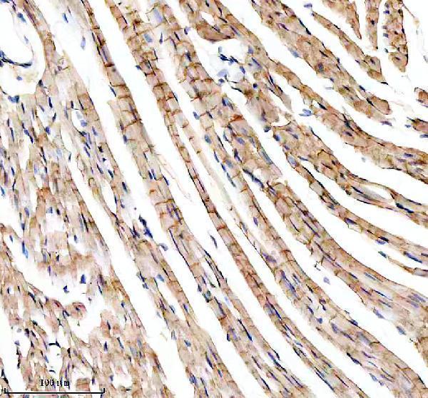
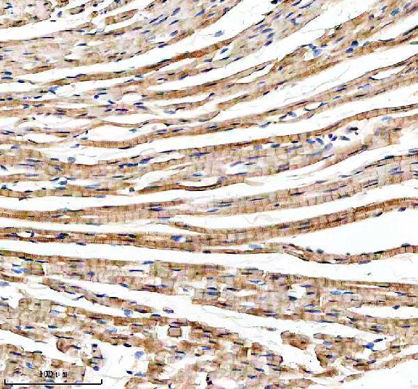
A03012-1 has IHC images from paraffin-embedded mouse and rat heart sections (catalog image captions). IF and ICC are listed applications, but no IF image is supplied (catalog applications; IF image alts).
A03012-1 will render with its mouse heart paraffin-section IHC figure (catalog card caption). Its catalog also shows rat heart paraffin-section IHC and lists Human, Mouse and Rat reactivity plus IF and ICC applications (catalog image captions; catalog applications and reactivity).
Which to pick: Choose A03012-1 for paraffin-section tissue IHC: its mouse and rat heart captions describe EDTA retrieval at pH 8.0 and primary antibody at 1:100 (catalog image captions). The sections’ fixative is unreported (catalog image captions). For IF/ICC or work across the listed Human, Mouse and Rat species, A03012-1 is the available rabbit polyclonal option; IF/ICC support comes from the application list, with no IF image supplied (catalog dilution_raw; catalog applications and reactivity; IF image alts).