YWHAG / 14-3-3 protein gamma · IHC design guide

Design Immunohistochemistry for YWHAG

Plan paraffin section IHC for YWHAG using its cytoplasmic and nuclear tissue staining profile (HPA tissue IHC). This guide covers tissue controls, antibody dilution, detection and interpretation using the catalog antibody’s IHC evidence (datasheet A04148-2).

Evidence assembled Oct 2026 · For research use; verify linked source records and product datasheet before use
Immunohistochemistry protocol sheet for YWHAG (IHC for YWHAG): expected localisation Cytoplasmic and nuclear tissue staining (HPA tissue IHC), antibody A04148-2, validated IHC image, and IHC protocol steps
Printable YWHAG IHC protocol sheet — expected localisation Cytoplasmic and nuclear tissue staining (HPA tissue IHC), antibody A04148-2, controls and protocol steps. Open the full YWHAG IHC guide →

YWHAG Immunohistochemistry Experimental Design Guide

Expected localisation, validated protocols, controls and antibodies — the at-a-glance facts below, then the full design guide.

Must know before staining
Expected localisation Cytoplasmic and nuclear tissue staining (HPA tissue IHC)
Staining pattern Cytoplasmic and nuclear staining across many cell types (HPA tissue IHC)
Antigen retrieval EDTA pH 8.0 HIER, heat-mediated (datasheet A04148-2)
Positive control ⓘ Caudate+4 more · see all
Negative control ⓘ None in HPA (detected in all 45 tissues); use no-primary + isotype controls
Important caveats
Reasons your staining may differ from the expected pattern.
Fixation Keep tissue fixation consistent across sections. (standard IHC practice; not target-specific)
Caveat Lung macrophage DAB staining may include endogenous peroxidase (HPA tissue IHC; standard IHC practice)
Regulation High in brain, skeletal muscle and heart (UniProt)
Isoform / epitope 0 annotated isoforms; chains start at residue 1 or 2 (UniProt)
Section 1

Recommended YWHAG IHC & IF Protocols

Compare the catalog antibody’s FFPE IHC protocol (datasheet A04148-2) with published YWHAG staining methods for human LSCC and mouse brain (PMC13619832; PMC10039879).

Recommended immunohistochemistry (IHC-P) protocol parameters
SampleParaffin-embedded human esophageal squamous carcinoma tissue; fixative not specified (datasheet A04148-2)
FixationImage fixative and duration unreported (datasheet A04148-2); verify before use.
Sectioning4–5 µm sections on charged slides (standard)
DeparaffinisationXylene, graded ethanol series to water (standard)
Antigen retrievalHeat retrieval: EDTA pH 8.0 (datasheet A04148-2); 20 min, 95–100 °C (standard)
Peroxidase block3% H2O2, 10 min, room temperature (standard)
Blocking10% goat serum (datasheet A04148-2)
Primary antibodyRabbit anti-YWHAG, 2-5 μg/ml (datasheet A04148-2)
Primary incubationOvernight at 4 °C (datasheet A04148-2)
DetectionHRP-conjugated secondary, DAB chromogen (datasheet A04148-2)
CounterstainHematoxylin, blue, dehydrate and mount (standard)
Expected resultYWHAG-positive staining in glial cells of caudate (HPA tissue IHC: High). HPA tissue profile: Ubiquitous cytoplasmic and nuclear expression. No signal in the no-primary control.
💡Decision noteStart with EDTA pH 8.0 heat retrieval for the catalog antibody (datasheet A04148-2); the LSCC study used citrate pH 6.0 (PMC13619832).
Section 2

What Is the Expected YWHAG Staining Pattern?

YWHAG is primarily cytosolic, with mitochondrial-matrix translocation reported after induction of MALM; it has no transmembrane segment (UniProt P61981). In paraffin-section IHC, expect widespread cytoplasmic and nuclear staining, with high staining in selected neuronal, glial, stromal, macrophage and glandular cells (HPA tissue IHC). HPA rates its tissue IHC profile Approved, with medium RNA–staining consistency and external verification pending (HPA tissue IHC).

What am I looking at on my slide?
Cytoplasmic staining, sometimes accompanied by nuclear staining, in the expected cells.This fits the ubiquitous cytoplasmic and nuclear tissue profile (HPA tissue IHC). Score the named cell population and compartment together: high staining is reported in cerebral-cortex and hippocampal neurons, caudate glia, and cerebellar granular-layer cells (HPA tissue IHC). A nuclear component on a tissue slide is compatible with HPA IHC, even though supported ICC-IF localisation is cytosolic (HPA tissue IHC; HPA subcellular ICC-IF).
Predominantly sharp plasma-membrane or extracellular deposits, with little intracellular staining.Treat this as a localisation mismatch and investigate an artefact or nonspecific signal; YWHAG has no transmembrane segment, and its annotated principal location is cytosolic (UniProt P61981; HPA subcellular ICC-IF). Its participation in a keratinocyte membrane-localisation complex does not make a membrane-only YWHAG pattern the expected tissue result (UniProt P61981; HPA tissue IHC).
Strong stain in an unexpected cell type while the expected cells remain weak.Assess cell identity before calling the slide positive. HPA reports high staining in lung macrophages, endometrial stromal cells, placental decidual cells and prostate glandular cells, so their signal can be expected in those tissues (HPA tissue IHC). In a different population, consider antibody cross-reactivity or endogenous chromogen-generating activity as general IHC possibilities; neither is established for this specimen by HPA (standard IHC practice).
Diffuse colour across cells, connective tissue and blank areas, with poor cell boundaries.This does not establish YWHAG localisation. Compare tissue morphology and a matched negative detection control; incomplete blocking, excess detection reagent or inadequate washing can produce diffuse background (standard IHC practice). Interpret a result only when intracellular staining can be distinguished from background in a plausible cell population (HPA tissue IHC; standard IHC practice).
No discernible stain in a selected high-staining tissue.Target-specific fixation effects are not established by the supplied assay evidence. Verify with a matched IHC source before attributing a result to fixation.
💡Expected YWHAG appearanceCall a positive result when identifiable cells show predominantly intracellular cytoplasmic staining, with possible nuclear staining, at an intensity consistent with their HPA cell-level category; high-staining examples include cortical neurons and lung macrophages, whereas isolated extracellular or membrane-only colour is suspect (HPA tissue IHC; UniProt P61981).
How each factor affects the staining
Cell population and tissue choice (HPA tissue IHC)HPA lists high staining in specific cells across brain, lung, endometrium, placenta and prostate; seminal-vesicle glandular cells, vaginal squamous epithelium and smooth-muscle cells are listed as low (HPA tissue IHC). Select controls by the named cells, and treat low as low rather than absent; HPA supplies no negative tissue category (HPA tissue IHC).
Compartment and cellular context (UniProt P61981; HPA tissue IHC)UniProt annotates cytosol and mitochondrial matrix, with matrix translocation following MALM induction (UniProt P61981). HPA tissue IHC describes cytoplasmic and nuclear staining (HPA tissue IHC). These sources support assessing the full intracellular pattern; they do not make mitochondrial puncta a required feature of routine paraffin-section IHC (UniProt P61981; HPA tissue IHC).
Processing and isoform interpretation (UniProt P61981)UniProt lists full-length and N-terminally processed chains and no annotated isoforms or signal peptide (UniProt P61981). The supplied record gives no antibody epitope, so it cannot predict whether N-terminal processing changes staining with a particular reagent (UniProt P61981).
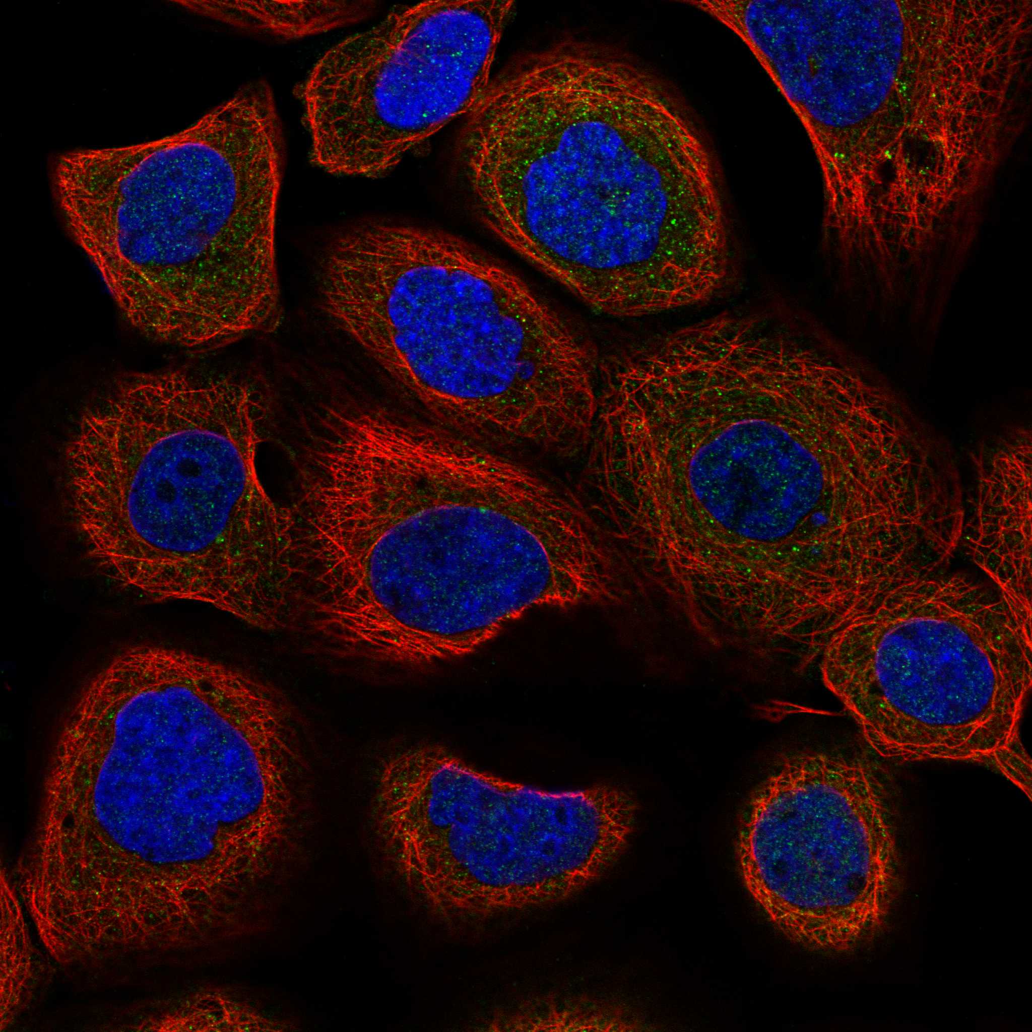
What should IF/ICC show? (HPA subcellular ICC-IF)Mainly cytosolic signal is supported; additional vesicle localisation is uncertain (HPA subcellular ICC-IF). This IF/ICC observation helps interpret compartment differences, but the tissue IHC profile separately allows nuclear staining (HPA tissue IHC).
Why is my staining missing, weak or wrong?
SituationLikely causeNext action
A high-staining reference tissue is blank.The relevant cells may be absent from the section, or an IHC workflow step may have failed (HPA tissue IHC; standard IHC practice).Confirm the expected cell population, then review the catalog antibody's IHC-P dilution and retrieval instructions and verify detection with a suitable control (standard IHC practice). Do not infer a YWHAG-specific fixation effect from this result (HPA tissue IHC).
The slide is uniformly brown with no clear cell-level pattern.Background from blocking, washing or detection chemistry can obscure the intracellular profile (standard IHC practice).Inspect a matched negative detection control, review blocking and wash steps, and score only resolved cellular signal (standard IHC practice; HPA tissue IHC).
Colour is confined to tissue edges, empty spaces or extracellular material.The distribution conflicts with the reported intracellular pattern and may reflect a staining artefact (HPA tissue IHC; standard IHC practice).Compare central and edge regions, check tissue morphology and a negative detection control, and avoid calling extracellular deposits YWHAG-positive cells (standard IHC practice).
Only a cell population listed as low appears intensely positive.Cell identification, nonspecific binding or endogenous detection activity may explain the mismatch; HPA's low category is not a negative control (HPA tissue IHC; standard IHC practice).Recheck morphology and detection controls, then compare staining in a named high-staining population on a suitable section (HPA tissue IHC; standard IHC practice).
Nuclear staining appears alongside cytoplasmic staining.The combination can match tissue IHC even though supported ICC-IF localisation is cytosolic (HPA tissue IHC; HPA subcellular ICC-IF).Record nuclear and cytoplasmic components separately and compare their distribution with cell identity and background controls before judging specificity (HPA tissue IHC; standard IHC practice).
Different antibodies or sections show inconsistent intensity.HPA rates the tissue profile Approved but notes medium RNA–staining consistency and pending external verification (HPA tissue IHC).Compare the same named cell population and compartment across sections; document antibody identity and matched conditions before interpreting a biological difference (HPA tissue IHC; standard IHC practice).

Sample controls for YWHAG IHC & IF

🧪Run cerebral cortex first: neuronal cells should show strong staining (HPA: High in cerebral cortex neuronal cells). HPA detects YWHAG in all 45 scored tissues, so there is no supported negative tissue; no-primary and isotype controls carry the negative, while cells without specific cytoplasmic staining on the positive slide are only internal background comparators, not validated YWHAG-negative cells (HPA: no negative tissue rows; UniProt P61981: cytosol).
Positive control tissue: Caudate (Glial cells, HPA High)
Negative control tissue: None in HPA: YWHAG is detected in all 45 scored tissues. Use a no-primary (secondary-only) and an isotype control instead.
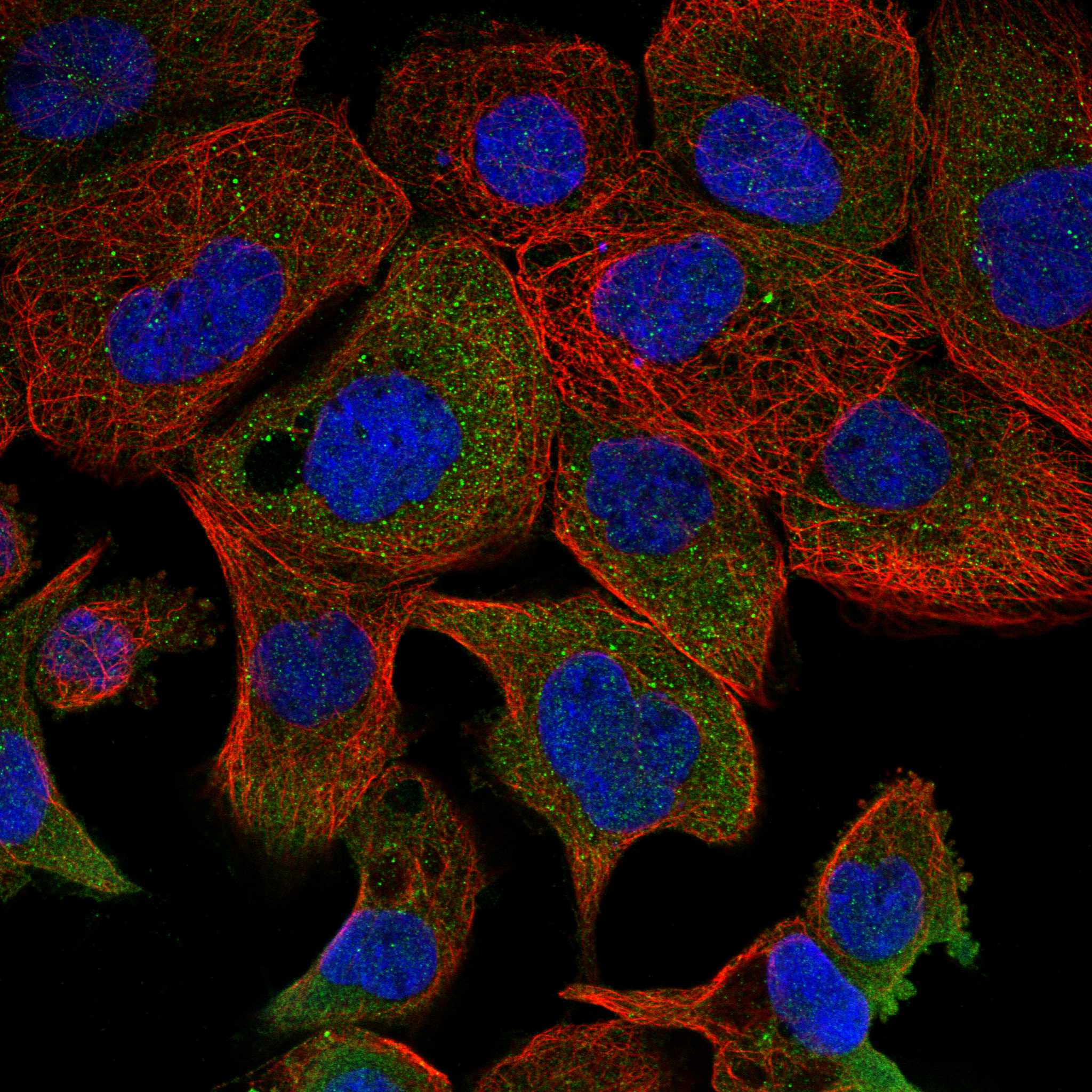
ICC-IF cell lines (HPA subcellular resource): HPA ICC-IF images show YWHAG in A-431, U2OS, A-549, HEK293, ASC52telo, RPTEC/TERT1, hTERT-RPE1 (serum starved), KOLF2.1J, with annotated localisation: Cytosol (supported) (HPA subcellular).
Technical controls: Include a no-primary, secondary-only section and a concentration-matched rabbit IgG isotype control, matching clonality where known (selected-SKU caption: rabbit primary; standard IHC practice). A comparably processed YWHAG knockout cell pellet would provide a biological negative if available; quench endogenous peroxidase for DAB detection, and distinguish brain pigment from specific staining (standard IHC practice; selected-SKU caption: HRP/DAB).
⚠️Feasibility: No target-specific fixation window or fixation effect is reported in the supplied evidence, and the selected-SKU paraffin-section caption does not state a fixative (selected-SKU caption: fixative unreported). That caption uses heat retrieval in EDTA, pH 8.0, but it does not establish that retrieval is required for every specimen (selected-SKU caption: EDTA retrieval). The evidence does not establish whether frozen sections or IF are easier; ICC-IF images support mainly cytosolic localization, while brain lipofuscin can complicate fluorescence interpretation (HPA: cytosol supported; standard IF practice).

HPA tissue IHC evidence for YWHAG

Comprehensive Human Protein Atlas IHC scoring per tissue (reliability: Approved — Medium consistency between antibody staining and RNA expression data. Pending external verification.). Rows are taken directly from the HPA tissue chart — click any row's HPA link to view the source.

Positive expression · recommended positive controls

TissueCell typeLevelEvidenceSource
Caudate Glial cells High Protein (IHC) HPA →
Cerebellum Cells in granular layer High Protein (IHC) HPA →
Cerebral cortex Neuronal cells High Protein (IHC) HPA →
Endometrium Cells in endometrial stroma High Protein (IHC) HPA →
Hippocampus Neuronal cells High Protein (IHC) HPA →

Undetected expression · recommended negative controls

TissueCell typeLevelEvidenceSource
None in HPA: YWHAG is detected in all 45 scored tissues. Use a no-primary (secondary-only) and an isotype control instead.
Section 3

Advanced YWHAG IHC Tips

Troubleshoot YWHAG staining in paraffin sections by checking retrieval, compartment, antibody specificity, and controls before comparing staining intensity.

Which antigen retrieval should I start with for YWHAG paraffin sections?
Start with heat-mediated retrieval in EDTA at pH 8.0 for paraffin sections (datasheet A04148-2). The selected image used this retrieval before 10% goat serum blocking and 2 μg/ml catalog antibody overnight at 4°C (datasheet A04148-2). If staining is weak, first compare retrieved and unretrieved sections while keeping antibody concentration and detection conditions constant (standard IHC practice). Excessive retrieval can damage section morphology or increase diffuse staining, so inspect both tissue integrity and cytoplasmic signal (standard IHC practice; UniProt P61981 localisation). The caption does not report a heating duration, so establish that parameter with a small retrieval series (datasheet A04148-2; standard IHC practice).
Could fixation explain weak or patchy YWHAG staining?
The selected paraffin-section caption does not state its fixative, and target-specific fixation sensitivity is unknown (datasheet A04148-2). Record the fixative, fixation duration, section age, and processing history for each specimen before attributing weak staining to YWHAG abundance (standard IHC practice). Compare sections processed together using the same EDTA pH 8.0 retrieval and antibody conditions to isolate specimen preparation as a variable (datasheet A04148-2; standard IHC practice). Assess morphology alongside staining because poorly preserved tissue can create patchy or misleading chromogenic signal (standard IHC practice). Do not infer fixation tolerance from tissue expression patterns or protein modifications (HPA tissue IHC; UniProt P61981 processing; standard IHC practice).
How should I assess cytoplasmic and nuclear YWHAG staining?
Prioritize interpretable cytoplasmic staining: UniProt places YWHAG in the cytosol, and HPA supports cytosolic localisation in cell imaging (UniProt P61981 localisation; HPA subcellular). HPA tissue IHC also reports ubiquitous cytoplasmic and nuclear expression, so nuclear colour alone does not establish an artefact or a specific YWHAG signal (HPA tissue IHC; standard IHC practice). Score cytoplasm and nucleus separately, using matched negative controls and consistent counterstaining to judge compartment boundaries (standard IHC practice). Mitochondrial matrix localisation follows induction of MALM, so routine diffuse cytoplasmic DAB cannot establish mitochondrial recruitment (UniProt P61981 localisation; standard IHC practice). Check whether apparent nuclear staining tracks section edges or damaged areas before interpreting it biologically (standard IHC practice).
Can processing or related 14-3-3 proteins affect antibody interpretation?
UniProt lists 0 YWHAG isoforms and describes both a 1–247 chain and an N-terminally processed 2–247 chain (UniProt P61981 processing). It also records N-terminal acetylation and phosphorylation at residues 71 and 133, without identifying this antibody’s epitope (UniProt P61981 modified residues; datasheet A04148-2). Consequently, do not assign a staining difference to processing or phosphorylation unless epitope information and appropriate controls support that inference (standard IHC practice). YWHAG can form heterodimers with other 14-3-3 proteins, but that interaction does not establish antibody cross-reactivity (UniProt P61981 subunit; standard IHC practice). Confirm specificity using a validated loss-of-target control where available and score only the staining pattern it removes (standard IHC practice).
How can IF help verify a YWHAG pattern seen by chromogenic IHC?
Use IF as a separate localisation check, with a marker identifying the expected cell type in the same specimen (standard IF practice). For example, HPA reports high YWHAG staining in lung macrophages and cerebral-cortex neuronal cells; choose a cell marker appropriate to the tissue examined (HPA tissue IHC; standard IF practice). Place the weaker signal in a fluorophore channel with low tissue autofluorescence, and include unstained and single-stain controls when multiplexing (standard IF practice). Because YWHAG is mainly cytosolic and has no transmembrane segment, use permeabilisation suited to an intracellular epitope, while checking that it preserves cell morphology (UniProt P61981 topology and localisation; standard IF practice). Compare compartment patterns with IHC without treating IF intensity as a direct DAB measurement (standard IHC/IF practice).
What should I check when DAB staining appears widespread?
First inspect a no-primary control for endogenous peroxidase activity and secondary-reagent background, particularly in specimens with pigment or inflammatory cells (standard IHC practice). The selected image used 10% goat serum blocking, a peroxidase-conjugated secondary, and DAB development; its caption does not specify a peroxidase-block step (datasheet A04148-2). Use a peroxidase block and control DAB development as general chromogenic workflow measures, then compare sections under identical conditions (standard IHC practice). Widespread signal alone is insufficient evidence of background because HPA describes ubiquitous cytoplasmic and nuclear YWHAG expression (HPA tissue IHC). Evaluate whether staining follows intact cells and expected compartments instead of edges, folds, or necrotic areas (standard IHC practice; UniProt P61981 localisation).
How should I quantify YWHAG staining across paraffin sections? ⚠ ANSWER MARKED FOR VERIFICATION
Define the tissue region and cell population before scoring, then keep retrieval, DAB development, imaging, and threshold settings consistent (standard IHC practice). An H-score combines the percentage of cells at each intensity; alternatively, report percent positive cells or positive-cell density per mm² (standard IHC practice). Normalise counts to the number of eligible intact cells or measured viable tissue area, and exclude folds, edges, and necrosis by prespecified rules (standard IHC practice). Score cytoplasmic and nuclear compartments separately because both appear in HPA tissue IHC, while UniProt identifies cytosol as a principal location (HPA tissue IHC; UniProt P61981 localisation). Report the scoring threshold and control performance with each comparison (standard IHC practice).
When does a YWHAG-positive IHC pattern warrant biological interpretation?
Look for reproducible signal within intact cells and a plausible cytoplasmic distribution, consistent with UniProt and supported HPA cell imaging (UniProt P61981 localisation; HPA subcellular). Check the cell population: HPA reports high staining in lung macrophages, cerebellar granular-layer cells, and cerebral-cortex neuronal cells, among others (HPA tissue IHC). Treat isolated staining at tissue edges, in necrosis, or in a no-primary control as possible processing or detection artefact (standard IHC practice). HPA describes its tissue-IHC reliability as Approved with medium agreement between staining and RNA data, pending external verification (HPA tissue IHC). Confirm unexpected nuclear-only or mitochondrial-looking patterns with compartment controls before assigning a signalling or MALM interpretation (UniProt P61981 localisation and function; standard IHC practice).
Boster reagents

Best YWHAG / 14-3-3 protein gamma IHC Antibodies

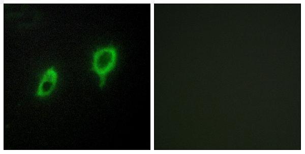
Both antibodies have human paraffin-section IHC images (catalog images: A04148, A04148-2); A04148 also has an IF image of COS7 cells (catalog image: A04148).

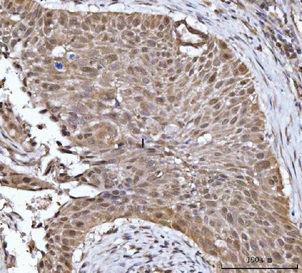
Real IHC data IHC analysis of 14-3-3 gamma/YWHAG using anti-14-3-3 gamma/YWHAG antibody (A04148-2). 14-3-3 gamma/YWHAG was detected in a paraffin-embedded section of human esophageal squamous carcinoma tissue. Heat mediated antigen retrieval was performed in EDTA buffer (pH 8.0, epitope retrieval solution). The tissue section was blocked with 10% goat serum. The tissue section was then incubated with 2 μg/ml rabbit anti-14-3-3 gamma/YWHAG Antibody (A04148-2) overnight at 4°C. Peroxidase Conjugated Goat Anti-rabbit IgG was used as secondary antibody and incubated for 30 minutes at 37°C. The tissue section was developed using HRP Conjugated Rabbit IgG Super Vision Assay Kit (Catalog # SV0002) with DAB as the chromogen.
Anti-14-3-3 gamma/YWHAG Antibody ®
Cat # A04148-2
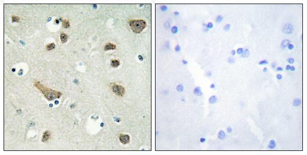
Real IHC data Immunohistochemistry analysis of paraffin-embedded human brain tissue, using 14-3-3 gamma Antibody. The picture on the right is blocked with the synthesized peptide.
Anti-14-3-3 gamma YWHAG Antibody
Cat # A04148

A04148 will render with a paraffin-section IHC image of human brain; its catalog also lists IF/ICC and shows IF in COS7 cells (A04148 image captions; catalog applications). A04148-2 will render with a paraffin-section IHC image of human esophageal squamous carcinoma (A04148-2 image caption).

Which to pick: For tissue IHC, choose A04148-2 when its documented human carcinoma conditions are useful: EDTA retrieval at pH 8.0 and 2 μg/ml primary antibody (A04148-2 IHC image caption). For IF/ICC, choose the polyclonal A04148: both applications are listed, and its IF image shows COS7 cells; its own IHC image shows paraffin-section human brain (A04148 catalog applications, dilution data and image captions). Both list Human, Mouse and Rat reactivity, but the supplied IHC images show human tissue only; neither IHC caption reports a fixative (catalog reactivity; A04148 and A04148-2 IHC image captions).

Each figure is that product's own IHC / IF validation image from its datasheet.

References

  1. UniProt Consortium. UniProt entry P61981 (1433G_HUMAN, 14-3-3 protein gamma).
  2. Human Protein Atlas. YWHAG tissue IHC expression (reliability: Approved).
  3. Human Protein Atlas. YWHAG subcellular location (ICC-IF): Mainly localized to the cytosol. In addition localized to vesicles..
  4. Human Protein Atlas. YWHAG antibody validation summary (4 antibodies).
  5. YWHAG overexpression correlates with an immune-desert microenvironment and poor prognosis in laryngeal squamous cell carcinoma. Frontiers in immunology 2026 — PMC13619832.
  6. YWHAG promotes bladder cancer metastasis by regulating TMOD3 to activate ERK1/2 and JNK phosphorylation in the MAPK pathway. Journal of translational medicine 2024 — PMC11687020.
  7. YWHAG promotes the progression of lung adenocarcinoma through the JAK2/STAT3 pathway. Cancer cell international 2025 — PMC11929182.
  8. Distribution of phosphorylated alpha-synuclein in non-diseased brain implicates olfactory bulb mitral cells in synucleinopathy pathogenesis. NPJ Parkinson's disease 2023 — PMC10039879.
  9. PubMed PMID:10433554 — UniProt-cited evidence.
  10. PubMed PMID:10486217 — UniProt-cited evidence.
  11. PubMed PMID:12853948 — UniProt-cited evidence.