ZFP36L2 / mRNA decay activator protein ZFP36L2 · IHC design guide

Design Immunohistochemistry for ZFP36L2

Plan paraffin-section ZFP36L2 IHC around the cytoplasmic staining reported in most tissues (HPA tissue IHC). Use high-staining small-intestine glandular cells as a positive control and interpret intensity cautiously because staining and RNA expression show low consistency (HPA tissue IHC).

Evidence assembled Oct 2026 · For research use; verify linked source records and product datasheet before use
Immunohistochemistry protocol sheet for ZFP36L2 (IHC for ZFP36L2): expected localisation Cytoplasmic tissue staining; nuclear shuttling is possible (HPA tissue IHC; UniProt), antibody A05565, validated IHC image, and IHC protocol steps
Printable ZFP36L2 IHC protocol sheet — expected localisation Cytoplasmic tissue staining; nuclear shuttling is possible (HPA tissue IHC; UniProt), antibody A05565, controls and protocol steps. Open the full ZFP36L2 IHC guide →

ZFP36L2 Immunohistochemistry Experimental Design Guide

Expected localisation, validated protocols, controls and antibodies — the at-a-glance facts below, then the full design guide.

Must know before staining
Expected localisation Cytoplasmic tissue staining; nuclear shuttling is possible (HPA tissue IHC; UniProt)
Staining pattern Cytoplasmic staining in most tissues (HPA tissue IHC)
Antigen retrieval Citrate pH 6.0 HIER, heat-mediated (datasheet A05565)
Positive control ⓘ Epididymis+4 more · see all
Negative control ⓘ Caudate+4 more · see all
Important caveats
Reasons your staining may differ from the expected pattern.
Fixation Keep fixation conditions consistent across sections (standard IHC practice; not target-specific)
Caveat Staining and RNA expression show low consistency (HPA tissue IHC)
Regulation Basal epidermis exceeds suprabasal layers (UniProt)
Isoform / epitope No annotated isoforms; one 1–494 chain (UniProt)
Section 1

Recommended ZFP36L2 IHC & IF Protocols

The catalog antibody uses citrate pH 6.0 heat retrieval (datasheet A05565). The published IHC protocols below cover cervical cancer, pancreatic ductal adenocarcinoma, and heart sections (PMC10432607; PMC5276842; PMC8206151).

Recommended immunohistochemistry (IHC-P) protocol parameters
SampleParaffin-embedded human small intestinal carcinoma tissue; fixative not specified (datasheet A05565)
FixationImage fixative and duration unreported (datasheet A05565); verify before use.
Sectioning4–5 µm sections on charged slides (standard)
DeparaffinisationXylene, graded ethanol series to water (standard)
Antigen retrievalHeat retrieval: Citrate pH 6.0, 20min (datasheet A05565)
Peroxidase block3% H2O2, 10 min, room temperature (standard)
Blocking10% normal serum of the secondary host, 30 min, room temperature (standard)
Primary antibodyRabbit anti-ZFP36L2, 1:100-1:300 (datasheet A05565)
Primary incubationOvernight at 4 °C (standard)
DetectionHRP-polymer secondary, DAB chromogen 5–10 min (standard)
CounterstainHematoxylin, blue, dehydrate and mount (standard)
Expected resultZFP36L2-positive staining in glandular cells of epididymis (HPA tissue IHC: High). HPA tissue profile: Cytoplasmic expression in most tissues. No signal in the no-primary control.
💡Decision noteStart with citrate pH 6.0 heat retrieval for the catalog antibody (datasheet A05565); the cervical cancer article mentions antigen repair without specifying its method (PMC10432607).
Section 2

What Is the Expected ZFP36L2 Staining Pattern?

ZFP36L2 can localise to the cytoplasm and nucleus and shuttle between them; it has no transmembrane segment (UniProt P47974). In tissue IHC, expect predominantly cytoplasmic staining, including in epididymal glandular cells, smooth muscle cells and lymph node non-germinal center cells (HPA: tissue IHC). Treat that pattern as provisional: HPA rates the antibody Approved but reports low consistency between staining and RNA expression, pending external verification (HPA: reliability).

What am I looking at on my slide?
Clear cytoplasmic stain in epididymal glandular cells, smooth muscle cells or lymph node non-germinal center cells (HPA: High).This matches cell populations scored High by HPA and its predominantly cytoplasmic tissue profile (HPA: tissue IHC). Compare stained cells with nearby structures on the same section; HPA's Approved rating still carries a low staining–RNA consistency caveat (HPA: reliability).
Some nuclei stain along with the cytoplasm, while cell boundaries remain interpretable.Nuclear signal can be biologically plausible because ZFP36L2 shuttles between nucleus and cytoplasm (UniProt P47974). HPA describes the overall tissue pattern as cytoplasmic, so assess nuclear staining alongside a credible cell distribution and controls rather than requiring every positive cell to have an identical compartment pattern (HPA: tissue IHC).
Stain is predominantly membranous or extracellular, with little convincing intracellular signal.That distribution conflicts with the nuclear and cytoplasmic localisation and absence of a transmembrane segment reported for ZFP36L2 (UniProt P47974). Suspect nonspecific deposition or a detection artefact; the compartment mismatch alone cannot identify which reagent caused it (standard IHC practice).
Strong signal appears in ovarian stroma cells, cholangiocytes or chondrocytes.HPA scored these specific cell populations Not detected; this does not make the entire ovary, liver or soft tissue a negative control (HPA: tissue IHC). If the signal repeats, assess cross-reactivity or endogenous detection activity with appropriate controls before calling it ZFP36L2 (standard IHC practice).
A known-positive cell population has no stain, or diffuse colour obscures cells throughout the section.Absence in a population scored High by HPA calls for a run and control check; widespread haze prevents a reliable compartment call (HPA: tissue IHC; standard IHC practice). Neither appearance, on its own, establishes loss of ZFP36L2 or a target-specific fixation effect.
💡Expected ZFP36L2 appearanceCall a result positive when discrete, predominantly cytoplasmic signal is appreciable in a cell population scored High by HPA, with possible nuclear staining consistent with shuttling; isolated membranous or extracellular colour is suspect (HPA: tissue IHC; UniProt P47974).
How each factor affects the staining
Tissue and cell choiceHPA reports High stain in epididymal glandular cells, tonsil and lymph node non-germinal center cells, small-intestinal glandular cells, and smooth muscle cells (HPA: tissue IHC). Its Not detected calls apply to named cell populations, so select and score controls at cell level.
Compartment assignmentHPA's tissue profile is cytoplasmic, while UniProt lists nucleus and cytoplasm with XPO1/CRM1-dependent shuttling (HPA: tissue IHC; UniProt P47974). A nuclear component is therefore plausible, but neither source supplies an IHC nuclear-to-cytoplasmic ratio.
Strength of tissue evidenceThe listed IHC antibody is Approved, and HPA reports low consistency between staining and RNA expression with external verification pending (HPA: HPA047428 validation; HPA: reliability). Use the reported cell pattern as an expectation, not proof that every stained cell expresses the target.
IF/ICC expectation?UniProt permits nuclear and cytoplasmic localisation, but HPA provides no main ICC-IF location or cell-line images for this target (UniProt P47974; HPA: subcellular). Interpret an IF pattern with its own controls; these sources do not establish an IF staining benchmark or protocol.
Why is my staining missing, weak or wrong?
SituationLikely causeNext action
No signal in epididymal glandular cells or another HPA High population.The run may have insufficient detection or an unsuitable staining condition; HPA's High call is an observation, not a guarantee for every section (HPA: tissue IHC; standard IHC practice).Check the positive control, reagent preparation, retrieval conditions and detection steps, then repeat with a matched section if needed (standard IHC practice). Do not infer target-specific fixation sensitivity from this result.
Diffuse colour covers both cells and tissue spaces.Background from detection reagents, incomplete blocking or excess antibody can obscure a cellular pattern (standard IHC practice).Review a no-primary control, blocking and washes; adjust the primary-antibody dilution against a positive cell population while preserving cellular detail (standard IHC practice; HPA: tissue IHC).
Only nuclei stain strongly.Nuclear ZFP36L2 is possible, but an exclusively nuclear tissue pattern differs from HPA's cytoplasmic profile (UniProt P47974; HPA: tissue IHC).Check that the stain follows the expected cells and compare control sections; assess counterstain and chromogen separation before calling a compartment shift (HPA: tissue IHC; standard IHC practice).
Strong stain appears in ovarian stroma cells or cholangiocytes.Both populations are scored Not detected by HPA; cross-reactivity or endogenous detection activity is possible but unproven (HPA: tissue IHC; standard IHC practice).Confirm cell identity, inspect a no-primary control and compare a known-positive population in the same run before assigning the signal to ZFP36L2 (HPA: tissue IHC; standard IHC practice).
Signal tracks membranes, lumens or section edges instead of cell interiors.A predominantly extracellular or membranous pattern conflicts with ZFP36L2 localisation and topology; edge or luminal deposits can reflect staining artefact (UniProt P47974; standard IHC practice).Compare intact cells away from edges, review the no-primary control and repeat the stain if deposits obscure localisation (standard IHC practice).

Sample controls for ZFP36L2 IHC & IF

🧪Run small intestine first: its glandular cells should stain (HPA: High in small-intestinal glandular cells). Use liver cholangiocytes as the negative comparator (HPA: Not detected in liver cholangiocytes); on the positive slide, neighboring cells independently confirmed as negative should show counterstain and background only (standard IHC control practice).
Positive control tissue: Epididymis (Glandular cells, HPA High)
Negative control tissue: Caudate (HPA Not detected)
ICC-IF cell lines (HPA subcellular resource): HPA carries no ICC-IF cell line for ZFP36L2; derive a cell-line control from the positive tissue's cell type (Glandular cells) and confirm it by RNA or western blot first.
Technical controls: Include no-primary (secondary-only) and irrelevant antibody controls matched to the primary antibody’s host species, immunoglobulin class, and monoclonal or polyclonal format; use a ZFP36L2 knockout specimen or a validated immunizing-peptide block as a biological specificity control (standard IHC control practice). For chromogenic small-intestine sections, quench endogenous peroxidase and, if using avidin–biotin detection, check endogenous biotin background (standard IHC practice).
⚠️Feasibility: No target-specific fixation window or fixation effect is reported in the supplied evidence, and the selected A05565 paraffin-section caption does not state a fixative (caption: fixative unreported). The caption uses sodium citrate at pH 6.0 above 98°C for 20 minutes, but does not establish that retrieval is required or that the specimen was paraffin-section (caption: antigen retrieval conditions). The evidence does not establish whether frozen sections or IF/ICC are easier; check small-intestinal autofluorescence with an unstained control if using IF (HPA: no ICC-IF images; standard IF practice). The selected A05565 tissue-IHC caption documents paraffin sections, but does not specify the fixative (selected-SKU IHC image A05565).

HPA tissue IHC evidence for ZFP36L2

Comprehensive Human Protein Atlas IHC scoring per tissue (reliability: Approved — Low consistency between antibody staining and RNA expression data. Pending external verification.). Rows are taken directly from the HPA tissue chart — click any row's HPA link to view the source.

Positive expression · recommended positive controls

TissueCell typeLevelEvidenceSource
Epididymis Glandular cells High Protein (IHC) HPA →
Lymph node Non-germinal center cells High Protein (IHC) HPA →
Small intestine Glandular cells High Protein (IHC) HPA →
Smooth muscle Smooth muscle cells High Protein (IHC) HPA →
Tonsil Non-germinal center cells High Protein (IHC) HPA →

Undetected expression · recommended negative controls

TissueCell typeLevelEvidenceSource
Caudate Glial cells Not detected Protein (IHC) HPA →
Cerebellum Cells in granular layer Not detected Protein (IHC) HPA →
Cerebral cortex Endothelial cells Not detected Protein (IHC) HPA →
Hippocampus Glial cells Not detected Protein (IHC) HPA →
Liver Cholangiocytes Not detected Protein (IHC) HPA →
Section 3

Advanced ZFP36L2 IHC Tips

Troubleshoot ZFP36L2 staining in paraffin sections using the catalog antibody’s documented IHC conditions, compartment expectations, and cell level controls.

How should I adjust retrieval when ZFP36L2 staining is weak?
Start with heat mediated citrate retrieval at pH 6.0 (datasheet A05565). The catalog image used sodium citrate above 98°C for 20 minutes on paraffin embedded human small intestinal carcinoma, followed by primary antibody at 1:200 overnight at 4°C (caption A05565). If staining is weak, check section adhesion, heating consistency, and cooling before changing retrieval conditions (standard IHC practice). Compare any adjusted condition beside the documented one using the same tissue and detection run, since stronger staining alone does not establish specificity (standard IHC practice). Include a no primary control to reveal detection related staining (standard IHC practice).
Could fixation explain variable ZFP36L2 staining between paraffin blocks?
The selected image identifies paraffin embedded tissue but does not state its fixative, so target specific fixation sensitivity is unknown (caption A05565). Do not infer a ZFP36L2 fixation effect from tissue staining patterns or its lack of a transmembrane segment (HPA tissue IHC; UniProt P47974 topology). Record each block’s fixative, fixation duration, and processing history, then compare sections under identical retrieval and detection conditions (standard IHC practice). Use the documented citrate pH 6.0 retrieval and 1:200 primary dilution as a consistent starting point (caption A05565). Assess staining alongside preserved morphology and matched controls before attributing a difference to fixation (standard IHC practice).
Is nuclear ZFP36L2 staining credible when most tissue staining looks cytoplasmic?
ZFP36L2 is annotated in both nucleus and cytoplasm and shuttles between them through an XPO1/CRM1 dependent mechanism (UniProt P47974 subcellular). HPA describes predominantly cytoplasmic staining across tissues, so a nuclear pattern deserves careful comparison with cytoplasmic signal in the same cell population (HPA tissue IHC). Inspect nuclei against the counterstain and check whether signal respects cell borders rather than section folds or damaged edges (standard IHC practice). Score nuclear and cytoplasmic staining separately instead of treating either compartment as automatically nonspecific (UniProt P47974 subcellular; standard IHC practice). Confirm an unusual compartment pattern with an independent specificity control before interpreting it biologically (standard IHC practice).
How do epitope uncertainty and phosphorylation affect ZFP36L2 IHC interpretation?
The supplied record lists 0 isoforms and no transmembrane segment, but it does not locate this antibody’s epitope (UniProt P47974 isoforms and topology; caption A05565). ZFP36L2 has annotated phosphorylated residues including Ser57, Ser125, Thr238, Ser490, and Ser492 (UniProt P47974 modified residues). Whether those modifications alter binding by this antibody is unknown without an epitope map or direct comparison (UniProt P47974 modified residues; caption A05565). Keep retrieval and primary incubation constant when comparing conditions, using citrate pH 6.0 and the documented 1:200 overnight incubation as reference conditions (caption A05565). Treat a changed stain as an antibody signal change until an independent assay supports an epitope or modification explanation (standard IHC practice).
How can I assess ZFP36L2 by multiplex IF after observing chromogenic IHC staining?
Plan IF as a separate validation: the supplied antibody image documents chromogenic staining of a paraffin section, without IF fixation or permeabilisation conditions (caption A05565). Pair ZFP36L2 with a validated marker for the cell population under study; HPA reports high glandular cell staining in small intestine and high non germinal center cell staining in lymph node (HPA tissue IHC). Choose fluorophores after measuring tissue autofluorescence and include single label and no primary controls to check bleed through and background (standard IF practice). Because ZFP36L2 lacks a transmembrane segment and can occupy nucleus and cytoplasm, optimise permeabilisation for access to intracellular epitopes once the antibody epitope is known (UniProt P47974 topology and subcellular; standard IF practice). Do not transfer the IHC image’s retrieval or incubation conditions to IF without testing them (caption A05565; standard IF practice).
What should I check when diffuse brown staining obscures ZFP36L2 positive cells?
HPA describes cytoplasmic expression in most tissues, so diffuse cytoplasmic color alone cannot distinguish signal from background (HPA tissue IHC). Run a no primary control and inspect deposits around folds, damaged edges, pigment, and necrotic areas (standard IHC practice). For chromogenic peroxidase detection, apply a peroxidase block and check the detection reagents before interpreting DAB deposits as ZFP36L2 (standard IHC practice). Keep the documented 1:200 primary and 1:200 secondary dilutions as comparison conditions when adjusting incubation or washing (caption A05565). Compare cellular staining and background within the same run, since a lighter section can lose genuine signal as well as artefact (standard IHC practice).
How should I score ZFP36L2 across mixed cell populations? ⚠ ANSWER MARKED FOR VERIFICATION
Define the cell population and compartment before scoring because HPA reports cell dependent staining and UniProt places ZFP36L2 in nucleus and cytoplasm (HPA tissue IHC; UniProt P47974 subcellular). For intensity based comparisons, calculate an H score as 0–300 from the percentage of cells at each staining intensity (standard IHC practice). Alternatively report the percentage of positive cells, or positive cell density per mm² when cell abundance varies (standard IHC practice). Normalise each measure to the relevant viable cells or viable tissue area, excluding necrosis and section edges by a prespecified rule (standard IHC practice). Keep retrieval, detection, exposure to chromogen, and scoring thresholds consistent across the compared sections (standard IHC practice).
What evidence separates true ZFP36L2 staining from artefact?
A credible pattern should resolve within cells and be assessed against ZFP36L2’s nuclear and cytoplasmic annotation and HPA’s predominantly cytoplasmic tissue profile (UniProt P47974 subcellular; HPA tissue IHC). Check whether stained cells match the tissue context: HPA reports high staining in small intestinal glandular cells and non germinal center lymph node cells, with no detection in the listed cerebellar granular layer cells (HPA tissue IHC). Reject edge restricted color, necrotic deposits, and signal reproduced by a no primary control as evidence of specific antigen staining (standard IHC practice). In peroxidase based IHC, investigate endogenous enzyme activity if control sections develop DAB color (standard IHC practice). HPA labels its tissue staining approved but notes low agreement with RNA expression and pending external verification, so seek independent confirmation for consequential conclusions (HPA tissue IHC).
Boster reagents

Best ZFP36L2 / mRNA decay activator protein ZFP36L2 IHC Antibodies

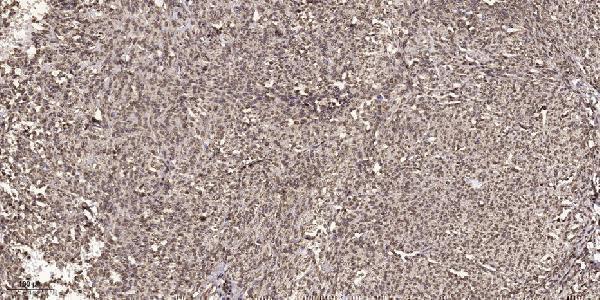
A05565 has a human paraffin-section IHC figure; IF is listed without a figure (A05565 image caption; catalog applications). Catalog reactivity covers human and mouse for both antibodies, plus rat for A05565-1 (catalog reactivity).

Real IHC data Immunohistochemical analysis of paraffin-embedded human small intestinal carcinoma tissue. 1, primary Antibody was diluted at 1:200 (4° overnight). 2, Sodium citrate pH 6.0 was used for antigen retrieval (>98°C, 20min). 3, Secondary antibody was diluted at 1:200
Anti-TIS11D ZFP36L2 Antibody
Cat # A05565

A05565 has an IHC figure from paraffin-embedded human small intestinal carcinoma tissue and lists IF for human and mouse, without an IF figure (A05565 image caption; catalog applications/reactivity). A05565-1 lists IHC for human, mouse and rat, but has no IHC or IF image caption (A05565-1 catalog applications/reactivity; image captions).

Which to pick: For paraffin-section tissue IHC, choose A05565 because its own figure documents that preparation; the fixative is unreported (A05565 image caption). For IF, choose A05565 because IF is listed at 1:50; ICC validation and an IF figure are unreported (A05565 catalog applications/dilution; image captions). For rat tissue IHC, consider A05565-1 because it lists rat reactivity and IHC; both antibodies are rabbit polyclonals, and A05565-1 has no processing details from an IHC figure (catalog reactivity/applications/clonality; A05565-1 image captions).

Each figure is that product's own IHC / IF validation image from its datasheet.

References

  1. UniProt Consortium. UniProt entry P47974 (TISD_HUMAN, mRNA decay activator protein ZFP36L2).
  2. Human Protein Atlas. ZFP36L2 tissue IHC expression (reliability: Approved).
  3. Human Protein Atlas. ZFP36L2 subcellular location (ICC-IF): Highest expression in THP-1: 202.5 nTPM.
  4. Human Protein Atlas. ZFP36L2 antibody validation summary (1 antibodies).
  5. MiR-520d-3p suppresses the proliferation and epithelial-mesenchymal transition of cervical cancer cells by targeting ZFP36L2. Heliyon 2023 — PMC10432607.
  6. ZFP36L2 promotes cancer cell aggressiveness and is regulated by antitumor microRNA-375 in pancreatic ductal adenocarcinoma. Cancer science 2017 — PMC5276842.
  7. The RNA binding proteins ZFP36L1 and ZFP36L2 are dysregulated in airway epithelium in human and a murine model of asthma. Frontiers in cell and developmental biology 2023 — PMC10624177.
  8. ZFP36L2 regulates myocardial ischemia/reperfusion injury and attenuates mitochondrial fusion and fission by LncRNA PVT1. Cell death & disease 2021 — PMC8206151.
  9. PubMed PMID:8545129 — UniProt-cited evidence.
  10. PubMed PMID:7835719 — UniProt-cited evidence.
  11. PubMed PMID:15815621 — UniProt-cited evidence.