This website uses cookies to ensure you get the best experience on our website.


- Table of Contents
Plan ZNF703 paraffin-section IHC around widespread nuclear tissue staining (HPA tissue IHC). This guide covers fixation consistency, controls using skin fibrohistiocytic cells with high staining, and interpretation across cell types (HPA tissue IHC).

Expected localisation, validated protocols, controls and antibodies — the at-a-glance facts below, then the full design guide.
| Expected localisation | Nuclear tissue staining (HPA tissue IHC); cytoplasm annotated (UniProt) | |
| Staining pattern | Widespread nuclear staining across cell types (HPA tissue IHC) | |
| Antigen retrieval | Tris-EDTA pH 9.0 HIER, 95–98 °C, 20 min (rule: nuclear antigen) | |
| Positive control | Skin+4 more · see all | |
| Negative control | None in HPA (detected in all 45 tissues); use no-primary + isotype controls |
| Fixation | Keep fixation consistent across sections (standard IHC practice; not target-specific) | |
| Caveat | Skin fibrohistiocytic cells stain high; other cell types vary (HPA tissue IHC) | |
| Regulation | Cell-type-dependent expression (HPA tissue IHC) | |
| Isoform / epitope | No isoforms annotated; chain spans residues 2–590 (UniProt) |
The catalog antibody’s IHC-P protocol is accompanied by published ZNF703 IHC protocols for breast and ovarian specimens (PMC8889678; PMC3753350; PMC7693506).
| Sample | Paraffin-embedded rat pancreas tissue; fixative not specified (datasheet A06570) |
| Fixation | Image fixative and duration unreported (datasheet A06570); verify before use. |
| Sectioning | 4–5 µm sections on charged slides (standard) |
| Deparaffinisation | Xylene, graded ethanol series to water (standard) |
| Antigen retrieval | Heat-induced epitope retrieval in Tris-EDTA buffer, pH 9.0, 20 min at 95–98 °C (standard rule: nuclear antigen) |
| Peroxidase block | 3% H2O2, 10 min, room temperature (standard) |
| Blocking | 10% normal serum of the secondary host, 30 min, room temperature (standard) |
| Primary antibody | Rabbit anti-ZNF703, 1:50-1:200 (datasheet A06570) |
| Primary incubation | Overnight at 4 °C (standard) |
| Detection | HRP-polymer secondary, DAB chromogen 5–10 min (standard) |
| Counterstain | Hematoxylin, blue, dehydrate and mount (standard) |
| Expected result | ZNF703-positive staining in fibrohistiocytic cells of skin (HPA tissue IHC: High). HPA tissue profile: Ubiquitous nuclear expression. No signal in the no-primary control. |
ZNF703 should appear predominantly nuclear in paraffin-section IHC, with possible cytoplasmic signal (HPA: ubiquitous nuclear expression; UniProt Q9H7S9: nucleus and cytoplasm). Skin fibrohistiocytic cells show high staining; several other listed cell populations show medium staining (HPA tissue IHC). Interpret these expectations with the reported Supported reliability, which reflects medium consistency between antibody staining and RNA data (HPA tissue IHC). ZNF703 has no transmembrane segment (UniProt Q9H7S9).
| Discrete nuclear stain in skin fibrohistiocytic cells, with visibly weaker staining in some other cell populations. | This fits the high skin signal and broadly nuclear profile; HPA reports medium signal in selected adipocytes, glandular cells, respiratory epithelium and glia (HPA tissue IHC). Compare cell types within the same section before assigning intensity; HPA’s categories describe its observations, not a universal scoring threshold (HPA tissue IHC). |
| Signal is confined to membranes, luminal material or extracellular deposits, with no convincing nuclear staining. | That compartment pattern conflicts with the observed nuclear profile and the lack of a transmembrane segment (HPA tissue IHC; UniProt Q9H7S9 topology). Check a known-positive cell population and detection controls before calling it ZNF703. Cytoplasmic staining alone deserves review rather than automatic rejection because UniProt also lists cytoplasm (UniProt Q9H7S9). |
| A cell population stains strongly where HPA reports low staining, while expected positive cells stain weakly. | Possible explanations include cross-reactivity, endogenous chromogen-generating activity or a technical mismatch between sections (general IHC practice). Treat the contrast as a reason to investigate, not proof of an artefact: HPA’s low categories are observations, not validated negatives, and its staining-to-RNA consistency is medium (HPA tissue IHC). |
| Brown precipitate is diffuse across tissue compartments or appears similarly in many unrelated cells. | Poorly localized signal can obscure whether nuclei contain genuine stain (general IHC practice). Inspect reagent-only and no-primary controls, morphology and local tissue background before scoring. HPA’s expected pattern is ubiquitous nuclear expression, which means widespread positive cells alone do not establish nonspecific staining (HPA tissue IHC). |
| Skin fibrohistiocytic cells lack nuclear signal despite adequate tissue morphology. | The absent high reference pattern warrants a run-level check (HPA tissue IHC). Review primary-antibody use, retrieval, detection and counterstain with the same run’s controls (general IHC practice). A failed reference does not establish that the specimen lacks ZNF703; HPA reports a supported pattern, not guaranteed positivity in every section (HPA tissue IHC). |
| Compartment used for scoring | HPA describes a ubiquitous nuclear IHC pattern, while UniProt lists both nucleus and cytoplasm (HPA tissue IHC; UniProt Q9H7S9). Prioritize interpretable nuclear staining and assess any cytoplasmic signal in its cellular context. |
| Reference cell population | Skin fibrohistiocytic cells are high; adipocytes, adrenal and appendix glandular cells, respiratory epithelial cells and specified neural cells are medium (HPA tissue IHC). HPA also lists low-staining populations, so these are comparison points rather than proven negative controls. |
| Strength of the tissue evidence | The tissue profile is Supported, with medium agreement between antibody staining and RNA expression (HPA tissue IHC). HPA reports tissue-enhanced RNA in skeletal muscle; that RNA category does not specify a skeletal-muscle IHC staining level (HPA tissue IHC). |
| Antibody and application evidence | HPA lists Supported IHC results for HPA023930 and CAB068249, but no ICC validation status for either (HPA antibodies). HPA provides no ICC-IF images or main subcellular location here, so an IF result needs its own controls and interpretation (HPA subcellular). |
| Protein processing and retrieval limits | UniProt lists no signal peptide, propeptide, transmembrane segment or annotated isoform for this record (UniProt Q9H7S9). The supplied sources give no antibody epitope or target-specific fixation effect; choose and verify retrieval conditions from the IHC-validated antibody’s instructions (general IHC practice). |
| Situation | Likely cause | Next action |
|---|---|---|
| No stain in the specimen or the skin reference. | The run may have failed at retrieval, primary-antibody incubation or chromogenic detection (general IHC practice). | Check the run’s positive and no-primary controls, reagent preparation and the IHC-validated antibody’s stated conditions before interpreting absence. Skin fibrohistiocytic cells provide an expected high reference, not an absolute guarantee (HPA tissue IHC). |
| Nuclei appear negative but cytoplasm stains. | UniProt includes cytoplasm, whereas HPA’s tissue pattern is nuclear; either a real compartment difference or nonspecific signal needs assessment (UniProt Q9H7S9; HPA tissue IHC). | Compare nuclear and cytoplasmic signal in a positive reference, inspect no-primary controls and report the observed compartments explicitly (general IHC practice). Do not label cytoplasm alone definitive without further support. |
| Only outlines of cells or extracellular areas are brown. | A membrane- or deposit-only pattern conflicts with the nuclear tissue profile and nonmembrane topology (HPA tissue IHC; UniProt Q9H7S9 topology). | Inspect morphology and no-primary controls, then confirm that a known-positive cell population develops nuclear stain in the same run (general IHC practice). |
| Background masks nuclear detail across the section. | Detection background or inadequate blocking can make a nuclear call unreliable (general IHC practice). | Compare no-primary and detection-only controls; optimize blocking, washing and chromogen development for the assay, then rescore nuclei only where cellular boundaries remain clear (general IHC practice). |
| Unexpected strong staining occurs in an HPA low-staining cell population. | HPA’s low category is not a validated negative; assay background or cross-reactivity is also possible (HPA tissue IHC; general IHC practice). | Confirm the cell identity and compartment, compare controls and a reported high population, and describe the discrepancy without converting the HPA low call into an absolute absence claim (HPA tissue IHC; general IHC practice). |
| An IF/ICC image seems to disagree with the IHC result. | The supplied HPA subcellular record has no ICC-IF images or main-location call, and the listed antibodies have no ICC status here (HPA subcellular; HPA antibodies). | Evaluate IF with its own controls and dedicated guide; use the paraffin-section nuclear pattern as context, without treating IHC support as IF validation (HPA tissue IHC; HPA antibodies). |
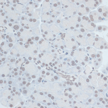
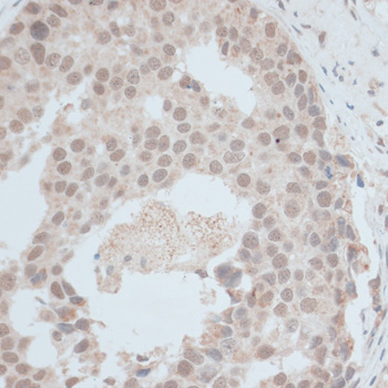
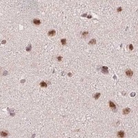
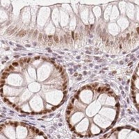


Comprehensive Human Protein Atlas IHC scoring per tissue (reliability: Supported — Medium consistency between antibody staining and RNA expression data.). Rows are taken directly from the HPA tissue chart — click any row's HPA link to view the source.
| Tissue | Cell type | Level | Evidence | Source |
|---|---|---|---|---|
| Skin | Fibrohistiocytic cells | High | Protein (IHC) | HPA → |
| Adipose tissue | Adipocytes | Medium | Protein (IHC) | HPA → |
| Adrenal gland | Glandular cells | Medium | Protein (IHC) | HPA → |
| Appendix | Glandular cells | Medium | Protein (IHC) | HPA → |
| Breast | Adipocytes | Medium | Protein (IHC) | HPA → |
| Tissue | Cell type | Level | Evidence | Source |
|---|---|---|---|---|
| None in HPA: ZNF703 is detected in all 45 scored tissues. Use a no-primary (secondary-only) and an isotype control instead. | ||||
Troubleshoot ZNF703 staining in paraffin sections by checking retrieval, nuclear localisation, controls, and scoring before interpreting chromogenic signal.
A06570 is listed for ZNF703 IHC and IF/ICC in human, mouse, and rat (catalog: applications and reactivity); its IHC images show paraffin sections (catalog: IHC image captions).
A06570 has IHC images from paraffin-embedded rat pancreas and human breast cancer, each stained at 1:100 (catalog: IHC image captions). It is listed for IF/ICC in human, mouse, and rat, but no IF image is supplied (catalog: applications, reactivity, IF image alts).
Which to pick: Choose A06570 for paraffin-section IHC because its own captions document rat pancreas and human breast cancer staining after microwave retrieval in 10 mM PBS, pH 7.2; the fixative is unreported (catalog: IHC image captions). For IF/ICC, A06570 is listed at 1:50–1:200, without an IF image in the payload (catalog: applications, IF dilution, IF image alts). It is the listed cross-species option for human, mouse, and rat; its host is rabbit and its clonality is unreported (catalog: reactivity, host, clone).