Click image to see more details
Product Info Summary
| SKU: | PA2076 |
|---|---|
| Size: | 100 μg/vial |
| Reactive Species: | Human |
| Host: | Rabbit |
| Application: | Flow Cytometry, IF, IHC, ICC, WB |
Customers Who Bought This Also Bought

Product info
Product Name
Anti-Annexin VII/ANXA7 Antibody
View all Annexin A7 Antibodies
SKU/Catalog Number
PA2076
Size
100 μg/vial
Form
Lyophilized
Description
Boster Bio Anti-Annexin VII/ANXA7 Antibody catalog # PA2076. Tested in Flow Cytometry, IF, IHC, ICC, WB applications. This antibody reacts with Human.
Storage & Handling
Store at -20˚C for one year from date of receipt. After reconstitution, at 4˚C for one month. It can also be aliquotted and stored frozen at -20˚C for six months. Avoid repeated freeze-thaw cycles.
Cite This Product
Anti-Annexin VII/ANXA7 Antibody (Boster Biological Technology, Pleasanton CA, USA, Catalog # PA2076)
Host
Rabbit
Contents
Each vial contains 5mg BSA, 0.9mg NaCl, 0.2mg Na2HPO4, 0.05mg Thimerosal, 0.05mg NaN3.
Clonality
Polyclonal
Isotype
Rabbit IgG
Immunogen
A synthetic peptide corresponding to a sequence at the C-terminus of human Annexin VII, different from the related mouse and rat sequences by one amino acid.
*Blocking peptide can be purchased. Costs vary based on immunogen length. Contact us for pricing.
Cross-reactivity
No cross-reactivity with other proteins
Reactive Species
PA2076 is reactive to ANXA7 in Human
Applications
PA2076 is guaranteed for Flow Cytometry, IF, IHC, ICC, WB Boster Guarantee
Observed Molecular Weight
50 kDa
Calculated molecular weight
52.739kDa
Background of Annexin A7
ANXA7 (Annexin A7), also known as ANX7 or SNX, is a protein that in humans is encoded by the ANXA7 gene. Annexin VII is a member of the annexin family of calcium-dependent phospholipid binding proteins. Shirvan et al. (1994) isolated genomic clones of the ANXA7 gene and assigned the gene to 10q21.1-q21.2 by study of somatic cell hybrids and by in situ hybridization. Srivastava et al. (2001) concluded that the ANX7 gene exhibits many biologic and genetic properties expected of a tumor suppressor gene and may play a role in prostate cancer progression. Caohuy et al. (1996) present experimental evidence, based on studies of recombinant human ANXA7 and isolated bovine chromaffin cells, that ANXA7 is a Ca (2+)-dependent GTP binding protein. ANXA7 was active in a chromaffin granule aggregation assays in the presence of Ca (2+) and GTP, and was deactivated upon GTP hydrolysis.
Antibody Validation
Boster validates all antibodies on WB, IHC, ICC, Immunofluorescence, and ELISA with known positive control and negative samples to ensure specificity and high affinity, including thorough antibody incubations.
Innovating Scientists Reward
If you are the first to review this product, or if you have results for a special sample, species or application this product is not validated in, share your results with us and receive product credits you can use towards any Boster products! Applicable to all scientists worldwide.
Submit A Review
Assay dilution & Images
Reconsitution
Add 0.2ml of distilled water will yield a concentration of 500ug/ml.
Assay Dilutions Recommendation
The recommendations below provide a starting point for assay optimization. The actual working concentration varies and should be decided by the user.
Western blot, 0.1-0.5μg/ml, Human
Immunohistochemistry (Paraffin-embedded Section), 0.5-1μg/ml, Human, By Heat
Immunocytochemistry/Immunofluorescence, 5μg/ml, Human
Flow Cytometry, 1-3μg/1x106 cells, Human
Validation Images & Assay Conditions
Click image to see more details
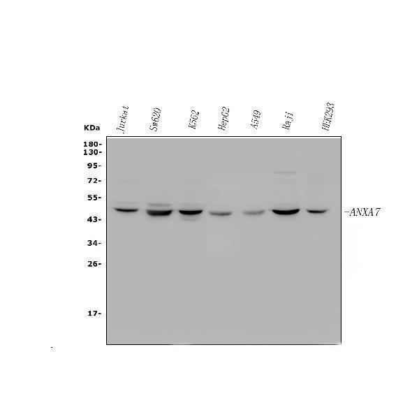
Figure 1. Western blot analysis of ANXA7 using anti-ANXA7 antibody (PA2076).
Electrophoresis was performed on a 5-20% SDS-PAGE gel at 70V (Stacking gel) / 90V (Resolving gel) for 2-3 hours. The sample well of each lane was loaded with 30 ug of sample under reducing conditions.
Lane 1: human Jurkat whole cell lysates,
Lane 2: human SW620 whole cell lysates,
Lane 3: human K562 whole cell lysates,
Lane 4: human HepG2 whole cell lysates,
Lane 5: human A549 whole cell lysates,
Lane 6: human Raji whole cell lysates,
Lane 7: human HEK293 whole cell lysates.
After electrophoresis, proteins were transferred to a nitrocellulose membrane at 150 mA for 50-90 minutes. Blocked the membrane with 5% non-fat milk/TBS for 1.5 hour at RT. The membrane was incubated with rabbit anti-ANXA7 antigen affinity purified polyclonal antibody (Catalog # PA2076) at 0.5 μg/mL overnight at 4°C, then washed with TBS-0.1%Tween 3 times with 5 minutes each and probed with a goat anti-rabbit IgG-HRP secondary antibody at a dilution of 1:5000 for 1.5 hour at RT. The signal is developed using an Enhanced Chemiluminescent detection (ECL) kit (Catalog # EK1002) with Tanon 5200 system. A specific band was detected for ANXA7 at approximately 50 kDa. The expected band size for ANXA7 is at 53 kDa.

Click image to see more details
Figure 2. IHC analysis of ANXA7 using anti-ANXA7 antibody (PA2076).
ANXA7 was detected in a paraffin-embedded section of Human Mammary Cancer tissue. Heat mediated antigen retrieval was performed in EDTA buffer (pH 8.0, epitope retrieval solution). The tissue section was blocked with 10% goat serum. The tissue section was then incubated with 1 μg/ml rabbit anti-ANXA7 Antibody (PA2076) overnight at 4°C. Biotinylated goat anti-rabbit IgG was used as secondary antibody and incubated for 30 minutes at 37°C. The tissue section was developed using Strepavidin-Biotin-Complex (SABC) (Catalog # SA1022) with DAB as the chromogen.

Click image to see more details
Figure 3. IF analysis of ANXA7 using anti- ANXA7 antibody (PA2076).
ANXA7 was detected in immunocytochemical section of A431 cells. Enzyme antigen retrieval was performed using IHC enzyme antigen retrieval reagent (AR0022) for 15 mins. The cells were blocked with 10% goat serum. And then incubated with 5μg/mL rabbit anti- ANXA7 Antibody (PA2076) overnight at 4°C. DyLight®594 Conjugated Goat Anti-Rabbit IgG (BA1142) was used as secondary antibody at 1:100 dilution and incubated for 30 minutes at 37°C. The section was counterstained with DAPI. Visualize using a fluorescence microscope and filter sets appropriate for the label used.

Click image to see more details
Figure 4. Flow Cytometry analysis of HepG2 cells using anti-ANXA7 antibody (PA2076).
Overlay histogram showing HepG2 cells stained with PA2076 (Blue line).The cells were blocked with 10% normal goat serum. And then incubated with rabbit anti-ANXA7 Antibody (PA2076, 1μg/1x106 cells) for 30 min at 20°C. DyLight®488 conjugated goat anti-rabbit IgG (BA1127, 5-10μg/1x106 cells) was used as secondary antibody for 30 minutes at 20°C. Isotype control antibody (Green line) was rabbit IgG (1μg/1x106) used under the same conditions. Unlabelled sample (Red line) was also used as a control.
Protein Target Info & Infographic
Gene/Protein Information For ANXA7 (Source: Uniprot.org, NCBI)
Gene Name
ANXA7
Full Name
Annexin A7
Weight
52.739kDa
Superfamily
annexin family
Alternative Names
Annexin A7; ANX7; ANXA7; SNX; Synexin ANXA7 ANX7, SNX, SYNEXIN annexin A7 annexin A7|annexin VII|annexin-7
*If product is indicated to react with multiple species, protein info is based on the gene entry specified above in "Species".For more info on ANXA7, check out the ANXA7 Infographic

We have 30,000+ of these available, one for each gene! Check them out.
In this infographic, you will see the following information for ANXA7: database IDs, superfamily, protein function, synonyms, molecular weight, chromosomal locations, tissues of expression, subcellular locations, post-translational modifications, and related diseases, research areas & pathways. If you want to see more information included, or would like to contribute to it and be acknowledged, please contact [email protected].
Specific Publications For Anti-Annexin VII/ANXA7 Antibody (PA2076)
Hello CJ!
PA2076 has been cited in 1 publications:
*The publications in this section are manually curated by our staff scientists. They may differ from Bioz's machine gathered results. Both are accurate. If you find a publication citing this product but is missing from this list, please let us know we will issue you a thank-you coupon.
Zhou Sn, Li Cs, Liu Lq, Li Y, Wang Xf, Shen L. Histol Histopathol. 2011 May;26(5):571-9. Increased Expression Of Annexin A7 In Temporal Lobe Tissue Of Patients With Refractory Epilepsy.
Recommended Resources
Here are featured tools and databases that you might find useful.
- Boster's Pathways Library
- Protein Databases
- Bioscience Research Protocol Resources
- Data Processing & Analysis Software
- Photo Editing Software
- Scientific Literature Resources
- Research Paper Management Tools
- Molecular Biology Software
- Primer Design Tools
- Bioinformatics Tools
- Phylogenetic Tree Analysis
Customer Reviews
Have you used Anti-Annexin VII/ANXA7 Antibody?
Submit a review and receive an Amazon gift card.
- $30 for a review with an image
Be the first to review Anti-Annexin VII/ANXA7 Antibody
*The first user to submit a review for a product is eligible for Boster's Innovating Scientists Reward, which gives product credits. This is in addition to the gift card reward.
Customer Q&As
Have a question?
Find answers in Q&As, reviews.
Can't find your answer?
Submit your question
4 Customer Q&As for Anti-Annexin VII/ANXA7 Antibody
Question
Will anti-Annexin VII/ANXA7 antibody PA2076 work for IHC with liver?
Verified Customer
Verified customer
Asked: 2019-03-05
Answer
According to the expression profile of liver, ANXA7 is highly expressed in liver. So, it is likely that anti-Annexin VII/ANXA7 antibody PA2076 will work for IHC with liver.
Boster Scientific Support
Answered: 2019-03-05
Question
Will PA2076 anti-Annexin VII/ANXA7 antibody work on parafin embedded sections? If so, which fixation method do you recommend we use (PFA, paraformaldehyde, other)?
O. Patel
Verified customer
Asked: 2016-12-07
Answer
As indicated on the product datasheet, PA2076 anti-Annexin VII/ANXA7 antibody as been tested on IHC. It is best to use PFA for fixation because it has better tissue penetration ability. PFA needs to be prepared fresh before use. Long term stored PFA turns into formalin, as the PFA molecules congregate and become formalin.
Boster Scientific Support
Answered: 2016-12-07
Question
I see that the anti-Annexin VII/ANXA7 antibody PA2076 works with IHC, what is the protocol used to produce the result images on the product page?
Z. Jha
Verified customer
Asked: 2016-04-26
Answer
You can find protocols for IHC on the "support/technical resources" section of our navigation menu. If you have any further questions, please send an email to [email protected]
Boster Scientific Support
Answered: 2016-04-26
Question
We are currently using anti-Annexin VII/ANXA7 antibody PA2076 for rat tissue, and we are happy with the IHC results. The species of reactivity given in the datasheet says human, mouse, rat. Is it possible that the antibody can work on pig tissues as well?
T. Miller
Verified customer
Asked: 2016-03-09
Answer
The anti-Annexin VII/ANXA7 antibody (PA2076) has not been tested for cross reactivity specifically with pig tissues, though there is a good chance of cross reactivity. We have an innovator award program that if you test this antibody and show it works in pig you can get your next antibody for free. Please contact me if I can help you with anything.
Boster Scientific Support
Answered: 2016-03-09
