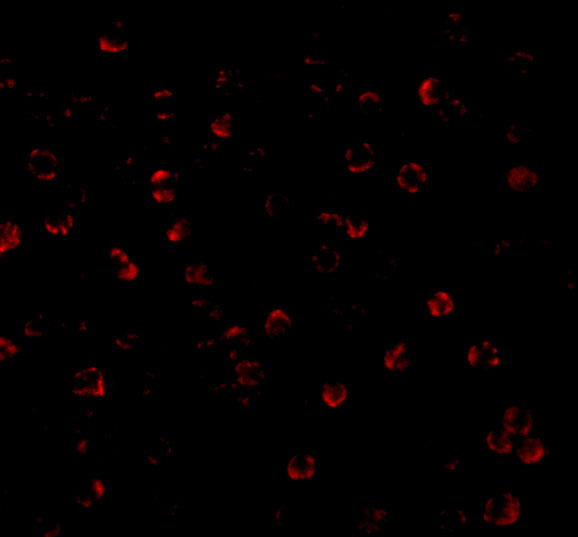
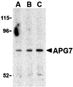
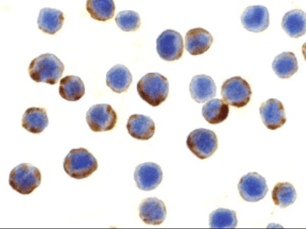
ATG7 antibody (A00346-1) performs well in ELISA, IF, ICC, WB and shows reactivity with Human, Mouse.
  • Validated for inter-lot consistency, specificity, and reproducibility
  • Committed to quality since 1993
Filter:
a00346 1 atg7 primary antibodies wb testing 1a00346 1 atg7 primary antibodies icc testing 2a00346 1 atg7 primary antibodies if testing 3

Anti-APG7 ATG7 Antibody

ATG7 antibody

Reactive SpeciesHostApplication
Human, MouseRabbitELISA, IF, ICC, WB

SKU: A00346-1

Size: 0.1 mg

Bundle to save: I do not want free secondary ab
Price:
$481/0.1 mg

In stock. Ships Next Business Day.

Quantity: 1

1
Get A Quote
Guarantee / Product Promise

Recommend Dilution

Application
Dilution
Species
APG7 antibody can be used for the detection of APG7 by Western blot at 0.5 - 2 μg/mL. Antibody can also be used for immunocytochemistry starting at 10 μg/mL. For immunofluorescence start at 20 μg/mL.
Antibody validated: Western Blot in human samples; Immunocytochemistry in human samples and Immunofluorescence in human samples. All other applications and species not yet tested. Optimal dilutions for each application should be determined by the researcher.

* It is recommended to optimize the amount of this reagent in each experimental system to achieve the best results.

Product Information

Product Name
Anti-APG7 ATG7 Antibody
View all ATG7 Antibodies
Form
Liquid
Uniprot
Gene ID
Storage & Handling
APG7 antibody can be stored at 4°C, stable for up to one year. Avoid repeated freeze-thaw cycles. Antibodies should not be exposed to prolonged high temperatures.
Host
Rabbit
Contents
APG7 Antibody is supplied in PBS containing ≤0.09% sodium azide.
Clonality
Polyclonal
Isotype
IgG
Immunogen
APG7 antibody was raised against a 17 amino acid synthetic peptide from near the carboxy terminus of human APG7. The immunogen is located within amino acids 590 - 640 of APG7.
Cross-Reactivity
At least three isoforms of APG7 are known to exist; this antibody will detect all three isoforms. APG7 antibody is predicted not to cross-react with other ATG family proteins.
Observed Molecular Weight
68 kDa
Calculated Molecular Weight

78.0 kDa

Background of ATG7
Autophagy, the process of bulk degradation of cellular proteins through an autophagosomic-lysosomal pathway is important for normal growth control and may be defective in tumor cells. It is involved in the preservation of cellular nutrients under starvation conditions as well as the normal turnover of cytosolic components. This process is negatively regulated by TOR (Target of rapamycin) through phosphorylation of autophagy protein APG1. Another member of the autophagy family of proteins is APG7 which was identified in yeast as a ubiquitin-E1-like enzyme; this function is conserved in the mammalian homolog. In mammalian cells, APG7 is essential for autophagy conjugation systems, autophagosome formation, starvation-induced bulk degradation of proteins and organelles. It has been suggested that caspase-8 may alter APG7 levels and thus the APG7 program of autophagic cell death.
Cite This Product
Anti-APG7 ATG7 Antibody (Boster Biological Technology, Pleasanton CA, USA, Catalog # A00346-1)

Review

Have you used Anti-APG7 ATG7 Antibody?

Submit a review and receive an Amazon gift card.

$30 for a review with an image

Submit A Review

Product Promise

We promise Anti-APG7 ATG7 Antibody performs excellently in relevant applications for ELISA, IF, ICC, WB. If it doesn't perform as promised, we will refund the product. Learn more


In stock
Order Product
A00346-1
Buy one primary antibody get one 0.5ml HRP or Biotin secondary antibody for free.
*Sample sizes are prepared on demand and will take extra lead time. (cannot be conjugated)
$481.00

Troubleshooting

troubleshooting-box-image

Download troubleshooting handbooks for IHC, Western blot and ELISA for FREE.

Download Free PDFs Now

Boster Guarantee

Guaranteed product quality

Guaranteed product quality

We promise all of our products perform as described in datasheets.

This product is for Research Use Only. Not for use in diagnostic procedures.

Update date: Sep 16, 2026