Click image to see more details
-

-
-
-
-
+3
Product Info Summary
| SKU: | A05997-2 |
|---|---|
| Size: | 100 μg/vial |
| Reactive Species: | Human, Mouse, Rat |
| Host: | Rabbit |
| Application: | ELISA, Flow Cytometry, IF, IHC, ICC, WB |
Customers Who Bought This Also Bought


Product info
Product Name
Anti-BRSK1 Antibody Picoband™
SKU/Catalog Number
A05997-2
Size
100 μg/vial
Form
Lyophilized
Description
Boster Bio Anti-BRSK1 Antibody Picoband™ catalog # A05997-2. Tested in ELISA, Flow Cytometry, IF, IHC, ICC, WB applications. This antibody reacts with Human, Mouse, Rat.
Storage & Handling
At -20°C for one year from date of receipt. After reconstitution, at 4°C for one month. It can also be aliquotted and stored frozen at -20°C for six months. Avoid repeated freezing and thawing.
Cite This Product
Anti-BRSK1 Antibody Picoband™ (Boster Biological Technology, Pleasanton CA, USA, Catalog # A05997-2)
Host
Rabbit
Contents
Each vial contains 4 mg Trehalose, 0.9 mg NaCl, 0.2 mg Na2HPO4.
Clonality
Polyclonal
Isotype
Rabbit IgG
Immunogen
E.coli-derived human BRSK1 recombinant protein (Position: E269-E321).
*Blocking peptide can be purchased. Costs vary based on immunogen length. Contact us for pricing.
Cross-reactivity
No cross-reactivity with other proteins.
Reactive Species
A05997-2 is reactive to BRSK1 in Human, Mouse, Rat
Applications
A05997-2 is guaranteed for ELISA, Flow Cytometry, IF, IHC, ICC, WB Boster Guarantee
Observed Molecular Weight
90 kDa
Calculated molecular weight
85.087kDa
Background of BRSK1
BR serine/threonine kinase 1 is an enzyme that in humans is encoded by the BRSK1 gene. BRSK1 was initially identified as a mammalian homolog to the fission yeast S. pombe Cdr2, a mitosis-regulatory kinase and also shows significant homology to the C. elegans neuronal cell polarity regulator SAD1. BRSK1 is unbiquitously expressed, with highest levels of expression in the brain and testes. Similar to its yeast homolog, BRSK1 is thought to be involved in stress-induced cell cycle arrest. Overexpression of this protein leads to the G2/M arrest in HeLa S2 cells and UV-induced G2/M arrest could be partially abrogated by reduced expression of BRSK1 through the use of siRNA, indicating its role in DNA damage checkpoint function. More recently, it has been shown that both BRSK1 and the related protein BRSK2 are required for mammalian neuronal polarization. While BRSK1- and BRSK2-null mice were viable, double-mutant mice died within two hours of birth. Neurons from these mice showed uniformly-sized neurites as opposed to the normal long axon and multiple shorter dendrites. These neurites also displayed both axonal and dendritic markers. At least two isoforms of BRSK1 are known to exist.
Antibody Validation
Boster validates all antibodies on WB, IHC, ICC, Immunofluorescence, and ELISA with known positive control and negative samples to ensure specificity and high affinity, including thorough antibody incubations.
Assay dilution & Images
Reconsitution
Adding 0.2 ml of distilled water will yield a concentration of 500 μg/ml.
Assay Dilutions Recommendation
The recommendations below provide a starting point for assay optimization. The actual working concentration varies and should be decided by the user.
Western blot, 0.1-0.25 μg/ml, Human
Immunohistochemistry(Paraffin-embedded Section), 2-5 μg/ml, Human, Mouse, Rat
Immunocytochemistry/Immunofluorescence, 5 μg/ml, Human
Flow Cytometry, 1-3 μg/1x106 cells, Human
Direct ELISA, 0.1-0.5 μg/ml, Human
Validation Images & Assay Conditions
Click image to see more details
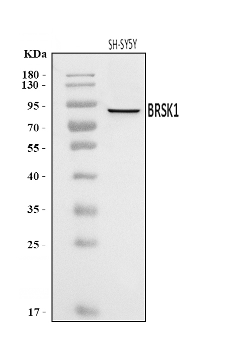
Figure 1. Western blot analysis of BRSK1 using anti-BRSK1 antibody (A05997-2).
Electrophoresis was performed on a 5-20% SDS-PAGE gel at 70V (Stacking gel) / 90V (Resolving gel) for 2-3 hours. The sample well of each lane was loaded with 30 ug of sample under reducing conditions.
Lane 1: human SH-SY5Y whole cell lysates.
After electrophoresis, proteins were transferred to a nitrocellulose membrane at 150 mA for 50-90 minutes. Blocked the membrane with 5% non-fat milk/TBS for 1.5 hour at RT. The membrane was incubated with rabbit anti-BRSK1 antigen affinity purified polyclonal antibody (Catalog # A05997-2) at 0.25 μg/mL overnight at 4°C, then washed with TBS-0.1%Tween 3 times with 5 minutes each and probed with a goat anti-rabbit IgG-HRP secondary antibody at a dilution of 1:5000 for 1.5 hour at RT. The signal is developed using an Enhanced Chemiluminescent detection (ECL) kit (Catalog # EK1002) with Tanon 5200 system. A specific band was detected for BRSK1 at approximately 90 kDa. The expected band size for BRSK1 is at 90 kDa.

Click image to see more details
Figure 2. IHC analysis of BRSK1 using anti-BRSK1 antibody (A05997-2).
BRSK1 was detected in a paraffin-embedded section of human laryngeal carcinoma tissue. Heat mediated antigen retrieval was performed in EDTA buffer (pH 8.0, epitope retrieval solution). The tissue section was blocked with 10% goat serum. The tissue section was then incubated with 2 μg/ml rabbit anti-BRSK1 Antibody (A05997-2) overnight at 4°C. Biotinylated goat anti-rabbit IgG was used as secondary antibody and incubated for 30 minutes at 37°C. The tissue section was developed using Strepavidin-Biotin-Complex (SABC) (Catalog # SA1022) with DAB as the chromogen.

Click image to see more details
Figure 3. IHC analysis of BRSK1 using anti-BRSK1 antibody (A05997-2).
BRSK1 was detected in a paraffin-embedded section of human placenta tissue. Heat mediated antigen retrieval was performed in EDTA buffer (pH 8.0, epitope retrieval solution). The tissue section was blocked with 10% goat serum. The tissue section was then incubated with 2 μg/ml rabbit anti-BRSK1 Antibody (A05997-2) overnight at 4°C. Biotinylated goat anti-rabbit IgG was used as secondary antibody and incubated for 30 minutes at 37°C. The tissue section was developed using Strepavidin-Biotin-Complex (SABC) (Catalog # SA1022) with DAB as the chromogen.

Click image to see more details
Figure 4. IHC analysis of BRSK1 using anti-BRSK1 antibody (A05997-2).
BRSK1 was detected in a paraffin-embedded section of mouse brain tissue. Heat mediated antigen retrieval was performed in EDTA buffer (pH 8.0, epitope retrieval solution). The tissue section was blocked with 10% goat serum. The tissue section was then incubated with 2 μg/ml rabbit anti-BRSK1 Antibody (A05997-2) overnight at 4°C. Biotinylated goat anti-rabbit IgG was used as secondary antibody and incubated for 30 minutes at 37°C. The tissue section was developed using Strepavidin-Biotin-Complex (SABC) (Catalog # SA1022) with DAB as the chromogen.

Click image to see more details
Figure 5. IHC analysis of BRSK1 using anti-BRSK1 antibody (A05997-2).
BRSK1 was detected in a paraffin-embedded section of rat brain tissue. Heat mediated antigen retrieval was performed in EDTA buffer (pH 8.0, epitope retrieval solution). The tissue section was blocked with 10% goat serum. The tissue section was then incubated with 2 μg/ml rabbit anti-BRSK1 Antibody (A05997-2) overnight at 4°C. Biotinylated goat anti-rabbit IgG was used as secondary antibody and incubated for 30 minutes at 37°C. The tissue section was developed using Strepavidin-Biotin-Complex (SABC) (Catalog # SA1022) with DAB as the chromogen.

Click image to see more details
Figure 6. IF analysis of BRSK1 using anti-BRSK1 antibody (A05997-2).
BRSK1 was detected in an immunocytochemical section of A431 cells. Enzyme antigen retrieval was performed using IHC enzyme antigen retrieval reagent (AR0022) for 15 mins. The cells were blocked with 10% goat serum. And then incubated with 5 μg/mL rabbit anti-BRSK1 Antibody (A05997-2) overnight at 4°C. DyLight®488 Conjugated Goat Anti-Rabbit IgG (BA1127) was used as secondary antibody at 1:100 dilution and incubated for 30 minutes at 37°C. The section was counterstained with DAPI. Visualize using a fluorescence microscope and filter sets appropriate for the label used.

Click image to see more details
Figure 7. Flow Cytometry analysis of CACO-2 cells using anti-BRSK1 antibody (A05997-2).
Overlay histogram showing CACO-2 cells stained with A05997-2 (Blue line). The cells were blocked with 10% normal goat serum. And then incubated with rabbit anti-BRSK1 Antibody (A05997-2, 1 μg/1x106 cells) for 30 min at 20°C. DyLight®488 conjugated goat anti-rabbit IgG (BA1127, 5-10 μg/1x106 cells) was used as secondary antibody for 30 minutes at 20°C. Isotype control antibody (Green line) was rabbit IgG (1 μg/1x106) used under the same conditions. Unlabelled sample (Red line) was also used as a control.
Protein Target Info & Infographic
Gene/Protein Information For BRSK1 (Source: Uniprot.org, NCBI)
Gene Name
BRSK1
Full Name
Serine/threonine-protein kinase BRSK1
Weight
85.087kDa
Superfamily
protein kinase superfamily
Alternative Names
BR serine/threonine kinase 1; EC 2.7.11; EC 2.7.11.1; FLJ43009; KIAA1811BR serine/threonine-protein kinase 1; protein kinase SAD1A; SAD1 kinase; SAD1; Serine/threonine-protein kinase SAD-B BRSK1 hSAD1 BR serine/threonine kinase 1 serine/threonine-protein kinase BRSK1|BR serine/threonine-protein kinase 1|SAD1 homolog|SAD1 kinase|brain-selective kinase 1|brain-specific serine/threonine-protein kinase 1|protein kinase SAD1A|serine/threonine-protein kinase SAD-B|synapses of Amphids Defective homolog 1
*If product is indicated to react with multiple species, protein info is based on the gene entry specified above in "Species".For more info on BRSK1, check out the BRSK1 Infographic

We have 30,000+ of these available, one for each gene! Check them out.
In this infographic, you will see the following information for BRSK1: database IDs, superfamily, protein function, synonyms, molecular weight, chromosomal locations, tissues of expression, subcellular locations, post-translational modifications, and related diseases, research areas & pathways. If you want to see more information included, or would like to contribute to it and be acknowledged, please contact [email protected].
Specific Publications For Anti-BRSK1 Antibody Picoband™ (A05997-2)
Hello CJ!
No publications found for A05997-2
*Do you have publications using this product? Share with us and receive a reward. Ask us for more details.
Recommended Resources
Here are featured tools and databases that you might find useful.
- Boster's Pathways Library
- Protein Databases
- Bioscience Research Protocol Resources
- Data Processing & Analysis Software
- Photo Editing Software
- Scientific Literature Resources
- Research Paper Management Tools
- Molecular Biology Software
- Primer Design Tools
- Bioinformatics Tools
- Phylogenetic Tree Analysis
Customer Reviews
Have you used Anti-BRSK1 Antibody Picoband™?
Submit a review and receive an Amazon gift card.
- $30 for a review with an image
0 Reviews For Anti-BRSK1 Antibody Picoband™
Customer Q&As
Have a question?
Find answers in Q&As, reviews.
Can't find your answer?
Submit your question
