Click image to see more details
-

-
-
-
-
+3
Product Info Summary
| SKU: | A02285-2 |
|---|---|
| Size: | 100 μg/vial |
| Reactive Species: | Human, Monkey, Mouse, Rat |
| Host: | Rabbit |
| Application: | ELISA, IF, IHC, WB |
Customers Who Bought This Also Bought


Product info
Product Name
Anti-Mannose Receptor/MRC1 Picoband™ Antibody
View all MMR/CD206/Mannose Receptor Antibodies
SKU/Catalog Number
A02285-2
Size
100 μg/vial
Form
Lyophilized
Description
Boster Bio Anti-Mannose Receptor/MRC1 Picoband™ Antibody catalog # A02285-2. Tested in ELISA, IF, IHC, WB applications. This antibody reacts with Human, Monkey, Mouse, Rat.
Storage & Handling
Store at -20˚C for one year from date of receipt. After reconstitution, at 4˚C for one month. It can also be aliquotted and stored frozen at -20˚C for six months. Avoid repeated freeze-thaw cycles.
Cite This Product
Anti-Mannose Receptor/MRC1 Picoband™ Antibody (Boster Biological Technology, Pleasanton CA, USA, Catalog # A02285-2)
Host
Rabbit
Contents
Each vial contains 4mg Trehalose, 0.9mg NaCl, 0.2mg Na2HPO4, 0.01mg NaN3.
Clonality
Polyclonal
Isotype
Rabbit IgG
Immunogen
E.coli-derived human Mannose Receptor/MRC1 recombinant protein (Position: D21-A1140).
*Blocking peptide can be purchased. Costs vary based on immunogen length. Contact us for pricing.
Cross-reactivity
No cross-reactivity with other proteins.
Reactive Species
A02285-2 is reactive to MRC1 in Human, Monkey, Mouse, Rat
Applications
A02285-2 is guaranteed for ELISA, IF, IHC, WB Boster Guarantee
Observed Molecular Weight
190-200 kDa
Calculated molecular weight
166.012kDa
Background of MMR/CD206/Mannose Receptor
The mannose receptor (Cluster of Differentiation 206, CD206) is a C-type lectin primarily present on the surface of macrophages, immature dendritic cells and liver sinusoidal endothelial cells, but is also expressed on the surface of skin cells such as human dermal fibroblasts and keratinocytes. It is mapped to 10p12.33. The recognition of complex carbohydrate structures on glycoproteins is an important part of several biological processes, including cell-cell recognition, serum glycoprotein turnover, and neutralization of pathogens. The protein encoded by this gene is a type I membrane receptor that mediates the endocytosis of glycoproteins by macrophages. The protein has been shown to bind high-mannose structures on the surface of potentially pathogenic viruses, bacteria, and fungi so that they can be neutralized by phagocytic engulfment.
Antibody Validation
Boster validates all antibodies on WB, IHC, ICC, Immunofluorescence, and ELISA with known positive control and negative samples to ensure specificity and high affinity, including thorough antibody incubations.
Innovating Scientists Reward
If you are the first to review this product, or if you have results for a special sample, species or application this product is not validated in, share your results with us and receive product credits you can use towards any Boster products! Applicable to all scientists worldwide.
Submit A Review
Assay dilution & Images
Reconsitution
Add 0.2ml of distilled water will yield a concentration of 500ug/ml.
Assay Dilutions Recommendation
The recommendations below provide a starting point for assay optimization. The actual working concentration varies and should be decided by the user.
Western blot, 0.25-0.5μg/ml, Human, Monkey, Mouse, Rat
Immunohistochemistry (Paraffin-embedded Section), 2-5μg/ml, Human, Mouse, Rat
Immunofluorescence, 5 μg/ml, Human, Mouse
Direct ELISA, 0.1-0.5μg/ml, Human
Validation Images & Assay Conditions
Click image to see more details
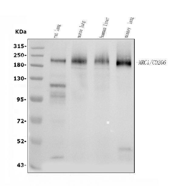
Figure 1. Western blot analysis of MRC1 using anti-MRC1 antibody (A02285-2).
Electrophoresis was performed on a 5-20% SDS-PAGE gel at 70V (Stacking gel) / 90V (Resolving gel) for 2-3 hours. The sample well of each lane was loaded with 30 ug of sample under reducing conditions.
Lane 1: rat lung tissue lysates,
Lane 2: mouse lung tissue lysates,
Lane 3: human liver tissue lysates,
Lane 4: monkey lung tissue lysates.
After electrophoresis, proteins were transferred to a nitrocellulose membrane at 150 mA for 50-90 minutes. Blocked the membrane with 5% non-fat milk/TBS for 1.5 hour at RT. The membrane was incubated with rabbit anti-MRC1 antigen affinity purified polyclonal antibody (Catalog # A02285-2) at 0.5 μg/mL overnight at 4°C, then washed with TBS-0.1%Tween 3 times with 5 minutes each and probed with a goat anti-rabbit IgG-HRP secondary antibody at a dilution of 1:5000 for 1.5 hour at RT. The signal is developed using an Enhanced Chemiluminescent detection (ECL) kit (Catalog # EK1002) with Tanon 5200 system. A specific band was detected for MRC1 at approximately 190-200 kDa. The expected band size for MRC1 is at 166 kDa.

Click image to see more details
Figure 2. IHC analysis of MRC1 using anti-MRC1 antibody (A02285-2).
MRC1 was detected in a paraffin-embedded section of human liver cancer tissue. Heat mediated antigen retrieval was performed in EDTA buffer (pH 8.0, epitope retrieval solution). The tissue section was blocked with 10% goat serum. The tissue section was then incubated with 2 μg/ml rabbit anti-MRC1 Antibody (A02285-2) overnight at 4°C. Peroxidase Conjugated Goat Anti-rabbit IgG was used as secondary antibody and incubated for 30 minutes at 37°C. The tissue section was developed using HRP Conjugated Rabbit IgG Super Vision Assay Kit (Catalog # SV0002) with DAB as the chromogen.

Click image to see more details
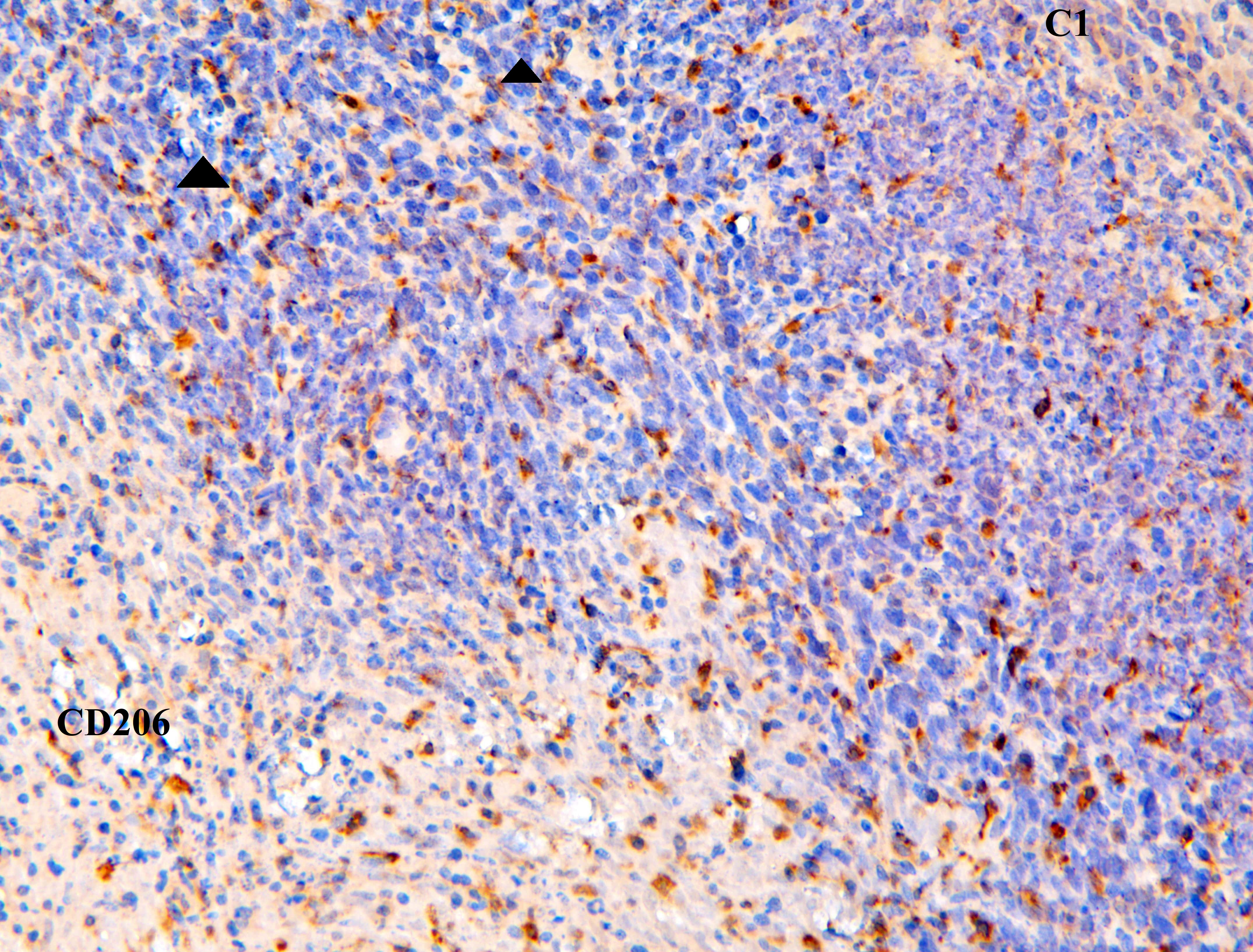
Figure 3. IHC analysis of MRC1 using anti-MRC1 antibody (A02285-2).
MRC1 was detected in a paraffin-embedded section of human tonsil tissue. Heat mediated antigen retrieval was performed in EDTA buffer (pH 8.0, epitope retrieval solution). The tissue section was blocked with 10% goat serum. The tissue section was then incubated with 2 μg/ml rabbit anti-MRC1 Antibody (A02285-2) overnight at 4°C. Peroxidase Conjugated Goat Anti-rabbit IgG was used as secondary antibody and incubated for 30 minutes at 37°C. The tissue section was developed using HRP Conjugated Rabbit IgG Super Vision Assay Kit (Catalog # SV0002) with DAB as the chromogen.

Click image to see more details
Figure 4. IHC analysis of MRC1 using anti-MRC1 antibody (A02285-2).
MRC1 was detected in a paraffin-embedded section of mouse liver tissue. Heat mediated antigen retrieval was performed in EDTA buffer (pH 8.0, epitope retrieval solution). The tissue section was blocked with 10% goat serum. The tissue section was then incubated with 2 μg/ml rabbit anti-MRC1 Antibody (A02285-2) overnight at 4°C. Peroxidase Conjugated Goat Anti-rabbit IgG was used as secondary antibody and incubated for 30 minutes at 37°C. The tissue section was developed using HRP Conjugated Rabbit IgG Super Vision Assay Kit (Catalog # SV0002) with DAB as the chromogen.

Click image to see more details
Figure 5. IHC analysis of MRC1 using anti-MRC1 antibody (A02285-2).
MRC1 was detected in a paraffin-embedded section of rat liver tissue. Heat mediated antigen retrieval was performed in EDTA buffer (pH 8.0, epitope retrieval solution). The tissue section was blocked with 10% goat serum. The tissue section was then incubated with 2 μg/ml rabbit anti-MRC1 Antibody (A02285-2) overnight at 4°C. Peroxidase Conjugated Goat Anti-rabbit IgG was used as secondary antibody and incubated for 30 minutes at 37°C. The tissue section was developed using HRP Conjugated Rabbit IgG Super Vision Assay Kit (Catalog # SV0002) with DAB as the chromogen.

Click image to see more details
Figure 6. IF analysis of MRC1 using anti-MRC1 antibody (A02285-2).
MRC1 was detected in a paraffin-embedded section of human tonsil tissue. Heat mediated antigen retrieval was performed in EDTA buffer (pH 8.0, epitope retrieval solution). The tissue section was blocked with 10% goat serum. The tissue section was then incubated with 5 μg/mL rabbit anti-MRC1 Antibody (A02285-2) overnight at 4°C. DyLight®550 Conjugated Goat Anti-Rabbit IgG (BA1135) was used as secondary antibody at 1:500 dilution and incubated for 30 minutes at 37°C. The section was counterstained with DAPI. Visualize using a fluorescence microscope and filter sets appropriate for the label used.

Click image to see more details
Figure 7. IF analysis of MRC1 using anti-MRC1 antibody (A02285-2).
MRC1 was detected in a paraffin-embedded section of mouse liver tissue. Heat mediated antigen retrieval was performed in EDTA buffer (pH 8.0, epitope retrieval solution). The tissue section was blocked with 10% goat serum. The tissue section was then incubated with 5 μg/mL rabbit anti-MRC1 Antibody (A02285-2) overnight at 4°C. DyLight®550 Conjugated Goat Anti-Rabbit IgG (BA1135) was used as secondary antibody at 1:500 dilution and incubated for 30 minutes at 37°C. The section was counterstained with DAPI. Visualize using a fluorescence microscope and filter sets appropriate for the label used.
Protein Target Info & Infographic
Gene/Protein Information For MRC1 (Source: Uniprot.org, NCBI)
Gene Name
MRC1
Full Name
Macrophage mannose receptor 1
Weight
166.012kDa
Alternative Names
CD206; CLEC13D; CLEC13Dmacrophage mannose receptor 1; C-type lectin domain family 13 member D; mannose receptor, C type 1; MMR; MMRCD206 antigen; MRC1 MRC1 CD206, CLEC13D, CLEC13DL, MMRL1, bA541I19.1, hMR, MRC1 mannose receptor C-type 1 macrophage mannose receptor 1|C-type lectin domain family 13 member D|human mannose receptor|macrophage mannose receptor 1-like protein 1|mannose receptor, C type 1-like 1
*If product is indicated to react with multiple species, protein info is based on the gene entry specified above in "Species".For more info on MRC1, check out the MRC1 Infographic

We have 30,000+ of these available, one for each gene! Check them out.
In this infographic, you will see the following information for MRC1: database IDs, superfamily, protein function, synonyms, molecular weight, chromosomal locations, tissues of expression, subcellular locations, post-translational modifications, and related diseases, research areas & pathways. If you want to see more information included, or would like to contribute to it and be acknowledged, please contact [email protected].
Specific Publications For Anti-Mannose Receptor/MRC1 Picoband™ Antibody (A02285-2)
Hello CJ!
A02285-2 has been cited in 8 publications:
*The publications in this section are manually curated by our staff scientists. They may differ from Bioz's machine gathered results. Both are accurate. If you find a publication citing this product but is missing from this list, please let us know we will issue you a thank-you coupon.
IL-1β pre-stimulation enhances the therapeutic effects of endometrial regenerative cells on experimental colitis
Protective Effects of Thalidomide on High-Glucose-Induced Podocyte Injury through In Vitro Modulation of Macrophage M1/M2 Differentiation
Protective Effects of Two Safflower Derived Compounds, Kaempferol and Hydroxysafflor Yellow A, on Hyperglycaemic Stress-Induced Podocyte Apoptosis via Modulating of Macrophage M1/M2 Polarization
Near infrared light-triggered on-demand Cur release from Gel-PDA@Cur composite hydrogel for antibacterial wound healing
Species: Rat
Yu D,Zhao Y,Wang H,Kong D,Jin W,Hu Y,Qin Y,Zhang B,Li X,Hao J,Li G,Wang H.IL-1β pre-stimulation enhances the therapeutic effects of endometrial regenerative cells on experimental colitis. Stem Cell Res Ther.2021 Jun 5;12(1):324.doi:10.1186/s13287-021-02392-9.PMID:34090510.
Species: Human,Mouse
A02285-2 usage in article: APP:IHC, SAMPLE:COLON TISSUE, DILUTION:1:100
Rong Ji,Lixiang Ma,Xinyu Chen et al. INCB24360 Suppresses M1-like Macrophage Formation and NLRP3 Expression Whereas Increases IL-1β Secretion in RAW264.7 and BV-2, 23 December 2020, PREPRINT (Version 1) available at Research Square [https://doi.org/10.212
Species: Mouse
A02285-2 usage in article: APP:IF, SAMPLE:RAW264.7 CELL AND BV-2 CELL, DILUTION:1:200
Liu H,Chen W,Zhao B,Quan W,Zhang Y,Zhou Y,Wan Z,Zhang X,Xue G,Li J,Luo S,Wang J,Liu Y,Zhen M,Zhao Y.Autologous bionic tissue for inguinal hernia repair.J Biomed Mater Res A.2020 Jun; 108(6):1351-1368.doi:10.1002/jbm.a.36907.Epub 2020 Mar 2.PMID:32090432.
Species: New Zealand white Rabbit
A02285-2 usage in article: APP:IHC, SAMPLE:MSCS, DILUTION:NA
Liao H,Li Y,Zhang X,Zhao X,Zheng D,Shen D,Li R. Protective Effects of Thalidomide on High-Glucose-Induced Podocyte Injury through In Vitro Modulation of Macrophage M1/M2 Differentiation.J Immunol Res.2020 Aug 27;2020:8263598.doi:10.1155/2020/ 8263598.PMID
Species: Mouse
A02285-2 usage in article: APP:WB, SAMPLE:MACROPHAGES, DILUTION:1:500
Recommended Resources
Here are featured tools and databases that you might find useful.
- Boster's Pathways Library
- Protein Databases
- Bioscience Research Protocol Resources
- Data Processing & Analysis Software
- Photo Editing Software
- Scientific Literature Resources
- Research Paper Management Tools
- Molecular Biology Software
- Primer Design Tools
- Bioinformatics Tools
- Phylogenetic Tree Analysis
Customer Reviews
Have you used Anti-Mannose Receptor/MRC1 Picoband™ Antibody?
Submit a review and receive an Amazon gift card.
- $30 for a review with an image
1 Reviews For Anti-Mannose Receptor/MRC1 Picoband™ Antibody
0
Immunohistochemistry for Anti-Mannose Receptor/MRC1
Excellent
| Application | Immunohistochemistry (paraffin-embedded) |
|---|---|
| Blocking step | 5% BSA as a blocking agent for 30 min at 37°C |
| Sample | Mouse skin |
| Fixative | Fixed with 4% paraformaldehyde |
| Primary Ab Incubation | 37°C for 30 minutes |
| Primary Ab Incubation diluent | 5% BSA in TBS |
| Primary Ab Concentration | 1ug/ml |
| Secondary Antibody | SABC kit from Boster Bio, (SA1022) |
| Secondary Ab Dilution | The kit was ready to use, no dilution needed |
| Secondary Ab Incubation | at 37°C for 30 min |
Verified Customer
Verified customer
Submitted 2020-02-06
Customer Q&As
Have a question?
Find answers in Q&As, reviews.
Can't find your answer?
Submit your question
