Click image to see more details
-

-
-
-
-
+3
Product Info Summary
| SKU: | A05052-1 |
|---|---|
| Size: | 100 μg/vial |
| Reactive Species: | Human, Mouse, Rat |
| Host: | Rabbit |
| Application: | IF, IHC, ICC, WB |
Customers Who Bought This Also Bought

Product info
Product Name
Anti-Musashi 1/Msi1 Antibody Picoband®
SKU/Catalog Number
A05052-1
Size
100 μg/vial
Form
Lyophilized
Description
Boster Bio Anti-Musashi 1/Msi1 Antibody Picoband® catalog # A05052-1. Tested in IF, IHC, ICC, WB applications. This antibody reacts with Human, Mouse, Rat. The brand Picoband indicates this is a premium antibody that guarantees superior quality, high affinity, and strong signals with minimal background in Western blot applications. Only our best-performing antibodies are designated as Picoband, ensuring unmatched performance.
Storage & Handling
Store at -20˚C for one year from date of receipt. After reconstitution, at 4˚C for one month. It can also be aliquotted and stored frozen at -20˚C for six months. Avoid repeated freeze-thaw cycles.
Cite This Product
Anti-Musashi 1/Msi1 Antibody Picoband® (Boster Biological Technology, Pleasanton CA, USA, Catalog # A05052-1)
Host
Rabbit
Contents
Each vial contains 5mg BSA, 0.9mg NaCl, 0.2mg Na2HPO4, 0.01mg NaN3.
Clonality
Polyclonal
Isotype
Rabbit IgG
Immunogen
A synthetic peptide corresponding to a sequence at the N-terminus of human Musashi 1/Msi1, identical to the related mouse and rat sequences.
*Blocking peptide can be purchased. Costs vary based on immunogen length. Contact us for pricing.
Cross-reactivity
No cross-reactivity with other proteins
Reactive Species
A05052-1 is reactive to MSI1 in Human, Mouse, Rat
Reconstitution
Add 0.2ml of distilled water will yield a concentration of 500ug/ml.
Observed Molecular Weight
39 kDa
Calculated molecular weight
39125 MW
Background of Musashi-1
RNA-binding protein Musashi homolog 1 is a protein that in humans is encoded by the MSI1 gene. This gene encodes a protein containing two conserved tandem RNA recognition motifs. Similar proteins in other species function as RNA-binding proteins and play central roles in posttranscriptional gene regulation. Expression of this gene has been correlated with the grade of the malignancy and proliferative activity in gliomas and melanomas. A pseudogene for this gene is located on chromosome 11q13.
Antibody Validation
Boster validates all antibodies on WB, IHC, ICC, Immunofluorescence, and ELISA with known positive control and negative samples to ensure specificity and high affinity, including thorough antibody incubations.
Application & Images
Applications
A05052-1 is guaranteed for IF, IHC, ICC, WB Boster Guarantee
Assay Dilutions Recommendation
The recommendations below provide a starting point for assay optimization. The actual working concentration varies and should be decided by the user.
Western blot, 0.1-0.5μg/ml, Human, Mouse, Rat
Immunohistochemistry (Paraffin-embedded Section), 0.5-1μg/ml, Human, Mouse, Rat, By Heat
Immunocytochemistry/Immunofluorescence, 5 μg/ml, Human
Positive Control
WB: human 293T whole cell, human T-47D whole cell, human Colo320 whole cell, rat brain tissue, mouse brain tissue
IHC: mouse intestine tissue, rat intestine tissue, rat brain tissue, human lung cancer tissue, human mammary cancer tissue
ICC/IF: MCF7 cell
FCM: CACO-2 cell
Validation Images & Assay Conditions
Click image to see more details
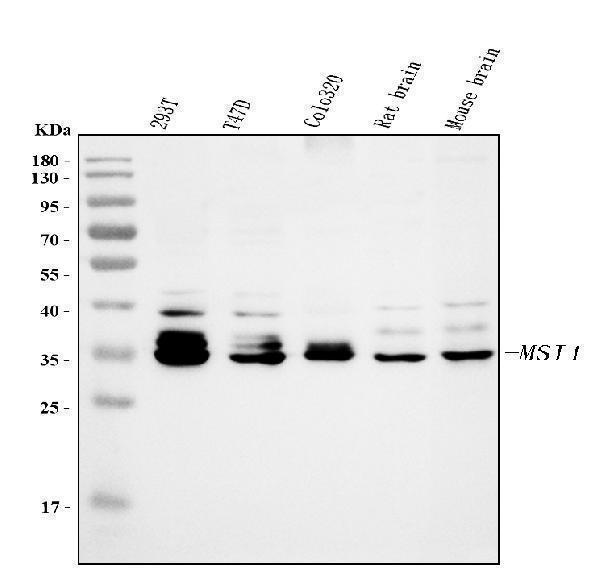
Figure 1. Western blot analysis of Musashi 1/Msi1 using anti-Musashi 1/Msi1 antibody (A05052-1).
Electrophoresis was performed on a 5-20% SDS-PAGE gel at 70V (Stacking gel) / 90V (Resolving gel) for 2-3 hours. The sample well of each lane was loaded with 30 ug of sample under reducing conditions.
Lane 1: human 293T whole cell lysates,
Lane 2: human T-47D whole cell lysates,
Lane 3: human Colo320 whole cell lysates,
Lane 4: rat brain tissue lysates,
Lane 5: mouse brain tissue lysates.
After electrophoresis, proteins were transferred to a nitrocellulose membrane at 150 mA for 50-90 minutes. Blocked the membrane with 5% non-fat milk/TBS for 1.5 hour at RT. The membrane was incubated with rabbit anti-Musashi 1/Msi1 antigen affinity purified polyclonal antibody (Catalog # A05052-1) at 0.5 μg/mL overnight at 4°C, then washed with TBS-0.1%Tween 3 times with 5 minutes each and probed with a goat anti-rabbit IgG-HRP secondary antibody at a dilution of 1:5000 for 1.5 hour at RT. The signal is developed using an Enhanced Chemiluminescent detection (ECL) kit (Catalog # EK1002) with Tanon 5200 system. A specific band was detected for Musashi 1/Msi1 at approximately 39 kDa. The expected band size for Musashi 1/Msi1 is at 39 kDa.

Click image to see more details
Figure 2. IHC analysis of Musashi 1/Msi1 using anti-Musashi 1/Msi1 antibody (A05052-1).
Musashi 1/Msi1 was detected in paraffin-embedded section of mouse intestine tissues. Heat mediated antigen retrieval was performed in citrate buffer (pH6, epitope retrieval solution) for 20 mins. The tissue section was blocked with 10% goat serum. The tissue section was then incubated with 1μg/ml rabbit anti-Musashi 1/Msi1 Antibody (A05052-1) overnight at 4°C. Biotinylated goat anti-rabbit IgG was used as secondary antibody and incubated for 30 minutes at 37°C. The tissue section was developed using Strepavidin-Biotin-Complex (SABC)(Catalog # SA1022) with DAB as the chromogen.

Click image to see more details
Figure 3. IHC analysis of Musashi 1/Msi1 using anti-Musashi 1/Msi1 antibody (A05052-1).
Musashi 1/Msi1 was detected in paraffin-embedded section of rat intestine tissues. Heat mediated antigen retrieval was performed in citrate buffer (pH6, epitope retrieval solution) for 20 mins. The tissue section was blocked with 10% goat serum. The tissue section was then incubated with 1μg/ml rabbit anti-Musashi 1/Msi1 Antibody (A05052-1) overnight at 4°C. Biotinylated goat anti-rabbit IgG was used as secondary antibody and incubated for 30 minutes at 37°C. The tissue section was developed using Strepavidin-Biotin-Complex (SABC)(Catalog # SA1022) with DAB as the chromogen.

Click image to see more details
Figure 4. IHC analysis of Musashi 1/Msi1 using anti-Musashi 1/Msi1 antibody (A05052-1).
Musashi 1/Msi1 was detected in paraffin-embedded section of rat brain tissues. Heat mediated antigen retrieval was performed in citrate buffer (pH6, epitope retrieval solution) for 20 mins. The tissue section was blocked with 10% goat serum. The tissue section was then incubated with 1μg/ml rabbit anti-Musashi 1/Msi1 Antibody (A05052-1) overnight at 4°C. Biotinylated goat anti-rabbit IgG was used as secondary antibody and incubated for 30 minutes at 37°C. The tissue section was developed using Strepavidin-Biotin-Complex (SABC)(Catalog # SA1022) with DAB as the chromogen.

Click image to see more details
Figure 5. IHC analysis of Musashi 1/Msi1 using anti-Musashi 1/Msi1 antibody (A05052-1).
Musashi 1/Msi1 was detected in paraffin-embedded section of human lung cancer tissues. Heat mediated antigen retrieval was performed in citrate buffer (pH6, epitope retrieval solution) for 20 mins. The tissue section was blocked with 10% goat serum. The tissue section was then incubated with 1μg/ml rabbit anti-Musashi 1/Msi1 Antibody (A05052-1) overnight at 4°C. Biotinylated goat anti-rabbit IgG was used as secondary antibody and incubated for 30 minutes at 37°C. The tissue section was developed using Strepavidin-Biotin-Complex (SABC)(Catalog # SA1022) with DAB as the chromogen.

Click image to see more details
Figure 6. IHC analysis of Musashi 1/Msi1 using anti-Musashi 1/Msi1 antibody (A05052-1).
Musashi 1/Msi1 was detected in paraffin-embedded section of human mammary cancer tissues. Heat mediated antigen retrieval was performed in citrate buffer (pH6, epitope retrieval solution) for 20 mins. The tissue section was blocked with 10% goat serum. The tissue section was then incubated with 1μg/ml rabbit anti-Musashi 1/Msi1 Antibody (A05052-1) overnight at 4°C. Biotinylated goat anti-rabbit IgG was used as secondary antibody and incubated for 30 minutes at 37°C. The tissue section was developed using Strepavidin-Biotin-Complex (SABC)(Catalog # SA1022) with DAB as the chromogen.

Click image to see more details
Figure 7. IF analysis of Musashi 1/Msi1 using anti-Musashi 1/Msi1 antibody (A05052-1).
Musashi 1/Msi1 was detected in an immunocytochemical section of MCF7 cells. Enzyme antigen retrieval was performed using IHC enzyme antigen retrieval reagent (AR0022) for 15 mins. The cells were blocked with 10% goat serum. And then incubated with 5 μg/mL rabbit anti-Musashi 1/Msi1 Antibody (A05052-1) overnight at 4°C. DyLight®488 Conjugated Goat Anti-Rabbit IgG (BA1127) was used as secondary antibody at 1:500 dilution and incubated for 30 minutes at 37°C. The section was counterstained with DAPI. Visualize using a fluorescence microscope and filter sets appropriate for the label used.
Protein Target Info & Infographic
Gene/Protein Information For MSI1 (Source: Uniprot.org, NCBI)
Gene Name
MSI1
Full Name
RNA-binding protein Musashi homolog 1
Weight
39125 MW
Superfamily
Musashi family
Alternative Names
RNA-binding protein Musashi homolog 1;Musashi-1;MSI1; MSI1 musashi RNA binding protein 1 RNA-binding protein Musashi homolog 1|musashi-1|musashi1
*If product is indicated to react with multiple species, protein info is based on the gene entry specified above in "Species".For more info on MSI1, check out the MSI1 Infographic

We have 30,000+ of these available, one for each gene! Check them out.
In this infographic, you will see the following information for MSI1: database IDs, superfamily, protein function, synonyms, molecular weight, chromosomal locations, tissues of expression, subcellular locations, post-translational modifications, and related diseases, research areas & pathways. If you want to see more information included, or would like to contribute to it and be acknowledged, please contact [email protected].
Specific Publications For Anti-Musashi 1/Msi1 Antibody Picoband® (A05052-1)
Hello CJ!
No publications found for A05052-1
*Do you have publications using this product? Share with us and receive a reward. Ask us for more details.
Recommended Resources
Here are featured tools and databases that you might find useful.
- Boster's Pathways Library
- Protein Databases
- Bioscience Research Protocol Resources
- Data Processing & Analysis Software
- Photo Editing Software
- Scientific Literature Resources
- Research Paper Management Tools
- Molecular Biology Software
- Primer Design Tools
- Bioinformatics Tools
- Phylogenetic Tree Analysis
Customer Reviews
Have you used Anti-Musashi 1/Msi1 Antibody Picoband®?
Submit a review and receive an Amazon gift card.
- $30 for a review with an image
0 Reviews For Anti-Musashi 1/Msi1 Antibody Picoband®
Customer Q&As
Have a question?
Find answers in Q&As, reviews.
Can't find your answer?
Submit your question
12 Customer Q&As for Anti-Musashi 1/Msi1 Antibody Picoband®
Question
We are interested in to test anti-Musashi 1/Msi1 antibody A05052-1 on mouse fetal brain for research purposes, then I may be interested in using anti-Musashi 1/Msi1 antibody A05052-1 for diagnostic purposes as well. Is the antibody suitable for diagnostic purposes?
Verified Customer
Verified customer
Asked: 2020-03-30
Answer
The products we sell, including anti-Musashi 1/Msi1 antibody A05052-1, are only intended for research use. They would not be suitable for use in diagnostic work. If you have the means to develop a product into diagnostic use, and are interested in collaborating with us and develop our product into an IVD product, please contact us for more discussions.
Boster Scientific Support
Answered: 2020-03-30
Question
Would anti-Musashi 1/Msi1 antibody A05052-1 work on goat IHC with fetal brain?
Verified Customer
Verified customer
Asked: 2020-03-16
Answer
Our lab technicians have not validated anti-Musashi 1/Msi1 antibody A05052-1 on goat. You can run a BLAST between goat and the immunogen sequence of anti-Musashi 1/Msi1 antibody A05052-1 to see if they may cross-react. If the sequence homology is close, then you can perform a pilot test. Keep in mind that since we have not validated goat samples, this use of the antibody is not covered by our guarantee. However we have an innovator award program that if you test this antibody and show it works in goat fetal brain in IHC, you can get your next antibody for free.
Boster Scientific Support
Answered: 2020-03-16
Question
I see that the anti-Musashi 1/Msi1 antibody A05052-1 works with WB, what is the protocol used to produce the result images on the product page?
Verified Customer
Verified customer
Asked: 2020-02-13
Answer
You can find protocols for WB on the "support/technical resources" section of our navigation menu. If you have any further questions, please send an email to [email protected]
Boster Scientific Support
Answered: 2020-02-13
Question
We appreciate helping with my inquiry over the phone. Here are the WB image, lot number and protocol we used for fetal brain using anti-Musashi 1/Msi1 antibody A05052-1. Let me know if you need anything else.
Verified Customer
Verified customer
Asked: 2019-09-10
Answer
Thanks for the data. You have provided everything we needed. Our lab team are working to resolve your inquiry as quickly as possible, and we appreciate your patience and understanding! Please let me know if there is anything you need in the meantime.
Boster Scientific Support
Answered: 2019-09-10
Question
Is this A05052-1 anti-Musashi 1/Msi1 antibody reactive to the isotypes of MSI1?
Verified Customer
Verified customer
Asked: 2019-07-05
Answer
The immunogen of A05052-1 anti-Musashi 1/Msi1 antibody is A synthetic peptide corresponding to a sequence at the N-terminus of human Musashi 1/Msi1 (21-54aa KMFIGGLSWQTTQEGLREYFGQFGEVKECLVMRD), identical to the related mouse and rat sequences. Could you tell me which isotype you are interested in so I can help see if the immunogen is part of this isotype?
Boster Scientific Support
Answered: 2019-07-05
Question
See below the WB image, lot number and protocol we used for fetal brain using anti-Musashi 1/Msi1 antibody A05052-1. Please let me know if you require anything else.
Verified Customer
Verified customer
Asked: 2019-05-24
Answer
Thank you very much for the data. Our lab team are working to resolve this as quickly as possible, and we appreciate your patience and understanding! You have provided everything we needed. Please let me know if there is anything you need in the meantime.
Boster Scientific Support
Answered: 2019-05-24
Question
Would A05052-1 anti-Musashi 1/Msi1 antibody work on parafin embedded sections? If so, which fixation method do you recommend we use (PFA, paraformaldehyde, other)?
Verified Customer
Verified customer
Asked: 2018-07-13
Answer
As indicated on the product datasheet, A05052-1 anti-Musashi 1/Msi1 antibody as been tested on WB. It is best to use PFA for fixation because it has better tissue penetration ability. PFA needs to be prepared fresh before use. Long term stored PFA turns into formalin, as the PFA molecules congregate and become formalin.
Boster Scientific Support
Answered: 2018-07-13
Question
Do you have a BSA free version of anti-Musashi 1/Msi1 antibody A05052-1 available?
S. Parker
Verified customer
Asked: 2018-06-05
Answer
Thank you for your recent telephone inquiry. I can confirm that some lots of this anti-Musashi 1/Msi1 antibody A05052-1 are BSA free. For now, these lots are available and we can make a BSA free formula for you free of charge. It will take 3 extra days to prepare. If you require this antibody BSA free again in future, please do not hesitate to contact me and I will be pleased to check which lots we have in stock that are BSA free.
Boster Scientific Support
Answered: 2018-06-05
Question
Will anti-Musashi 1/Msi1 antibody A05052-1 work for WB with fetal brain?
Verified Customer
Verified customer
Asked: 2017-06-19
Answer
According to the expression profile of fetal brain, MSI1 is highly expressed in fetal brain. So, it is likely that anti-Musashi 1/Msi1 antibody A05052-1 will work for WB with fetal brain.
Boster Scientific Support
Answered: 2017-06-19
Question
I was wanting to use your anti-Musashi 1/Msi1 antibody for WB for mouse fetal brain on frozen tissues, but I want to know if it has been tested for this particular application. Has this antibody been tested and is this antibody a good choice for mouse fetal brain identification?
Verified Customer
Verified customer
Asked: 2017-06-08
Answer
As indicated on the product datasheet, A05052-1 anti-Musashi 1/Msi1 antibody has been validated for IHC, WB on human, mouse, rat tissues. We have an innovator award program that if you test this antibody and show it works in mouse fetal brain in IHC-frozen, you can get your next antibody for free.
Boster Scientific Support
Answered: 2017-06-08
Question
I have a question about product A05052-1, anti-Musashi 1/Msi1 antibody. I was wondering if it would be possible to conjugate this antibody with biotin. I would need it to be without BSA or sodium azide. I am planning on using a buffer exchange of sodium azide with PBS only. Would there be problems for me to conjugate the antibody and store it in -20 degrees in small aliquots?
D. Krishna
Verified customer
Asked: 2016-06-03
Answer
We do not recommend storing this antibody with PBS buffer only in -20 degrees. If you want to store it in -20 degrees it is best to add some cryoprotectant like glycerol. If you want carrier free A05052-1 anti-Musashi 1/Msi1 antibody, we can provide it to you in a special formula with trehalose and/or glycerol. These molecules will not interfere with conjugation chemistry and provide a good level of protection for the antibody from degradation. Please be sure to specify this in your purchase order.
Boster Scientific Support
Answered: 2016-06-03
Question
Is a blocking peptide available for product anti-Musashi 1/Msi1 antibody (A05052-1)?
S. Jones
Verified customer
Asked: 2015-05-29
Answer
We do provide the blocking peptide for product anti-Musashi 1/Msi1 antibody (A05052-1). If you would like to place an order for it please contact [email protected] and make a special request.
Boster Scientific Support
Answered: 2015-05-29
