Product Info Summary
| SKU: | M00729-2 |
|---|---|
| Size: | 100ul |
| Reactive Species: | Dog, Human, Mouse, Rabbit, Rat |
| Host: | Mouse |
| Application: | WB |
Customers Who Bought This Also Bought
Product info
Product Name
Anti-NCAM-L1 L1CAM Monoclonal Antibody
SKU/Catalog Number
M00729-2
Size
100ul
Form
Liquid
Description
Boster Bio Anti-NCAM-L1 L1CAM Monoclonal Antibody catalog # M00729-2. Tested in WB applications. This antibody reacts with Canine, Human, Mouse, Rabbit, Rat.
Storage & Handling
Store at -20°C for one year. For short term storage and frequent use, store at 4°C for up to one month. Avoid repeated freeze-thaw cycles.
Cite This Product
Anti-NCAM-L1 L1CAM Monoclonal Antibody (Boster Biological Technology, Pleasanton CA, USA, Catalog # M00729-2)
Host
Mouse
Contents
Purified mouse monoclonal in buffer containing 0.1M Tris-Glycine (pH 7.4, 150 mM NaCl) with 0.2% sodium azide, 50% glycerol.
Clonality
Monoclonal
Clone Number
1B6
Isotype
IgG
Immunogen
Purified recombinant human NCAM-L1 (N-terminus) protein fragments expressed in E.coli.
Cross-reactivity
No cross reactivity with other proteins.
Reactive Species
M00729-2 is reactive to L1CAM in Dog, Human, Mouse, Rabbit, Rat
Calculated molecular weight
140.0 kDa
Antibody Validation
Boster validates all antibodies on WB, IHC, ICC, Immunofluorescence, and ELISA with known positive control and negative samples to ensure specificity and high affinity, including thorough antibody incubations.
Application & Images
Applications
M00729-2 is guaranteed for WB Boster Guarantee
Recommend Dilution
WB 1:1000-1:2000
Anti-TRPC6 monoclonal antibody has been tested by ELISA, immunohistochemistry and western blotting. Expect a band approximately 30 kDa in size corresponding to the cytoplasmic domain of TRPC6 protein by western blotting in the appropriate cell lysate or extract. Specific conditions for reactivity should be optimized by the end user. Use formalin-fixed paraffin-embedded sections for immunohistochemistry. No pre-treatment of sample is required. Strong staining was observed in adrenal, Purkinje neurons, cortical neurons, heart, ganglion cells, renal tubules, Sertoli cells, hepatocytes, skeletal muscle, exocrine pancreas, and germinal centers of lymphoid follicles. Moderate staining was observed in colon epithelium and plasma cells, B-lymphocytes, and parafollicular cells of the thyroid. Faint staining was seen in respiratory epithelium. Prostate and placenta were negative for staining. The antibody produced minimal to no background staining and appeared very specific at 2.5 µg/mL.
Validation Images & Assay Conditions
Click image to see more details
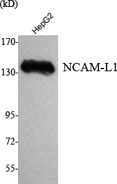
Western blotting validation for Anti-NCAM-L1 L1CAM Monoclonal Antibody M00729-2
Western Blot (WB) analysis using NCAM-L1 Monoclonal Antibody against HepG2 cell lysate.Electrophoresis was performed on a SDS-PAGE gel. To determine SDS-PAGE gel concentration
Specific Publications For Anti-NCAM-L1 L1CAM Monoclonal Antibody (M00729-2)
Loading publications
Recommended Resources
Here are featured tools and databases that you might find useful.
- Boster's Pathways Library
- Protein Databases
- Bioscience Research Protocol Resources
- Data Processing & Analysis Software
- Photo Editing Software
- Scientific Literature Resources
- Research Paper Management Tools
- Molecular Biology Software
- Primer Design Tools
- Bioinformatics Tools
- Phylogenetic Tree Analysis
Customer Reviews
Have you used Anti-NCAM-L1 L1CAM Monoclonal Antibody?
Share your experimental results or join a short interview to earn up to $1,000 in product credits or other rewards.
0 Reviews For Anti-NCAM-L1 L1CAM Monoclonal Antibody
Customer Q&As
Have a question?
Find answers in Q&As, reviews.
Can't find your answer?
Submit your question