Click image to see more details
Product Info Summary
| SKU: | A09633-3 |
|---|---|
| Size: | 100 μg/vial |
| Reactive Species: | Human |
| Host: | Rabbit |
| Application: | ELISA, Flow Cytometry, IHC, WB |
Customers Who Bought This Also Bought


Product info
Product Name
Anti-Nectin 3/NECTIN3 Antibody Picoband™
View all Nectin-3/PVRL3 Antibodies
SKU/Catalog Number
A09633-3
Size
100 μg/vial
Form
Lyophilized
Description
Boster Bio Anti-Nectin 3/NECTIN3 Antibody Picoband™ catalog # A09633-3. Tested in ELISA, Flow Cytometry, IHC, WB applications. This antibody reacts with Human.
Storage & Handling
Store at -20˚C for one year from date of receipt. After reconstitution, at 4˚C for one month. It can also be aliquotted and stored frozen at -20˚C for six months. Avoid repeated freeze-thaw cycles.
Cite This Product
Anti-Nectin 3/NECTIN3 Antibody Picoband™ (Boster Biological Technology, Pleasanton CA, USA, Catalog # A09633-3)
Host
Rabbit
Contents
Each vial contains 4mg Trehalose, 0.9mg NaCl, 0.2mg Na2HPO4, 0.05mg NaN3.
Clonality
Polyclonal
Isotype
Rabbit IgG
Immunogen
E.coli-derived human Nectin 3/NECTIN3 recombinant protein (Position: E92-N296).
*Blocking peptide can be purchased. Costs vary based on immunogen length. Contact us for pricing.
Cross-reactivity
No cross-reactivity with other proteins.
Reactive Species
A09633-3 is reactive to NECTIN3 in Human
Applications
A09633-3 is guaranteed for ELISA, Flow Cytometry, IHC, WB Boster Guarantee
Observed Molecular Weight
80 kDa
Calculated molecular weight
61.002kDa
Background of Nectin-3/PVRL3
Nectin-3, also known as poliovirus receptor-related 3, is a 95 kDa type I transmembrane glycoprotein belonging to the Nectin family of Ig superfamily proteins. It is mapped to 3q13.13. This gene encodes a member of the nectin family of proteins, which function as adhesion molecules at adherens junctions. This family member interacts with other nectin-like proteins and with afadin, a filamentous actin-binding protein involved in the regulation of directional motility, cell proliferation and survival. This gene plays a role in ocular development involving the ciliary body. Mutations in this gene are believed to result in congenital ocular defects. Alternative splicing results in multiple transcript variants.
Antibody Validation
Boster validates all antibodies on WB, IHC, ICC, Immunofluorescence, and ELISA with known positive control and negative samples to ensure specificity and high affinity, including thorough antibody incubations.
Assay dilution & Images
Reconsitution
Add 0.2ml of distilled water will yield a concentration of 500ug/ml.
Assay Dilutions Recommendation
The recommendations below provide a starting point for assay optimization. The actual working concentration varies and should be decided by the user.
Western blot, 0.25-0.5μg/ml, Human
Immunohistochemistry (Paraffin-embedded Section), 0.5-1μg/ml, Human
Flow Cytometry, 1-3μg/1x106 cells, Human
Direct ELISA, 0.1-0.5μg/ml, Human
Validation Images & Assay Conditions
Click image to see more details
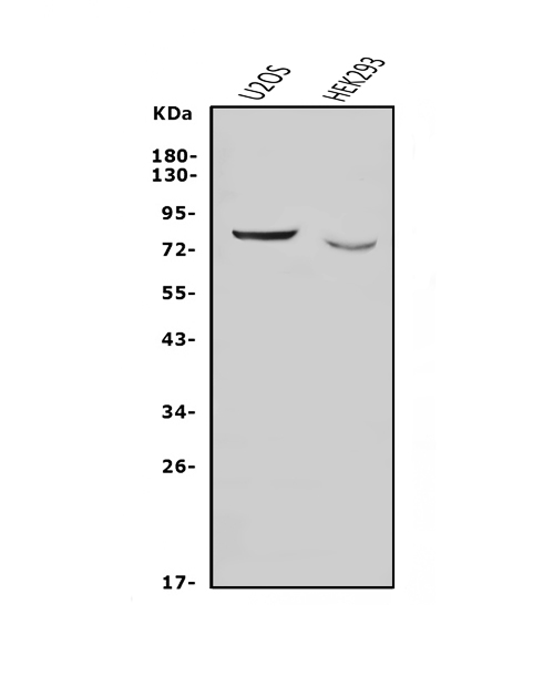
Figure 1. Western blot analysis of Nectin 3/NECTIN3 using anti-Nectin 3/NECTIN3 antibody (A09633-3).
Electrophoresis was performed on a 5-20% SDS-PAGE gel at 70V (Stacking gel) / 90V (Resolving gel) for 2-3 hours. The sample well of each lane was loaded with 50ug of sample under reducing conditions.
Lane 1: human U20S whole cell lysates.
Lane 2: human HEK293 whole cell lysates,
After Electrophoresis, proteins were transferred to a Nitrocellulose membrane at 150mA for 50-90 minutes. Blocked the membrane with 5% Non-fat Milk/ TBS for 1.5 hour at RT. The membrane was incubated with rabbit anti-Nectin 3/NECTIN3 antigen affinity purified polyclonal antibody (Catalog # A09633-3) at 0.5 μg/mL overnight at 4°C, then washed with TBS-0.1%Tween 3 times with 5 minutes each and probed with a goat anti-rabbit IgG-HRP secondary antibody at a dilution of 1:10000 for 1.5 hour at RT. The signal is developed using an Enhanced Chemiluminescent detection (ECL) kit (Catalog # EK1002) with Tanon 5200 system. A specific band was detected for Nectin 3/NECTIN3 at approximately 80KD. The expected band size for Nectin 3/NECTIN3 is at 61KD.

Click image to see more details
Figure 2. IHC analysis of Nectin 3/NECTIN3 using anti-Nectin 3/NECTIN3 antibody (A09633-3).
Nectin 3/NECTIN3 was detected in paraffin-embedded section of human rectal cancer tissue. Heat mediated antigen retrieval was performed in EDTA buffer (pH8.0, epitope retrieval solution). The tissue section was blocked with 10% goat serum. The tissue section was then incubated with 1μg/ml rabbit anti-Nectin 3/NECTIN3 Antibody (A09633-3) overnight at 4°C. Biotinylated goat anti-rabbit IgG was used as secondary antibody and incubated for 30 minutes at 37°C. The tissue section was developed using Strepavidin-Biotin-Complex (SABC) (Catalog # SA1022) with DAB as the chromogen.

Click image to see more details
Figure 3. Flow Cytometry analysis of A431 cells using anti-Nectin 3/NECTIN3 antibody (A09633-3).
Overlay histogram showing A431 cells stained with A09633-3 (Blue line).The cells were blocked with 10% normal goat serum. And then incubated with rabbit anti-Nectin 3/NECTIN3 Antibody (A09633-3,1μg/1x106 cells) for 30 min at 20°C. DyLight®488 conjugated goat anti-rabbit IgG (BA1127, 5-10μg/1x106 cells) was used as secondary antibody for 30 minutes at 20°C. Isotype control antibody (Green line) was rabbit IgG (1μg/1x106) used under the same conditions. Unlabelled sample (Red line) was also used as a control.
Protein Target Info & Infographic
Gene/Protein Information For NECTIN3 (Source: Uniprot.org, NCBI)
Gene Name
NECTIN3
Full Name
Nectin-3
Weight
61.002kDa
Superfamily
nectin family
Alternative Names
CD113 antigen; CD113; CDw113FLJ90624; DKFZp566B0846; nectin 3; Nectin3; Nectin-3; poliovirus receptor-related 3; poliovirus receptor-related protein 3; PPR3; PRR3CD113; PVRL3; PVRR3 NECTIN3 CD113, CDW113, NECTIN-3, PPR3, PRR3, PVRL3, PVRR3 nectin cell adhesion molecule 3 nectin-3|nectin 3|poliovirus receptor-related 3|poliovirus receptor-related protein 3
*If product is indicated to react with multiple species, protein info is based on the gene entry specified above in "Species".For more info on NECTIN3, check out the NECTIN3 Infographic

We have 30,000+ of these available, one for each gene! Check them out.
In this infographic, you will see the following information for NECTIN3: database IDs, superfamily, protein function, synonyms, molecular weight, chromosomal locations, tissues of expression, subcellular locations, post-translational modifications, and related diseases, research areas & pathways. If you want to see more information included, or would like to contribute to it and be acknowledged, please contact [email protected].
Specific Publications For Anti-Nectin 3/NECTIN3 Antibody Picoband™ (A09633-3)
Hello CJ!
No publications found for A09633-3
*Do you have publications using this product? Share with us and receive a reward. Ask us for more details.
Recommended Resources
Here are featured tools and databases that you might find useful.
- Boster's Pathways Library
- Protein Databases
- Bioscience Research Protocol Resources
- Data Processing & Analysis Software
- Photo Editing Software
- Scientific Literature Resources
- Research Paper Management Tools
- Molecular Biology Software
- Primer Design Tools
- Bioinformatics Tools
- Phylogenetic Tree Analysis
Customer Reviews
Have you used Anti-Nectin 3/NECTIN3 Antibody Picoband™?
Submit a review and receive an Amazon gift card.
- $30 for a review with an image
0 Reviews For Anti-Nectin 3/NECTIN3 Antibody Picoband™
Customer Q&As
Have a question?
Find answers in Q&As, reviews.
Can't find your answer?
Submit your question
