Click image to see more details
Product Info Summary
| SKU: | PB9793 |
|---|---|
| Size: | 100 μg/vial |
| Reactive Species: | Human, Mouse, Rat |
| Host: | Rabbit |
| Application: | IHC, WB |
Customers Who Bought This Also Bought

Product info
Product Name
Anti-RAG2 Antibody Picoband®
SKU/Catalog Number
PB9793
PB0841 is an alternative SKU for this antibody, used in previous lots.
Size
100 μg/vial
Form
Lyophilized
Description
Boster Bio Anti-RAG2 Antibody Picoband® catalog # PB9793. Tested in IHC, WB applications. This antibody reacts with Human, Mouse, Rat. The brand Picoband indicates this is a premium antibody that guarantees superior quality, high affinity, and strong signals with minimal background in Western blot applications. Only our best-performing antibodies are designated as Picoband, ensuring unmatched performance.
Storage & Handling
Store at -20˚C for one year from date of receipt. After reconstitution, at 4˚C for one month. It can also be aliquotted and stored frozen at -20˚C for six months. Avoid repeated freeze-thaw cycles.
Cite This Product
Anti-RAG2 Antibody Picoband® (Boster Biological Technology, Pleasanton CA, USA, Catalog # PB9793)
Host
Rabbit
Contents
Each vial contains antibody formulated with stabilizing components, 0.9 mg NaCl, 0.2 mg Na2HPO4, and 0.05 mg NaN3.
*This antibody is supplied in a stabilized formulation.
Compatibility with conjugation reactions depends on the chemistry of the conjugation method used.
For conjugation methods that are not compatible with the stabilizing components present in this formulation, a carrier-free antibody format is required.
Clonality
Polyclonal
Isotype
Rabbit IgG
Immunogen
A synthetic peptide corresponding to a sequence at the C-terminus of human RAG2, different from the related mouse sequence by three amino acids.
Cross-reactivity
No cross-reactivity with other proteins.
Reactive Species
PB9793 is reactive to RAG2 in Human, Mouse, Rat
Observed Molecular Weight
59 kDa
Calculated molecular weight
59.2 kDa
Background of RAG2
Recombination activating gene 2, also known as RAG-2, is a protein that in humans is encoded by the RAG2 gene. This gene encodes a protein that is involved in the initiation of V (D)J recombination during B and T cell development. This protein forms a complex with the product of the adjacent recombination activating gene 1, and this complex can form double-strand breaks by cleaving DNA at conserved recombination signal sequences. The recombination activating gene 1 component is thought to contain most of the catalytic activity, while the N-terminal of the recombination activating gene 2 component is thought to form a six-bladed propeller in the active core that serves as a binding scaffold for the tight association of the complex with DNA. A C-terminal plant homeodomain finger-like motif in this protein is necessary for interactions with chromatin components, specifically with histone H3 that is trimethylated at lysine 4. Mutations in this gene cause Omenn syndrome, a form of severe combined immunodeficiency associated with autoimmune-like symptoms.
Antibody Validation
Boster validates all antibodies on WB, IHC, ICC, Immunofluorescence, and ELISA with known positive control and negative samples to ensure specificity and high affinity, including thorough antibody incubations.
Application & Images
Applications
PB9793 is guaranteed for IHC, WB Boster Guarantee
Recommend Dilution
| Application | Dilution | Species |
|---|---|---|
| Immunohistochemistry (Paraffin-embedded Section) | 0.5-1μg/ml | Human, Mouse, Rat |
| Western blot | 0.1-0.5μg/ml | Human |
Tested application
Suggested blocking solution with 5% non-fat milk or BSA; (*)Recommended protein loading: 20-40 µg per lane
Use TE buffer pH 9.0 for antigen retrieval; (*) citrate buffer pH 6.0 is an alternative.
Validation Images & Assay Conditions
Click image to see more details
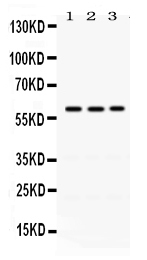
Western blot analysis of RAG2 using anti-RAG2 antibody (PB9793).
Electrophoresis was performed on a 5-20% SDS-PAGE gel at 70V (Stacking gel) / 90V (Resolving gel) for 2-3 hours. The sample well of each lane was loaded with 40 ug of sample under reducing conditions.
Lane 1: A549 Whole Cell Lysate,
Lane 2: 22RV1 Whole Cell Lysate,
Lane 3: U20S Whole Cell Lysate.
After electrophoresis, proteins were transferred to a nitrocellulose membrane at 150 mA for 50-90 minutes. Blocked the membrane with 5% non-fat milk/TBS for 1.5 hour at RT. The membrane was incubated with rabbit anti-RAG2 antigen affinity purified polyclonal antibody (Catalog # PB9793) at 0.5 μg/mL overnight at 4°C, then washed with TBS-0.1%Tween 3 times with 5 minutes each and probed with a goat anti-rabbit IgG-HRP secondary antibody at a dilution of 1:5000 for 1.5 hour at RT. The signal is developed using an Enhanced Chemiluminescent detection (ECL) kit (Catalog # EK1002) with Tanon 5200 system. A specific band was detected for RAG2 at approximately 59 kDa. The expected band size for RAG2 is at 59 kDa.

Click image to see more details
IHC analysis of RAG2 using anti-RAG2 antibody (PB9793).
RAG2 was detected in a paraffin-embedded section of mouse spleen tissue. Heat mediated antigen retrieval was performed in EDTA buffer (pH 8.0, epitope retrieval solution). The tissue section was blocked with 10% goat serum. The tissue section was then incubated with 1 μg/ml rabbit anti-RAG2 Antibody (PB9793) overnight at 4°C. Biotinylated goat anti-rabbit IgG was used as secondary antibody and incubated for 30 minutes at 37°C. The tissue section was developed using Strepavidin-Biotin-Complex (SABC) (Catalog # SA1022) with DAB as the chromogen.

Click image to see more details
IHC analysis of RAG2 using anti-RAG2 antibody (PB9793).
RAG2 was detected in a paraffin-embedded section of rat spleen tissue. Heat mediated antigen retrieval was performed in EDTA buffer (pH 8.0, epitope retrieval solution). The tissue section was blocked with 10% goat serum. The tissue section was then incubated with 1 μg/ml rabbit anti-RAG2 Antibody (PB9793) overnight at 4°C. Biotinylated goat anti-rabbit IgG was used as secondary antibody and incubated for 30 minutes at 37°C. The tissue section was developed using Strepavidin-Biotin-Complex (SABC) (Catalog # SA1022) with DAB as the chromogen.

Click image to see more details
IHC analysis of RAG2 using anti-RAG2 antibody (PB9793).
RAG2 was detected in a paraffin-embedded section of human tonsil tissue. Heat mediated antigen retrieval was performed in EDTA buffer (pH 8.0, epitope retrieval solution). The tissue section was blocked with 10% goat serum. The tissue section was then incubated with 1 μg/ml rabbit anti-RAG2 Antibody (PB9793) overnight at 4°C. Biotinylated goat anti-rabbit IgG was used as secondary antibody and incubated for 30 minutes at 37°C. The tissue section was developed using Strepavidin-Biotin-Complex (SABC) (Catalog # SA1022) with DAB as the chromogen.
Specific Publications For Anti-RAG2 Antibody Picoband® (PB9793)
Loading publications
Recommended Resources
Here are featured tools and databases that you might find useful.
- Boster's Pathways Library
- Protein Databases
- Bioscience Research Protocol Resources
- Data Processing & Analysis Software
- Photo Editing Software
- Scientific Literature Resources
- Research Paper Management Tools
- Molecular Biology Software
- Primer Design Tools
- Bioinformatics Tools
- Phylogenetic Tree Analysis
Customer Reviews
Have you used Anti-RAG2 Antibody Picoband®?
Share your experimental results or join a short interview to earn up to $1,000 in product credits or other rewards.
0 Reviews For Anti-RAG2 Antibody Picoband®
Customer Q&As
Have a question?
Find answers in Q&As, reviews.
Can't find your answer?
Submit your question
1 Customer Q&As for Anti-RAG2 Antibody Picoband®
Question
We are currently using anti-RAG2 antibody PB9793 for human tissue, and we are happy with the WB results. The species of reactivity given in the datasheet says human, mouse, rat. Is it possible that the antibody can work on zebrafish tissues as well?
Verified Customer
Verified customer
Asked: 2018-01-01
Answer
The anti-RAG2 antibody (PB9793) has not been validated for cross reactivity specifically with zebrafish tissues, but there is a good chance of cross reactivity. We have an innovator award program that if you test this antibody and show it works in zebrafish you can get your next antibody for free. Please contact me if I can help you with anything.
Boster Scientific Support
Answered: 2018-01-01
