Click image to see more details
Product Info Summary
| SKU: | PB9511 |
|---|---|
| Size: | 100 μg/vial |
| Reactive Species: | Human, Monkey, Mouse, Rat |
| Host: | Rabbit |
| Application: | IHC, WB |
Customers Who Bought This Also Bought


Product info
Product Name
Anti-STAT3 Antibody Picoband™
SKU/Catalog Number
PB9511
Size
100 μg/vial
Form
Lyophilized
Description
Boster Bio Anti-STAT3 Antibody Picoband™ catalog # PB9511. Tested in IHC, WB applications. This antibody reacts with Human, Monkey, Mouse, Rat.
Storage & Handling
Store at -20˚C for one year from date of receipt. After reconstitution, at 4˚C for one month. It can also be aliquotted and stored frozen at -20˚C for six months. Avoid repeated freeze-thaw cycles.
Cite This Product
Anti-STAT3 Antibody Picoband™ (Boster Biological Technology, Pleasanton CA, USA, Catalog # PB9511)
Host
Rabbit
Contents
Each vial contains 5mg BSA, 0.9mg NaCl, 0.2mg Na2HPO4, 0.05mg NaN3.
Clonality
Polyclonal
Isotype
Rabbit IgG
Immunogen
A synthetic peptide corresponding to a sequence at the N-terminus of human STAT3, identical to the related mouse and rat sequences.
*Blocking peptide can be purchased. Costs vary based on immunogen length. Contact us for pricing.
Cross-reactivity
No cross-reactivity with other proteins
Reactive Species
PB9511 is reactive to STAT3 in Human, Monkey, Mouse, Rat
Applications
PB9511 is guaranteed for IHC, WB Boster Guarantee
Observed Molecular Weight
83 kDa
Calculated molecular weight
88.068kDa
Background of STAT3
The transcription factor, signal transducer and activator of transcription-3 (STAT-3) is the most pleiotropic member of the signal transducer and activator of transcription (STAT) family of transcription factors and mediates pivotal responses for the cytokine family. The mouse STAT3 gene contains 24 exons and spans 30 kb. The translation initiation codon is in exon 2, and the stop codon is in exon 24. STAT3 is mapped to 17q21. It contributes to various physiological processes. Hepatic STAT-3 signaling is thus essential for normal glucose homeostasis and may provide new therapeutic targets for diabetes mellitus.
Antibody Validation
Boster validates all antibodies on WB, IHC, ICC, Immunofluorescence, and ELISA with known positive control and negative samples to ensure specificity and high affinity, including thorough antibody incubations.
Innovating Scientists Reward
If you are the first to review this product, or if you have results for a special sample, species or application this product is not validated in, share your results with us and receive product credits you can use towards any Boster products! Applicable to all scientists worldwide.
Submit A Review
Assay dilution & Images
Reconsitution
Add 0.2ml of distilled water will yield a concentration of 500ug/ml.
Assay Dilutions Recommendation
The recommendations below provide a starting point for assay optimization. The actual working concentration varies and should be decided by the user.
Western blot, 0.1-0.5 μg/ml, Human, Monkey, Mouse, Rat
Immunohistochemistry(Paraffin-embedded Section), 2-5 μg/ml, Human
Validation Images & Assay Conditions
Click image to see more details
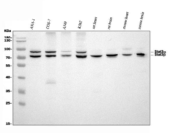
Figure 1. Western blot analysis of STAT3 using anti-STAT3 antibody (PB9511).
Electrophoresis was performed on a 5-20% SDS-PAGE gel at 70V (Stacking gel) / 90V (Resolving gel) for 2-3 hours. The sample well of each lane was loaded with 30 ug of sample under reducing conditions.
Lane 1: mouse ANA-1 whole cell lysates,
Lane 2: monkey COS-7 whole cell lysates,
Lane 3: human A549 whole cell lysates,
Lane 4: human K562 whole cell lysates,
Lane 5: rat heart tissue lysates,
Lane 6: rat brain tissue lysates,
Lane 7: mouse heart tissue lysates,
Lane 8: mouse brain tissue lysates.
After electrophoresis, proteins were transferred to a nitrocellulose membrane at 150 mA for 50-90 minutes. Blocked the membrane with 5% non-fat milk/TBS for 1.5 hour at RT. The membrane was incubated with rabbit anti-STAT3 antigen affinity purified polyclonal antibody (Catalog # PB9511) at 0.5 μg/mL overnight at 4°C, then washed with TBS-0.1%Tween 3 times with 5 minutes each and probed with a goat anti-rabbit IgG-HRP secondary antibody at a dilution of 1:5000 for 1.5 hour at RT. The signal is developed using an Enhanced Chemiluminescent detection (ECL) kit (Catalog # EK1002) with Tanon 5200 system. Specific bands were detected for STAT3α at approximately 86 kDa and for STAT3β at approximately 83 kDa. The expected band size for STAT3 is at 79 kDa.

Click image to see more details
Figure 2. IHC analysis of STAT3 using anti-STAT3 antibody (PB9511).
STAT3 was detected in a paraffin-embedded section of human breast cancer tissue. Heat mediated antigen retrieval was performed in EDTA buffer (pH 8.0, epitope retrieval solution). The tissue section was blocked with 10% goat serum. The tissue section was then incubated with 2 μg/ml rabbit anti-STAT3 Antibody (PB9511) overnight at 4°C. Peroxidase Conjugated Goat Anti-rabbit IgG was used as secondary antibody and incubated for 30 minutes at 37°C. The tissue section was developed using HRP Conjugated Rabbit IgG Super Vision Assay Kit (Catalog # SV0002) with DAB as the chromogen.
Protein Target Info & Infographic
Gene/Protein Information For STAT3 (Source: Uniprot.org, NCBI)
Gene Name
STAT3
Full Name
Signal transducer and activator of transcription 3
Weight
88.068kDa
Superfamily
transcription factor STAT family
Alternative Names
Acute-phase response factor; APRFMGC16063; DNA-binding protein APRF; FLJ20882; HIES; signal transducer and activator of transcription 3 (acute-phase responsefactor); signal transducer and activator of transcription 3; STAT3 STAT3 ADMIO, ADMIO1, APRF, HIES signal transducer and activator of transcription 3 signal transducer and activator of transcription 3|DNA-binding protein APRF|acute-phase response factor
*If product is indicated to react with multiple species, protein info is based on the gene entry specified above in "Species".For more info on STAT3, check out the STAT3 Infographic

We have 30,000+ of these available, one for each gene! Check them out.
In this infographic, you will see the following information for STAT3: database IDs, superfamily, protein function, synonyms, molecular weight, chromosomal locations, tissues of expression, subcellular locations, post-translational modifications, and related diseases, research areas & pathways. If you want to see more information included, or would like to contribute to it and be acknowledged, please contact [email protected].
Specific Publications For Anti-STAT3 Antibody Picoband™ (PB9511)
Hello CJ!
PB9511 has been cited in 18 publications:
*The publications in this section are manually curated by our staff scientists. They may differ from Bioz's machine gathered results. Both are accurate. If you find a publication citing this product but is missing from this list, please let us know we will issue you a thank-you coupon.
Zhang T,Ma L,Wu P,Li W,Li T,Gu R,Dan X,Li Z,Fan X,Xiao Z.Gallic acid has anticancer activity and enhances the anticancer effects of cisplatin in non‑small cell lung cancer A549 cells via the JAK/STAT3 signaling pathway.Oncol Rep.2019 Mar;41(3):1779-1788.doi:10.3892/or.2019.6976.Epub 2019 Jan 22.PMID:30747218.
Species: Human
PB9511 usage in article: APP:WB, SAMPLE:A549 CELL, DILUTION:1:1000
He J,Zhang W,Di T,Meng J,Qi Y,Li G,Zhang Y,Su H,Yan W.Water extract of sporoderm-broken spores of Ganoderma lucidum enhanced pd-l1 antibody efficiency through downregulation and relieved complications of pd-l1 monoclonal antibody.Biomed Pharmacother.2020
Species: Human,Mouse
PB9511 usage in article: APP:WB, SAMPLE:HOS CELL AND K7M2 CELL, DILUTION:1:500
Zhang Q,Duan HX,Li RL,Sun JY,Liu J,Peng W,Wu CJ,Gao YX. Inducing Apoptosis and Suppressing Inflammatory Reactions in Synovial Fibroblasts are Two Important Ways for Guizhi-Shaoyao-Zhimu Decoction Against Rheumatoid Arthritis. J Inflamm Res.2021 Jan 26;14:
Species: Human
PB9511 usage in article: APP:WB, SAMPLE:MH7A CELL, DILUTION:1:300
Protective effects of Tongxinluo on cerebral ischemia/reperfusion injury related to Connexin 43/Calpain II/Bax/Caspase-3 pathway in rat
Exposure to Concentrated Ambient Fine Particulate Matter Induces Vascular Endothelial Dysfunction via miR-21
IL-6 Inhibits Starvation-induced Autophagy via the STAT3/Bcl-2 Signaling Pathway
Delivery of the co-expression plasmid pEndo-Si-Stat3 by attenuatedSalmonellaserovartyphimuriumfor prostate cancer treatment
The effects of HIF-1alpha on gene expression profiles of NCI-H446 human small cell lung cancer cells
Prognostic role of STAT3 in solid tumors: a systematic review and meta-analysis
Hu T, Zhang C, Tang Q, Su Y, Li B, Chen L, Zhang Z, Cai T, Zhu Y. Bmc Cancer. 2013 May 22;13:251. Doi: 10.1186/1471-2407-13-251. Variant G6Pd Levels Promote Tumor Cell Proliferation Or Apoptosis Via The Stat3/5 Pathway In The Human Melanoma Xenogr...
Recommended Resources
Here are featured tools and databases that you might find useful.
- Boster's Pathways Library
- Protein Databases
- Bioscience Research Protocol Resources
- Data Processing & Analysis Software
- Photo Editing Software
- Scientific Literature Resources
- Research Paper Management Tools
- Molecular Biology Software
- Primer Design Tools
- Bioinformatics Tools
- Phylogenetic Tree Analysis
Customer Reviews
Have you used Anti-STAT3 Antibody Picoband™?
Submit a review and receive an Amazon gift card.
- $30 for a review with an image
Be the first to review Anti-STAT3 Antibody Picoband™
*The first user to submit a review for a product is eligible for Boster's Innovating Scientists Reward, which gives product credits. This is in addition to the gift card reward.
Customer Q&As
Have a question?
Find answers in Q&As, reviews.
Can't find your answer?
Submit your question
4 Customer Q&As for Anti-STAT3 Antibody Picoband™
Question
We are currently using anti-STAT3 antibody PB9511 for rat tissue, and we are happy with the WB results. The species of reactivity given in the datasheet says human, mouse, rat. Is it possible that the antibody can work on feline tissues as well?
Verified Customer
Verified customer
Asked: 2020-04-28
Answer
The anti-STAT3 antibody (PB9511) has not been validated for cross reactivity specifically with feline tissues, though there is a good chance of cross reactivity. We have an innovator award program that if you test this antibody and show it works in feline you can get your next antibody for free. Please contact me if I can help you with anything.
Boster Scientific Support
Answered: 2020-04-28
Question
I was wanting to use using your anti-STAT3 antibody for positive regulation of tyrosine phosphorylation of stat protein studies. Has this antibody been tested with western blotting on brain tissue? We would like to see some validation images before ordering.
Verified Customer
Verified customer
Asked: 2019-10-15
Answer
I appreciate your inquiry. This PB9511 anti-STAT3 antibody is validated on rat lung tissue, tissue lysate, brain tissue, human placenta tissue, hela whole cell lysate, panc whole cell lysate, hepa whole cell lysate. It is guaranteed to work for WB in human, mouse, rat. Our Boster guarantee will cover your intended experiment even if the sample type has not been be directly tested.
Boster Scientific Support
Answered: 2019-10-15
Question
We have seen staining in mouse upper lobe of lung. Any tips? Is anti-STAT3 antibody supposed to stain upper lobe of lung positively?
Verified Customer
Verified customer
Asked: 2019-09-03
Answer
Based on literature upper lobe of lung does express STAT3. Based on Uniprot.org, STAT3 is expressed in upper lobe of lung, placenta, kidney pancreas, liver, cervix carcinoma, erythroleukemia, among other tissues. Regarding which tissues have STAT3 expression, here are a few articles citing expression in various tissues:
Cervix carcinoma, Pubmed ID: 18669648, 18691976, 20068231
Erythroleukemia, Pubmed ID: 23186163
Kidney, and Pancreas, Pubmed ID: 15489334
Liver, Pubmed ID: 7701321, 24275569
Placenta, Pubmed ID: 7512451
Boster Scientific Support
Answered: 2019-09-03
Question
My boss were content with the WB result of your anti-STAT3 antibody. However we have seen positive staining in erythroleukemia cytoplasm. nucleus using this antibody. Is that expected? Could you tell me where is STAT3 supposed to be expressed?
B. Roberts
Verified customer
Asked: 2013-11-15
Answer
From what I have seen in literature, erythroleukemia does express STAT3. Generally STAT3 expresses in cytoplasm. nucleus. Regarding which tissues have STAT3 expression, here are a few articles citing expression in various tissues:
Cervix carcinoma, Pubmed ID: 18669648, 18691976, 20068231
Erythroleukemia, Pubmed ID: 23186163
Kidney, and Pancreas, Pubmed ID: 15489334
Liver, Pubmed ID: 7701321, 24275569
Placenta, Pubmed ID: 7512451
Boster Scientific Support
Answered: 2013-11-15
