Click image to see more details
-

-
-
-
-
+6
Product Info Summary
| SKU: | A01096-3 |
|---|---|
| Size: | 0.1 mg |
| Reactive Species: | Human, Mouse |
| Host: | Rabbit |
| Application: | ELISA, IF, IHC-P, WB |
Customers Who Bought This Also Bought

Product info
Product Name
Anti-Thymic stromal lymphopoietin TSLP Antibody
SKU/Catalog Number
A01096-3
Size
0.1 mg
Form
Liquid
Description
Boster Bio Anti-Thymic stromal lymphopoietin TSLP Antibody (Catalog # A01096-3). Tested in ELISA, WB, IHC-P, IF applications. This antibody reacts with Human, Mouse.
Storage & Handling
TSLP antibody can be stored at 4°C up to one year. Antibodies should not be exposed to prolonged high temperatures.
Cite This Product
Anti-Thymic stromal lymphopoietin TSLP Antibody (Boster Biological Technology, Pleasanton CA, USA, Catalog # A01096-3)
Host
Rabbit
Contents
TSLP Antibody is supplied in PBS containing 0.02% sodium azide.
Clonality
Polyclonal
Isotype
IgG
Immunogen
Anti-TSLP antibody was raised against a peptide corresponding to 19 amino acids near the center of human TSLP. The immunogen is located within the amino acids 40-90 of TSLP.
Reactive Species
A01096-3 is reactive to TSLP in Human, Mouse
Observed Molecular Weight
68 kDa
Calculated molecular weight
18.1 kDa
Background of TSLP
Thymic stromal lymphopoietin (TSLP) has recently been identified as an important factor capable of driving dendritic cell maturation and activation. TSLP is a four-helix-bundle cytokine that is expressed mainly by barrier epithelial cells and is a potent activator of several cell types such as myeloid dendritic cells. TSLP is involved in the positive selection of regulatory T cells, maintenance of peripheral CD4+ T cell homeostasis and the induction of CD4+ T cell-mediated allergic reaction. TSLP is also capable of supporting the growth of fetal liver and adult B cell progenitors and their differentiation to the IgM-positive stage of B cell development. Amino acid sequence analysis has shown poor homology between human and mouse TSLP although they exhibit similar biological functions and are expressed in similar tissues. At least two differentially spliced isoforms of TSLP are known to exist.
Antibody Validation
Boster validates all antibodies on WB, IHC, ICC, Immunofluorescence, and ELISA with known positive control and negative samples to ensure specificity and high affinity, including thorough antibody incubations.
Application & Images
Applications
A01096-3 is guaranteed for ELISA, IF, IHC-P, WB Boster Guarantee
Recommend Dilution
WB: 0.25 - 4 μg/mL; IHC-P: 1-2.5 μg/mL.
Antibody validated: Western Blot in human and mouse samples; Immunohistochemistry in human and mouse samples. All other applications and species not yet tested.
Validation Images & Assay Conditions
Click image to see more details
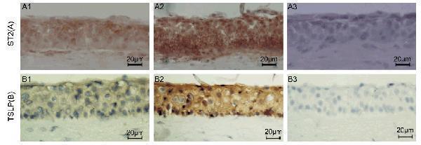
Induced Expression Validation of TSLP in Human Corneal Tissues (Lin et al., 2013)
Immunohistochemical images showing TSLP protein detected by anti-TSLP antibodies in ex vivo donor human corneal tissues without (B1) or with exposure to IL-33 (10ng/mL) (B2). An isotype IgG antibody (B3) was used as a negative control. Magnificaiton 400X. The staining confirms the increased level of TSLP after IL-33 treatment.

Click image to see more details
Induced Expression Validation of TSLP in Mesenchymal Stromal Cells (MSCs). (Allakhverdi et al., 2013)
Immunocytochemical images showing TSLP protein detected by anti-TSLP antibodies in BM-MSCs unstimulated (control) or stimulated with IL-1/TNF or supernatants of activated MCs (MCactSNT). Isotype is used as a negative control. The production of TSLP increased with the stimulated conditions.

Click image to see more details
Regulated Expression Validation of TSLP in COPD Patients. (Anzalone et al., 2018)
(A) shows NHBE cells, in the absence (Lane 2) or presence (Lane 3) of Tiotropium (100 nM), were stimulated with rhIL-17A (20 ng/ml) (n = 3).
(B) shows NHBE cells, with ISs from COPD patients untreated (Lane 2 and Lane 3) or treated (Lane 4) with anti-IL-17A antibody (n = 3).
(A) and (B) show TSLP expression decreases with the treatment of anti-cholinergic drugs.

Click image to see more details
Induced Expression Validation of TSLP in Human Corneal Epithelium (Lin et al., 2018)
Immunohistochemical images showing the
TSLP protein detected by anti-TSLP antibodies in donor corneal tissues without (Control) or after exposure to IL-32 (50 ng/ml) ex vivo; an isotype IgG antibody was used as a negative control. The production of TSLP was increased after IL-32 treatment.

Click image to see more details
Immunohistochemistry Validation of TSLP in Human Brain Tissue
Immunohistochemical analysis of paraffin-embedded Human Brain Tissue using anti-TSLP antibody (A01096-3) at 2.5 μg/ml. Tissue was fixed with formaldehyde and blocked with 10% serum for 1 h at RT; antigen retrieval was by heat mediation with a citrate buffer (pH6). Samples were incubated with primary antibody overnight at 4˚C. A goat anti-rabbit IgG H&L (HRP) at 1/250 was used as secondary. Counter stained with Hematoxylin.

Click image to see more details
Immunohistochemistry Validation of TSLP in Human Liver Tissue Immunohistochemical analysis of paraffin-embedded human liver tissue using anti-TSLP antibody (A01096-3) at 2 μg/ml. Tissue was fixed with formaldehyde and blocked with 10% serum for 1 h at RT; antigen retrieval was by heat mediation with a citrate buffer (pH6). Samples were incubated with primary antibody overnight at 4°C. A goat anti-rabbit IgG H&L (HRP) at 1/250 was used as secondary. Counter stained with Hematoxylin.

Click image to see more details
Immunohistochemistry Validation of TSLP in Mouse Liver Tissue Immunohistochemical analysis of paraffin-embedded mouse liver tissue using anti-TSLP antibody (A01096-3) at 2 μg/ml. Tissue was fixed with formaldehyde and blocked with 10% serum for 1 h at RT; antigen retrieval was by heat mediation with a citrate buffer (pH6). Samples were incubated with primary antibody overnight at 4°C. A goat anti-rabbit IgG H&L (HRP) at 1/250 was used as secondary. Counter stained with Hematoxylin.

Click image to see more details
Western Blot Validation in Mouse A-20 Cell Line
Loading: 15 μg of lysates per lane.
Antibodies: TSLP A01096-3 (A: 0.5 μg/mL, B: 1 μg/mL, C: 2 μg/mL), 1h incubation at RT in 5% NFDM/TBST.
Secondary: Goat anti-rabbit IgG HRP conjugate at 1:10000 dilution.

Click image to see more details
Western Blot Validation of TSLP with Human Recombinant protein
Loading: 30ng of TSLP partial human recombinant protein per lane. Antibodies: TSLP A01096-3 (Lane 1: 0.25 μg/mL, Lane 2: 0.5 μg/mL, Lane 3: 1 μg/mL), 1h incubation at RT in 5% NFDM/TBST.
Secondary: Goat anti-rabbit IgG HRP conjugate at 1:10000 dilution.
TLSP partial human recombinant protein: 15kD, the observed bands at 30kD and 45kD are the dimer and trimer, respectively.

Click image to see more details
Western Blot Validation in Human Heart (Lane 1) and Human Prostate (Lane 2)
Loading: 15 μg of lysates per lane.
Antibodies: TSLP A01096-3 (4 μg/mL), 1h incubation at RT in 5% NFDM/TBST.
Secondary: Goat anti-rabbit IgG HRP conjugate at 1:10000 dilution.
Specific Publications For Anti-Thymic stromal lymphopoietin TSLP Antibody (A01096-3)
Loading publications
Recommended Resources
Here are featured tools and databases that you might find useful.
- Boster's Pathways Library
- Protein Databases
- Bioscience Research Protocol Resources
- Data Processing & Analysis Software
- Photo Editing Software
- Scientific Literature Resources
- Research Paper Management Tools
- Molecular Biology Software
- Primer Design Tools
- Bioinformatics Tools
- Phylogenetic Tree Analysis
Customer Reviews
Have you used Anti-Thymic stromal lymphopoietin TSLP Antibody?
Share your experimental results or join a short interview to earn up to $1,000 in product credits or other rewards.
0 Reviews For Anti-Thymic stromal lymphopoietin TSLP Antibody
Customer Q&As
Have a question?
Find answers in Q&As, reviews.
Can't find your answer?
Submit your question
