Click image to see more details
Product Info Summary
| SKU: | A14951 |
|---|---|
| Size: | 100μl |
| Reactive Species: | Human, Mouse |
| Host: | Rabbit |
| Application: | ELISA, WB |
Customers Who Bought This Also Bought

Product info
Product Name
Anti-TNFAIP8L3 Antibody
SKU/Catalog Number
A14951
Size
100μl
Form
Liquid
Description
Boster Bio Anti-TNFAIP8L3 Antibody catalog # A14951. Tested in WB, ELISA applications. This antibody reacts with Human, Mouse.
Storage & Handling
Store at -20°C for one year. For short term storage and frequent use, store at 4°C for up to one month. Avoid repeated freeze-thaw cycles.
Cite This Product
Anti-TNFAIP8L3 Antibody (Boster Biological Technology, Pleasanton CA, USA, Catalog # A14951)
Host
Rabbit
Contents
Liquid in PBS containing 50% glycerol, 0.5% stabilizing protein and 0.02% sodium azide.
This antibody is supplied in a stabilized formulation.
Compatibility with conjugation reactions depends on the chemistry of the conjugation method used.
For conjugation methods that are not compatible with the stabilizing components present in this formulation, a carrier-free antibody format is required.
Clonality
Polyclonal
Isotype
IgG
Immunogen
Synthesized peptide derived from human TNFAIP8L3. at AA range: 15-45
Reactive Species
A14951 is reactive to TNFAIP8L3 in Human, Mouse
Calculated molecular weight
32.7 kDa
Antibody Validation
Boster validates all antibodies on WB, IHC, ICC, Immunofluorescence, and ELISA with known positive control and negative samples to ensure specificity and high affinity, including thorough antibody incubations.
Application & Images
Applications
A14951 is guaranteed for ELISA, WB Boster Guarantee
Recommend Dilution
WB 1:500-2000
ELISA 1:10000-20000
Validation Images & Assay Conditions
Click image to see more details
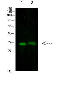
Western Blot analysis of 1, 293T 2, mouse-brain cells using primary antibody diluted at 1:1000 (4°C overnight). Secondary antibody:Goat Anti-rabbit IgG IRDye 800 ( diluted at 1:5000, 25°C, 1 hour)

Click image to see more details
T1: The expression and clinical significance of TIPE3 in PC. A Representative IHC staining of TIPE3. B IHC sum scores were applied to assess TIPE3 in PC specimens. C ) IHC staining of TIPE3 in PC tissues with or without lymph node metastasis. D IHC sum scores were applied to assess TIPE3 in PC specimens with or without lymph node metastasis. E TIPE3 expression in negative lymph node and metastatic lymph node. F Survival analysis according to TIPE3 expression in 188 PC patients. G Prognostic Nomogram of 188 PC patients. H Calibration curves of the OS nomogram. (I-K) DCA for the OS nomogram at 1-year I 2-year J and 3-year K . ***, P < 0.001. T2. The clinical significance of TIPE3 A IHC sum scores were applied to determine TIPE3 in PC. B IHC sum scores were applied to assess TIPE3 expression in PC specimens. C Survival analysis using TIPE3 in 66 PC patients. *, P < 0.05; ***, P < 0.001. T3. TIPE3 promoted malignant biological behaviors of PC cells. A Detection of TIPE3 mRNA expression using qRT-PCR. B TIPE3 expression were detected after TIPE3 silencing or overexpression using qRT-PCR. C CCK8 analysis was conducted after TIPE3 silencing in AsPC-1 and PANC-1 cells. D CCK8 analysis was conducted after TIPE3 overexpression in PC cells. E Trans-well assays were performed after TIPE3 silencing or overexpression in AsPC-1 cells. F Trans-well assays were performed after TIPE3 silencing or overexpression in PANC-1 cells. *, P < 0.05; **, P < 0.01; ***, P < 0.001. T4. TIPE3 promoted tumor progression and metastasis in mice. A Tumor growth curve of orthotopic xenograft mouse. B Representative pictures of primary tumors in pancreas. B Tumor volume in NC, shTIPE3 and TIPE3 group, respectively. D Tumor weight in NC, shTIPE3 and TIPE3 group, respectively. E Survival curves of different groups. F The representative of bioluminescent images in metastatic mouse model. G Number of liver metastatic foci were recorded. H Number of peritoneal metastatic tumors were recorded. *, P < 0.05; **, P < 0.01; ***, P < 0.001
Index in PubMed under a CC BY license. PMID: 35941647

Click image to see more details
T1: TIPE3 increased RAC1 in PC. A IHC staining of RAC1. B IHC sum scores were applied to determine RAC1 expression. C IHC sum scores of RAC1 in PC tissues with low or high TIPE3 expression. D Western-blot was conducted to detected RAC1 expression after TIPE3 silencing or overexpression. E Quantified results of western-blot for RAC1 expression in PC cells. F IHC staining of TIPE3, RAC1, RhoA and MMP9 in orthotopic xenograft tumors. G IHC sum scores of TIPE3, RAC1, RhoA and MMP9 expression in orthotopic xenograft tumors. *, P < 0.05; **, P < 0.01; ***, P < 0.001. T2. TIPE3 accelerated the malignant behaviors of PC cells in a RAC1-dependent manner. A NSC23766 (50 μM) was used in CCK8 analysis. B CCK8 assays was measurement after RAC1 silencing in PC cells. C Trans-well assays after treatment of NSC23766 (50 μM). D Trans-well assays were measurement after RAC1 silencing in PC cells. E CCK8 assays were measurement in TIPE3 silencing PC cells that pretreated with NSC23766 (50 μM) or transfection with RAC1 siRNA. F CCK8 assays were conducted in TIPE3 overexpressed PC cells that pretreated with NSC23766 (50 μM) or transfection with RAC1 siRNA. G Trans-well assays were conducted in TIPE3 silenced PC cells that pretreated with NSC23766 (50 μM). H Trans-well assays were conducted in TIPE3 silenced PC cells that transfected with RAC1 siRNA. I Trans-well assays were conducted in TIPE3 overexpressed PC cells that pretreated with NSC23766 (50 μM). J Trans-well migration and invasion assays were conducted in TIPE3 overexpressed PC cells that transfected with RAC1 siRNA
Index in PubMed under a CC BY license. PMID: 35941647
Specific Publications For Anti-TNFAIP8L3 Antibody (A14951)
Loading publications
Recommended Resources
Here are featured tools and databases that you might find useful.
- Boster's Pathways Library
- Protein Databases
- Bioscience Research Protocol Resources
- Data Processing & Analysis Software
- Photo Editing Software
- Scientific Literature Resources
- Research Paper Management Tools
- Molecular Biology Software
- Primer Design Tools
- Bioinformatics Tools
- Phylogenetic Tree Analysis
Customer Reviews
Have you used Anti-TNFAIP8L3 Antibody?
Share your experimental results or join a short interview to earn up to $1,000 in product credits or other rewards.
0 Reviews For Anti-TNFAIP8L3 Antibody
Customer Q&As
Have a question?
Find answers in Q&As, reviews.
Can't find your answer?
Submit your question
7 Customer Q&As for Anti-TNFAIP8L3 Antibody
Question
I see that the anti-TNFAIP8L3 antibody A14951 works with WB, what is the protocol used to produce the result images on the product page?
Verified Customer
Verified customer
Asked: 2020-03-03
Answer
You can find protocols for WB on the "support/technical resources" section of our navigation menu. If you have any further questions, please send an email to support@bosterbio.com
Boster Scientific Support
Answered: 2020-03-03
Question
My question regarding product A14951, anti-TNFAIP8L3 antibody. I was wondering if it would be possible to conjugate this antibody with biotin. I would need it to be without BSA or sodium azide. I am planning on using a buffer exchange of sodium azide with PBS only. Would there be problems for me to conjugate the antibody and store it in -20 degrees in small aliquots?
Verified Customer
Verified customer
Asked: 2019-10-18
Answer
We suggest not storing this antibody with PBS buffer only in -20 degrees. If you want to store it in -20 degrees it is best to add some cryoprotectant like glycerol. If you want carrier free A14951 anti-TNFAIP8L3 antibody, we can provide it to you in a special formula with trehalose and/or glycerol. These molecules will not interfere with conjugation chemistry and provide a good level of protection for the antibody from degradation. Please be sure to specify this in your purchase order.
Boster Scientific Support
Answered: 2019-10-18
Question
Would anti-TNFAIP8L3 antibody A14951 work for WB with left uterine tube?
Verified Customer
Verified customer
Asked: 2019-09-09
Answer
According to the expression profile of left uterine tube, TNFAIP8L3 is highly expressed in left uterine tube. So, it is likely that anti-TNFAIP8L3 antibody A14951 will work for WB with left uterine tube.
Boster Scientific Support
Answered: 2019-09-09
Question
Is this A14951 anti-TNFAIP8L3 antibody reactive to the isotypes of TNFAIP8L3?
Verified Customer
Verified customer
Asked: 2019-07-15
Answer
The immunogen of A14951 anti-TNFAIP8L3 antibody is Synthesized peptide derived from Human TNFAIP8L3 at AA range: 15-45. Could you tell me which isotype you are interested in so I can help see if the immunogen is part of this isotype?
Boster Scientific Support
Answered: 2019-07-15
Question
See below the WB image, lot number and protocol we used for left uterine tube using anti-TNFAIP8L3 antibody A14951. Please let me know if you require anything else.
Verified Customer
Verified customer
Asked: 2018-10-05
Answer
Thank you very much for the data. Our lab team are working to resolve this as quickly as possible, and we appreciate your patience and understanding! You have provided everything we needed. Please let me know if there is anything you need in the meantime.
Boster Scientific Support
Answered: 2018-10-05
Question
Would A14951 anti-TNFAIP8L3 antibody work on parafin embedded sections? If so, which fixation method do you recommend we use (PFA, paraformaldehyde, other)?
Verified Customer
Verified customer
Asked: 2017-10-26
Answer
It shows on the product datasheet, A14951 anti-TNFAIP8L3 antibody as been tested on WB. It is best to use PFA for fixation because it has better tissue penetration ability. PFA needs to be prepared fresh before use. Long term stored PFA turns into formalin, as the PFA molecules congregate and become formalin.
Boster Scientific Support
Answered: 2017-10-26
Question
I am looking for to test anti-TNFAIP8L3 antibody A14951 on human left uterine tube for research purposes, then I may be interested in using anti-TNFAIP8L3 antibody A14951 for diagnostic purposes as well. Is the antibody suitable for diagnostic purposes?
M. Krishna
Verified customer
Asked: 2013-02-14
Answer
The products we sell, including anti-TNFAIP8L3 antibody A14951, are only intended for research use. They would not be suitable for use in diagnostic work. If you have the means to develop a product into diagnostic use, and are interested in collaborating with us and develop our product into an IVD product, please contact us for more discussions.
Boster Scientific Support
Answered: 2013-02-14
