This website uses cookies to ensure you get the best experience on our website.


- Table of Contents
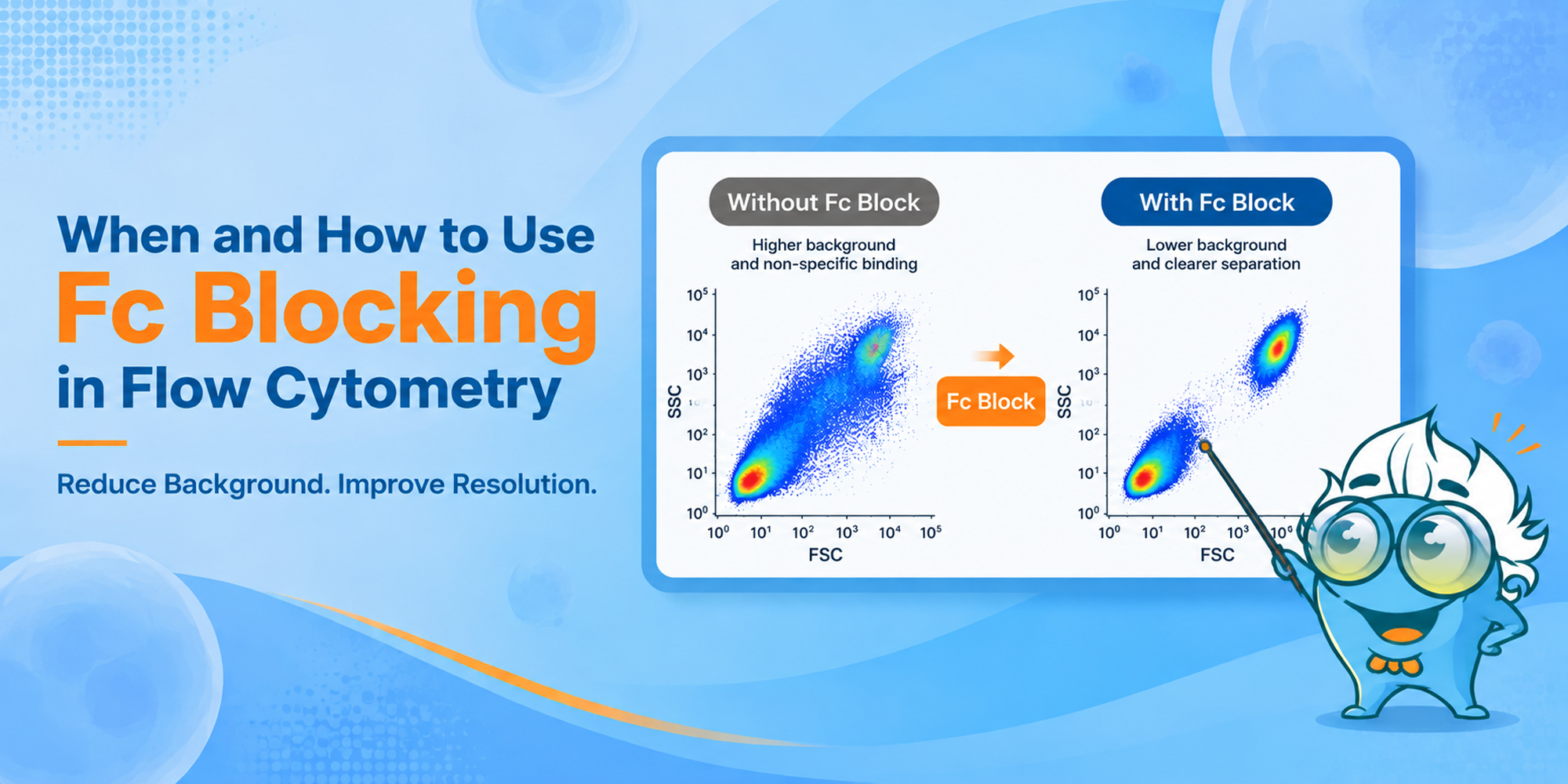
High background in flow cytometry is not always an antibody-specificity problem. Cells that express Fc receptors can capture antibodies through the Fc region, adding fluorescence that is unrelated to the target antigen. The result may be a higher negative population, poorer separation, or an apparently positive population that is difficult to interpret.
Fc blocking in flow cytometry is used to reduce that Fc receptor-mediated component before antibody staining. The more useful questions are when the step is likely to matter, how inadequate blocking appears in the data, and whether the blocker can interfere with a marker in the panel.
Those answers depend on the sample. Fc receptor expression varies with lineage and activation state, and blocker chemistry and antibody isotype also matter. For an unfamiliar preparation, a blocked-versus-unblocked comparison is often the quickest way to see whether Fc-mediated binding is affecting the assay.
Most flow cytometry antibodies contain an antigen-binding Fab region and an Fc region. The Fab region recognizes the target antigen; the Fc region can also interact with Fc receptors on leukocytes. When that second interaction contributes to staining, the measured fluorescence is no longer explained by antigen binding alone. [1,5,6]
Fc blocking reduces this unwanted binding by occupying or inhibiting Fc receptors before the staining antibodies are added. Depending on the system, the blocker may be a receptor-specific antibody, purified immunoglobulin, serum-derived immunoglobulin, or a commercial Fc receptor-blocking formulation. [1,5,6]
Fc-mediated binding can raise background, shift negative populations, and make dim or rare populations harder to resolve. In complex samples, it can compound other artifacts such as dead cells and doublets, particularly when profiling tumor-infiltrating immune cells. Fc blocking addresses the Fc-mediated component; viability and singlet controls still need to be handled separately. [1,2]
The need for Fc blocking follows the cells in the tube more than the name of the assay. Vendor protocols flag monocytes, macrophages, neutrophils, B cells, NK cells, and some T-cell subsets because they can express Fc gamma receptors. But the amount of nonspecific binding is not the same across all of these populations or across antibody isotypes. [1,5,6]
Fc receptor-rich myeloid cells deserve the most attention. Lymphocyte-only assays are more variable and should be judged in the context of the panel and sample.
Sample type is a useful shortcut, but cell composition is what matters. A tumor digest rich in macrophages, for example, is a different Fc-blocking problem from a purified T-cell preparation.
| Sample | Fc Blocking Consideration | Why |
|---|---|---|
| Whole blood | Strongly consider | Contains multiple Fc receptor-expressing leukocyte populations, including monocytes and granulocytes. |
| PBMCs | Often useful | Monocytes are retained, but granulocytes are largely absent after standard density separation; importance depends on the populations being analyzed. |
| Bone marrow | Often useful in mixed/myeloid-rich samples | Contains diverse developing and mature immune populations, including myeloid cells. |
| Spleen | Depends on populations analyzed | Mixed immune-cell composition can include substantial Fc receptor-positive populations. |
| Dissociated tumor | Strongly consider when myeloid-rich | Tumor-infiltrating myeloid cells can be particularly sensitive to Fc-mediated artifacts. [2] |
| Inflamed tissue | Often useful when FcR-rich | Inflammatory infiltrates can increase the proportion and activation state of Fc receptor-expressing leukocytes. |
| Purified monocytes/macrophages | Strongly consider | Directly enriched for populations in which Fc-mediated binding can be prominent. [1] |
| Purified conventional T cells | Usually lower priority | Fc-mediated background is commonly less prominent, but activation state and panel composition still matter. |
| Fc receptor-negative cultured cells | Usually low priority if truly FcR-negative | A blocker may add little if the cells do not express relevant Fc receptors and no other Fc-interacting component is present. |
For IgG-based staining, CD64 (Fc gamma RI), CD32 (Fc gamma RII), and CD16 (Fc gamma RIII) are the receptors most often encountered in this context. Their expression varies by lineage, differentiation state, and activation. BD and Thermo Fisher guidance discusses CD16/CD32-associated Fc-mediated binding across B cells, NK cells, granulocytes, monocytes, and macrophages. [5,6]
| Fc receptor | Commonly relevant populations | Note |
|---|---|---|
| CD64 / Fc gamma RI | Especially important in myeloid populations such as monocytes/macrophages | High-affinity Fc gamma receptor; blocker compatibility depends on reagent design. |
| CD32 / Fc gamma RII | B cells, monocytes, macrophages, granulocytes and other FcR-positive cells | A major target of common mouse anti-CD16/CD32 blocking antibodies. [6] |
| CD16 / Fc gamma RIII | NK cells, neutrophils, macrophage/monocyte subsets | Also targeted by common mouse CD16/CD32 Fc-blocking reagents. [6] |
Insufficient Fc blocking usually shows up as a pattern rather than a single diagnostic signal. Look for changes that make Fc-mediated binding plausible, then test that possibility by changing the blocking condition while leaving the rest of the stain unchanged.
A monocyte, macrophage, or neutrophil gate that is disproportionately bright compared with cleaner lymphocyte populations is a useful clue. Andersen et al. saw this cell-type dependence in their test system: monocytes and macrophages showed substantial nonspecific binding while the tested B, T, and NK cells did not. [1]
If Fc-mediated binding raises the negative population, a dim positive population can become harder to resolve. The effect matters most when the marker is already weak or the population of interest is rare. For dim or ambiguous populations in multicolor panels, FMO controls can help define gating boundaries.
A population expected to be negative or very low may shift into a weak-positive range. Before treating that shift as biology, compare Fc blocking conditions and review antibody titration, viability, and gating.
When antibodies against unrelated targets all become brighter in the same FcR-rich population, a shared technical interaction is more likely than several independent biological changes. Fc binding is one possibility; dead cells and dye-related interactions can produce similar patterns.
Keep the antibody panel unchanged and compare blocked with unblocked cells. If the suspicious background falls selectively, Fc-mediated binding was contributing to the signal. That comparison is more informative than trying to diagnose Fc binding from fluorescence intensity alone. For broader panel validation, review the appropriate flow cytometry experimental controls during assay development.
It can, although "over-blocking" is not always the best way to describe the problem. Once Fc-mediated binding is adequately suppressed, extra blocker does not necessarily cause a proportional loss of antigen-specific signal. Interference is more likely when the blocker conflicts with the marker, clone, or immunoglobulin readout being measured.
Not necessarily. A compatible blocker should reduce Fc-mediated binding without removing Fab-mediated recognition of an unrelated antigen. If the expected signal drops after blocking, check receptor overlap, clone competition, blocker composition, and staining order before attributing the change simply to dose.
Some Fc blockers are receptor-specific antibodies. Common mouse Fc blocks target CD16/CD32, and BD notes that the classic 2.4G2 reagent can also block CD64 through its Fc domain. That can complicate direct measurement of the same receptors or the use of certain second-step reagents. [3,6]
Compatibility is reagent- and clone-specific. Some commercial human Fc blockers are tested for use with selected anti-CD16, anti-CD32, and anti-CD64 clones, so the receptor name alone is not enough to decide whether a panel will work. [7]
Blocker composition can matter even when Fc receptors are not the target. Ardicli and colleagues compared FcR-blocking approaches for B-cell receptor immunoglobulin heavy-chain isotype profiling and found that human serum IgG-based blocking reagents could compromise detection, particularly for IgG subclasses. For assays that directly measure BCR IgH isotypes, compatibility should be established before using a human-derived FcR blocker. [4]
Fc-blocking products differ in mechanism, concentration, and incubation conditions, so the product data sheet should take precedence over a generic workflow. A typical surface-staining sequence is:
For example, Thermo Fisher lists 10–20 minute pre-incubation conditions for its mouse and human Fc-blocking options, while other commercial blockers use shorter validated incubations. These times belong to the individual products rather than to Fc blocking as a universal rule. [5,7]
Optimization is worthwhile when background still limits interpretation or when the panel contains a marker that might conflict with the blocker.
Start at the manufacturer's recommended condition. Fc blockers differ in affinity, composition, intended species, and receptor coverage, so a dose that works for one product should not be transferred automatically to another.
For unfamiliar tissues, myeloid-rich samples, rare populations, or a panel already limited by background, stain matched aliquots with and without Fc block. Keep cell number, antibody concentrations, incubation conditions, instrument settings, and gating strategy the same.
If a reproducible Fc-associated pattern remains at the recommended condition, a higher blocker concentration can be tested. Judge it by the change in background and population separation, not by the amount of reagent used.
High fluorescence can persist even when Fc receptors are fully blocked. Fc blocking does not correct excess antibody, dead-cell binding, autofluorescence, doublets, compensation or unmixing problems, or dye-specific interactions. In complex multicolor experiments, fluorophore choice and spectral overlap should also be reviewed during multicolor flow cytometry panel design. In the tumor study by Kuonen et al., Fc blocking improved the data, but dead-cell and doublet exclusion were also required to remove phenotypic artifacts. [2]
| Potential source of background | Will Fc blocking address it? | What to check instead / as well |
|---|---|---|
| Fc receptor-mediated antibody binding | Yes, if the blocker covers the relevant interaction | Blocker choice, dose, species and receptor coverage |
| Dead or damaged cells | No | Viability dye, sample handling, gating |
| Antibody concentration too high | No | Antibody titration |
| Autofluorescence | No | Unstained controls, fluorophore selection, gating/unmixing strategy |
| Cell doublets or aggregates | No | Singlet gating and sample preparation |
| Polymer/tandem/dye-specific interactions | Not necessarily | Use dye-compatible blocking/staining buffers where recommended. [5] |
A matched-sample comparison usually narrows this down:
If the marker of interest changes substantially after blocking, review compatibility with the receptor, detection clone, antibody format, or immunoglobulin readout.
Fc blocking matters most when samples contain FcR-rich myeloid cells. If background is concentrated in these populations, compare matched blocked and unblocked samples. A selective drop in suspicious signal supports an Fc-mediated contribution; if the marker of interest also changes, check blocker composition and clone compatibility before changing the dose.
Choose the blocker for the cells and markers in the assay, and confirm its effect under your own staining conditions. For broader guidance on sample preparation, staining, gating, optimization, and troubleshooting, see Boster's Flow Cytometry Technical Resource Center.
Often, especially when monocytes or other FcR-positive populations are part of the analysis. The benefit may be smaller in lymphocyte-focused panels; a blocked-versus-unblocked comparison can resolve the question during panel development.
Usually less than in myeloid-cell assays. Resting T-cell-focused panels are often lower priority, although subset and activation state can change the need for blocking.
Usually. Macrophages express Fc receptors and are among the cell types most prone to Fc-mediated nonspecific antibody binding.
Often, especially when the digest contains infiltrating myeloid cells or other FcR-rich populations. The need is driven by the cells in the sample rather than by the tumor label itself.
Not necessarily. Some protocols proceed directly to antibody staining, while others use product-specific steps. Follow the data sheet for the blocker in use.
It can, if the blocker conflicts with the assay. This is most relevant when measuring a receptor targeted by the blocker or when blocker composition interferes with immunoglobulin/BCR readouts. Check reagent and clone compatibility. [4,6]







