This website uses cookies to ensure you get the best experience on our website.
- Table of Contents
The house mouse (Mus musculus) has long been a cornerstone of scientific research, serving as a vital model organism that has propelled countless breakthroughs in biology and medicine. From its early adoption in the 20th century to its current status as a genetic and biomedical research powerhouse, the mouse has proven indispensable for scientists worldwide.
In this blog, we briefly describe the history of the mouse as a model organism, highlighting key milestones and discoveries that have shaped our understanding of human health and disease. We also delve into the advantages that make the mouse an ideal research model while also addressing its limitations and ethical considerations. Additionally, we discuss the various fields where the mouse has made major contributions, provide resources for researchers, and offer guiding questions to help determine if the mouse is the right model for your scientific studies. If you’re considering using mouse models for your research studies, this blog is for you!
Feel free to jump to a specific section about mouse:
The house mouse (Mus musculus) is native to various regions around the world and is commonly found in human habitats, fields, and laboratories. This small rodent, typically weighing between 20 to 30 grams, has become the backbone in biological and biomedical research. Adult Mus musculus measure about 6 to 10 centimeters in body length, with an additional 7 to 10 centimeters for the tail. They have a fur color that ranges from gray to brown, which can vary depending on the strain.
...
If you’re contemplating whether or not to use zebrafish (Danio rerio) as a model organism for your research studies, this guide is for you. In this blog, we highlight the key breakthroughs accomplished with zebrafish research, discuss the advantages and limitations of using zebrafish for research, and describe the research areas where the zebrafish has made significant contributions. Furthermore, we mention some resources and funding supporting zebrafish research, and provide guiding questions to help you decide if this model organism is right for your studies.
Feel free to jump to a specific section about zebrafish:
The zebrafish (Danio rerio) is a small freshwater fish from South Asia, including India, Bangladesh, and Myanmar. Adults are 2.5 to 4 cm long, with males having gold and blue stripes, and females blue and silver. Their transparent embryos and larvae make zebrafish ideal for developmental biology and genetics research.
Danio rerio is regularly used in research for its rapid development and transparency to directly observe internal processes. Zebrafish embryos develop outside the mother's body and can be examined under a microscope. The fish reach sexual maturity in about 3 to 4 months, with females laying hundreds of eggs per spawning event.
The complete Danio rerio genome sequenced in 2013 has offered a detailed genetic framework for studying gene function and disease. Zebrafish are also amenable to genetic manipulation, including the creation of transgenic and mutant lines. This makes them an excellent model for investigating gene function, disease mechanisms, and drug testing. Their use in research has led to significant discoveries in developmental biology, cancer research, and neurobiology.
The zebrafish (Danio rerio), originally known for its popularity in the aquarium trade, has become a cornerstone model organism in scientific research. Its journey from a hobbyist’s favorite to a vital research tool showcases the species' unique advantages in genetic and developmental studies. In this section, we briefly describe its history and key breakthroughs in scientific research.
The zebrafish's transformation into a model organism began in the 1970s, largely due to the work of George Streisinger, a pioneering geneticist at the University of Oregon who is also considered as the founding father of zebrafish research by his peers. Streisinger recognized that zebrafish embryos, with their transparency and rapid development, were ideal for directly observing developmental processes in real time.1 This feature enabled researchers to examine the formation of organs and tissues in real time, which would elucidate vertebrate development and mechanisms of embryogenesis, organogenesis, and cell differentiation. The species' high reproductive rate and ease of maintenance further highlighted its potential for large-scale genetic studies.
In 1981, Streisinger cloned the zebrafish and became the first person to clone a vertebrate.2,3 Streisinger and his colleagues also successfully conducted the first mutagenesis in zebrafish, creating mutant strains that could be used to study gene function.4 The development of in vitro fertilization methods and mutagenesis techniques during this period significantly contributed to the rise of zebrafish as a model organism.5 These early breakthroughs enabled researchers to perform systematic genetic screens and identify genes essential for development and disease.
The 1990s marked a period of rapid growth in zebrafish research. Large-scale mutagenesis screens and the establishment of a zebrafish mutant library allowed researchers to systematically explore the genetic underpinnings of various biological processes, leading to the discovery of many genes involved in embryonic development and disease.6 This period also saw the expansion of zebrafish research into fields such as neurobiology, toxicology, and regenerative medicine.7,8
Led by scientists at the Wellcome Trust Sanger Institute, the sequencing of the zebrafish genome was completed in 2013, which was a major milestone that further solidified the species' role in research.9 The availability of the Danio rerio’s complete genome, along with advanced genetic tools like CRISPR-Cas9, enhanced the zebrafish’s utility in studying gene function, regulatory networks, vertebrate development, and human disease models.
Zebrafish have been used to model human diseases, including cancer, cardiovascular disorders, and neurological conditions.10 Their rapid development, genetic tractability, and ability to exhibit disease phenotypes have made them a powerful tool for studying disease mechanisms and testing potential therapies. For instance, researchers have used zebrafish to investigate the effects of drugs on tumor growth and to screen pharmacological compounds.11
Zebrafish are renowned for their regenerative abilities, particularly in regenerating fins, heart, and spinal cord. Research on zebrafish has provided insights into the mechanisms of tissue regeneration and repair, with implications for regenerative medicine and therapeutic approaches to injury and degenerative diseases.12,13
Today, zebrafish models are used in research institutions globally. Their history as a model organism exemplifies the transition from a non-traditional organism to one of the most important models in modern biological research, particularly for studies involving vertebrate development, gene function, and disease modeling.
Zebrafish (Danio rerio) are a prominent model organism in scientific research due to their distinct features and versatility. Their transparent embryos, rapid development, and genetic similarity to humans (about 70% of their genes are similar) make them suitable for studying development, genetics, disease, and regenerative processes.
These advantages make zebrafish an essential model organism in various research fields, from developmental biology and genetics to drug discovery and regenerative medicine.
Although zebrafish have proven to be a valuable model organism for research, scientists should be aware of the limitations and challenges of working with zebrafish.
To alleviate the limitations of working with zebrafish, researchers can consider applying several approaches to the following challenges.
By addressing these challenges, researchers can maximize the utility of zebrafish as a model organism and enhance the reliability and relevance of their findings.
Zebrafish (Danio rerio) have emerged as a vital model organism across diverse research fields due to their transparent embryos, rapid development, and genetic manipulability. Researchers harness zebrafish to investigate a spectrum of topics, leveraging their unique attributes to advance understanding in both basic and applied sciences.
Zebrafish offer a versatile and powerful platform for a range of research areas. Their application in research continues to extend beyond developmental biology and disease modeling, uncovering knowledge of biological processes and encouraging exploration of new scientific frontiers.
Researchers working with zebrafish as a model organism have access to a range of vibrant communities, resources, conferences, and funding opportunities. We have listed some of the institutions and tools below.
Zebrafish Information Network (ZFIN): A comprehensive database for zebrafish research, including gene information, genetic tools, and community resources. Website: zfin.org
The Zebrafish Book: A free comprehensive guide to zebrafish biology and techniques, available online and through academic publishers and libraries. Website: zfin.org/zf_info/zfbook/zfbk.html
NCBI Genome Resource Consortium - Zebrafish: Provides information on the ongoing efforts to improve and maintain the zebrafish genome assembly, including updates on genome issues and data resources. Website: www.ncbi.nlm.nih.gov/grc/zebrafish
International Zebrafish Society (IZFS): An organization dedicated to supporting and promoting zebrafish research, including hosting conferences, providing resources, and presenting the George Streisinger Award. Website: www.izfs.org
Zebrafish Disease Models Society (ZDMS): Advances basic and clinical research using zebrafish disease models, fostering international collaboration and knowledge sharing. Website: www.zdmsociety.org
Boster Bio: In addition to off-the-shelf anti-zebrafish antibodies, Boster Bio also offers a deeply discounted $600 custom antibody service particularly for researchers working with model organisms like zebrafish.
Zebrafish Husbandry Association (ZHA): A non-profit organization dedicated to promoting and developing zebrafish husbandry standards through education, collaboration, and publication. Website: zhaonline.org
Zebrafish International Resource Center (ZIRC): A central repository for wild-type and mutant zebrafish strains, providing resources and information to support zebrafish research. Website: zebrafish.org/home/guide.php
European Zebrafish Resource Center (EZRC): Archives zebrafish lines and provides biomedical researchers with fish, plasmids, and screening services. Website: www.ezrc.kit.edu
Zebrafish Core Facilities: Many zebrafish core facilities established around the world provide specialized services, training, and support for zebrafish research.
International Zebrafish Conference: A conference hosted by IZFS that gathers researchers to discuss the latest advancements in zebrafish research across various fields. Website: www.izfs.org/education
Zebrafish Disease Models (ZDM): Hosted by the Zebrafish Disease Models Society, this is an annual conference focusing on the use of zebrafish in disease modeling and related research areas. Website: www.zdmsociety.org/home
The Allied Genetics Conference (TAGC): The Allied Genetics Conference (TAGC) is a flagship event by the Genetics Society of America that fosters collaboration across biological research communities, including researchers working with zebrafish, Drosophila, yeast, and more. Website: genetics-gsa.org/tagc/
European Zebrafish Society (EZS): Fosters zebrafish research by providing a platform for researchers and supporting grant funding for young scientists. Website: www.ezsociety.org
National Institutes of Health (NIH): Provides grants and funding opportunities specifically for research using zebrafish models through various institutes such as the National Institute of General Medical Sciences (NIGMS). Website: grants.nih.gov
National Science Foundation (NSF): Offers grants for research involving zebrafish in areas such as developmental biology and genetics. Website: nsf.gov
European Research Council (ERC): Supports zebrafish research through funding programs for projects in various scientific disciplines. Website: erc.europa.eu
These resources offer support and opportunities for scientists working with zebrafish models, facilitating advances in their research and fostering a collaborative scientific community.
Here are some guiding questions to consider if you are thinking about using zebrafish as a model organism in your research:
Research Goals:
Experimental Needs:
Genetic Tools:
Homology to Humans:
Ethical and Regulat

Drosophila melanogaster, commonly known as the fruit fly, has long been a cornerstone of genetic research. Its simplicity, rapid life cycle, and genetic tractability make it an invaluable model organism for scientists worldwide.
If you’re considering using Drosophila for your research studies, this guide is for you. In this blog, we delve into key breakthroughs that used Drosophila in research, explore the advantages and limitations of using Drosophila for research, and highlight the research areas where the fruit fly has made significant contributions. Additionally, we provide some resources and funding supporting Drosophila research, along with reflective questions to help you decide if this model organism is right for your studies.
Feel free to jump to a specific section about Drosophila:

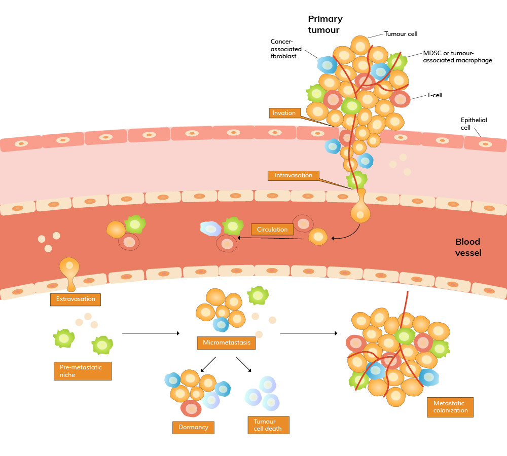
Immunotherapy, a treatment that uses someone’s own immune system to target and attack cancer cells is the next and best frontier of cancer treatment. CAR-T stands for Chimeric Antigen Receptor T-cell. It refers to a type of immunotherapy where T-cells are engineered to produce special receptors on their surface that help them target and kill cancer cells. Like all immunotherapy, CAR-T cell therapy harnesses the p


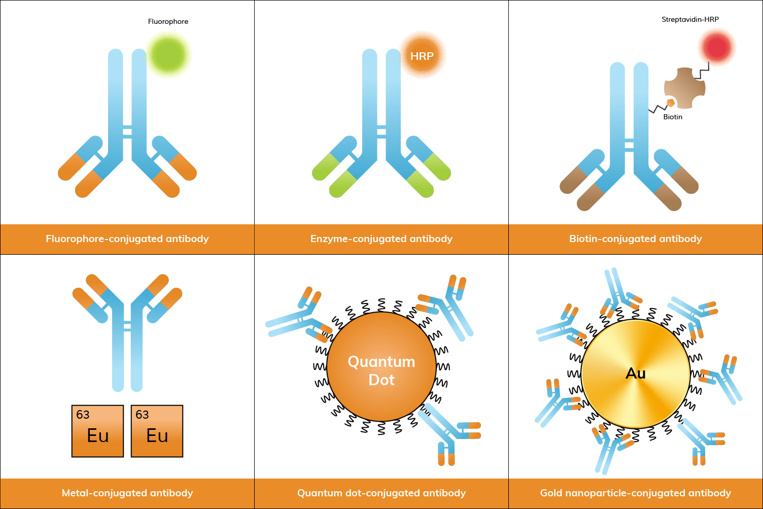
Antibody conjugates are essential tools in biological research, offering both specificity and sensitivity for detecting and quantifying proteins, cells, and other molecules. Below, we explore the most common types of antibody conjugates, their examples, applications, and popularity in research.
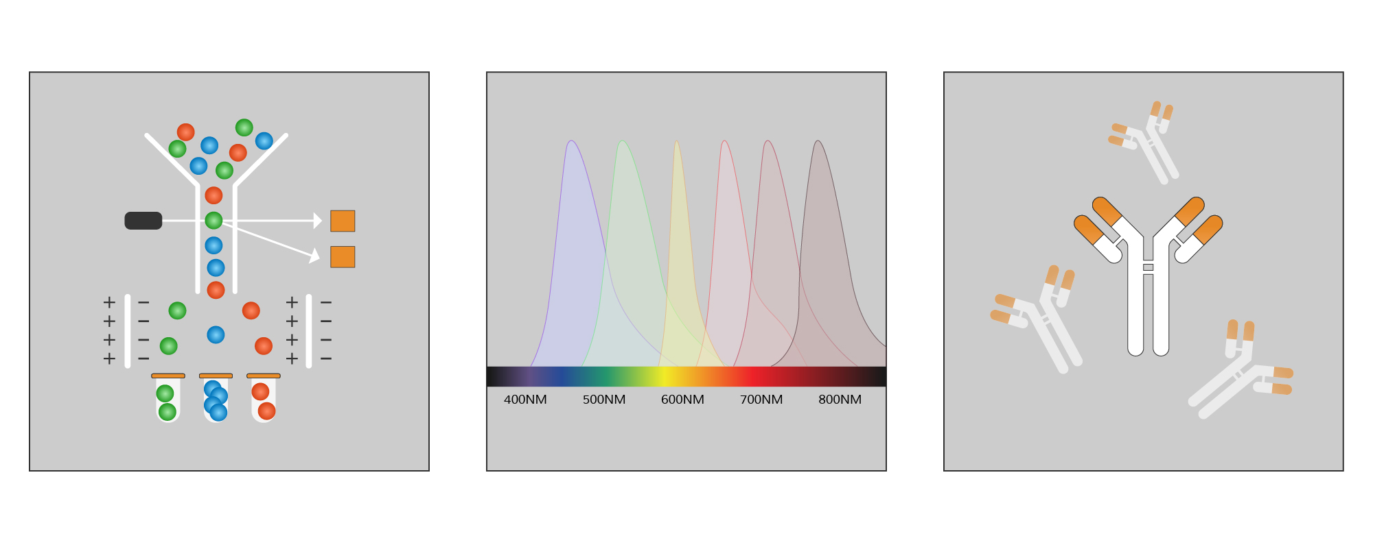
Antibody conjugation is the process of chemically linking an antibody to another molecule, such as a fluorescent dye, enzyme, biotin, or nanoparticle. This process enhances the antibody’s ability to detect specific targets by enabling visualization or measurement in various assays. Conjugated antibodies are widely used in research for applications like flow cytometry, ELISA, and immunofluorescence, where they facilitate the detection and analysis of specific proteins or cells in complex samples. In some experimental setups, especially those involving gene delivery or expression studies, related tools such as AAV Packaging Service may also be employed to introduce genetic material efficiently into target cells. These conjugates are often produced as part of comprehensive antibody production services, where antibodies are not only generated but also tailored with the appropriate labels to suit specific experimental needs.
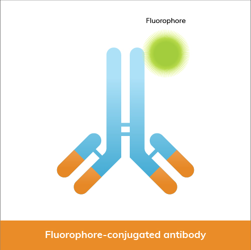
Among the most commonly used are fluorophore conjugates, which include dyes like fluorescein isothiocyanate (FITC), cyanine dyes, DyLight® dyes, allophycocyanin (APC), phycoerythrin (PE), R-phycoerythrin (R-PE), and iFluor® dyes.
Fluorophore-conjugated antibodies are widely used in:
Below, we have provided a table comparing key characteristics and uses of some of the most common fluorophore conjugates in research.
| Fluorophore | Color | Max Excitation (nm) | Max Emission (nm) | Extinction Coefficient (M⁻¹cm⁻¹) | Advantages | Applications |
|---|---|---|---|---|---|---|
| FITC | Green | 495 | 519 | 70,000 | Bright, photostable, common filter sets | Flow cytometry, immunofluorescence, microscopy |
| Cy3 | Orange | 552 | 570 | 150,000 | Bright, used in multiplexing | Flow cytometry, immunofluorescence, FISH |
| Cy5 | Red | 650 | 670 | 250,000 | Near-infrared, high sensitivity | Flow cytometry, imaging, FRET |
| DyLight® 488 | Green | 493 | 518 | 70,000 | Bright, photostable | Flow cytometry, immunofluorescence, microscopy |
| DyLight® 550 | Orange | 562 | 576 | 150,000 | High brightness, photostable | Western blotting, fluorescence microscopy, flow cytometry |
| DyLight® 594 | Red | 593 | 618 | 115,000 | Bright, minimal spectral overlap | Multicolor fluorescence imaging, flow cytometry |
| DyLight® 650 | Far-red | 652 | 672 | 250,000 | Near-infrared, reduced background | Flow cytometry, fluorescence imaging |
| DyLight® 800 | Near-IR | 783 | 800 | 270,000 | Near-infrared, minimal autofluorescence | In vivo imaging, Western blotting, NIR fluorescence imaging |
| iFluor® 488 | Green | 491 | 516 | 70,000 | Bright, photostable, FITC alternative | Flow cytometry, immunofluorescence, confocal microscopy |
| iFluor® 555 | Orange | 555 | 565 | 150,000 | High brightness, photostable | Fluorescence microscopy, flow cytometry, multicolor applications |
| iFluor® 594 | Red | 590 | 615 | 115,000 | Bright, minimal spectral overlap | Multicolor fluorescence imaging, flow cytometry |
| iFluor® 647 | Far-red | 650 | 665 | 250,000 | High brightness, photostable | Flow cytometry, fluorescence imaging, super-resolution microscopy |
| iFluor® 750 | Near-IR | 755 | 779 | 270,000 | Near-infrared, minimal autofluorescence | In vivo imaging, NIR fluorescence imaging |
| APC | Red | 650 | 660 | 700,000 | High quantum yield, photostable | Flow cytometry, imaging |
| PE | Orange | 480-565 | 575-590 | 1,960,000 | High brightness, quantum yield | Flow cytometry, fluorescence microscopy |
| R-PE | Red-orange | 488, 546, 565 | 575-585 | 1,960,000 | Extremely bright, multiple chromophores | Flow cytometry, high sensitivity applications |
Fluorophore conjugates are very popular due to their versatility, high sensitivity, and the variety of available dyes that allow multiplexing. When searching for primary antibodies and secondary antibodies at Boster, you’ll be able to select from a range of conjugation options, such as Cy3, DyLight® dyes, FITC, APC, PE, or iFluor® dyes. You can also request custom antibody conjugation with our antibody conjugation service, which offers more conjugate labels.
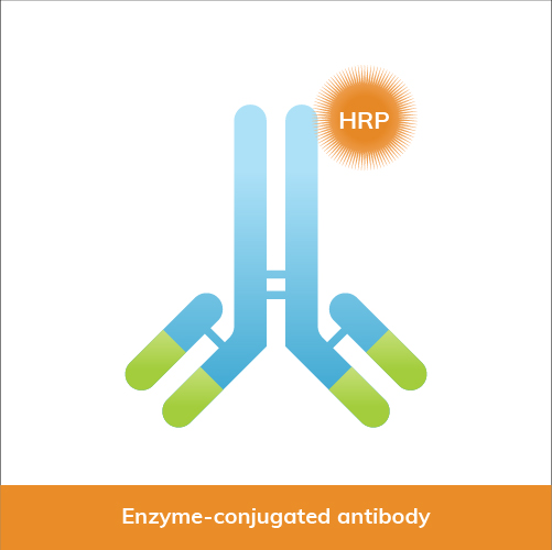
Enzyme conjugates, such as those linked to horseradish peroxidase (HRP) and alkaline phosphatase (AP), are also commonly used in research. These conjugates are crucial in assays like ELISA, WB, and IHC. In particular, enzyme-conjugated antibodies are widely utilized in sandwich ELISA formats, where the precise coordination between the capture and detection antibodies is essential for achieving optimal signal development and minimizing background interference. Antibody Pair Development Service develops matched antibody pairs for these assays involving careful selection to ensure that the antibodies bind to non-competing epitopes with high affinity and stability across varying assay conditions.
Enzyme-conjugated antibodies are used in:
Enzyme conjugates are highly popular in routine laboratory assays due to their robustness and ease of use. However, when assays demand superior specificity and minimal background noise, especially in enzyme-linked applications like ELISA and Western blotting, sourcing antibodies through specialized Rabbit Monoclonal Antibody Services can provide researchers with tailored solutions that consistently deliver reliable signal detection in complex biological samples. At Boster Bio, you can find primary antibodies and secondary antibodies conjugated to HRP, AP, and more. In addition, you can select specific conjugates for your antibodies with our custom antibody conjugation service.
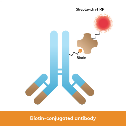
Biotin, a vitamin that can be easily bound by streptavidin, has proven to be another essential antibody conjugate in research. It provides significant advantages due to its amplification capabilities. Biotin-labeled antibodies, often paired with streptavidin-HRP or AP, are used by researchers in ELISA, Western blotting, and immunohistochemistry.
In research, biotin-conjugated antibodies are frequently used in:
Biotin conjugates are widely used due to their ability to provide amplification for applications that require high sensitivity. Boster Bio's catalog contains biotin-conjugated primary antibodies and secondary antibodies, and additional conjugate options. You can also learn more about our custom antibody conjugation service and book a meeting with us to discuss your project, so we can better serve your research needs. Submit an inquiry today!
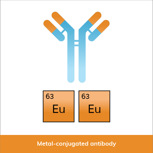
Metal conjugates, including lanthanide-chelated antibodies (e.g., Europium, Terbium) and metal isotope-tagged antibodies for mass cytometry (CyTOF), are gaining traction in advanced applications.
Metal-conjugated antibodies are used in:
Growing popularity of metal conjugates, especially in advanced applications like CyTOF, reflects their capability to provide comprehensive cellular analysis.
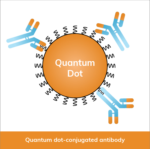
Quantum dot conjugates are semiconductor nanoparticles, including Qdot 525 and Qdot 655, known for their unique optical properties.
Quantum dot-conjugated antibodies are used in:
Although less common than traditional fluorophores, quantum dots (Qdots) are increasingly popular in imaging applications for their photostability and distinct spectral properties.

Gold nanoparticles (AuNPs) are widely employed in various diagnostics, biosensing, and imaging applications.
Gold nanoparticle-conjugated antibodies are used in:
Gold nanoparticle-conjugated antibodies are quite popular in diagnostics and increasingly in biosensing applications due to their practical utility and ease of detection.
Antibody conjugates play a vital role in modern research, with each type offering distinct advantages. Fluorophore and enzyme conjugates remain staples due to their broad applications and established protocols. Biotin conjugates are favored for applications requiring high sensitivity, while metal conjugates offer advanced analysis capabilities. Quantum dots and gold nanoparticles, though more specialized, are expanding in use as techniques and technologies improve. Selecting the appropriate conj...
Flow cytometry and Fluorescence-Activated Cell Sorting (FACS) are indispensable tools in biomedical research and clinical diagnostics. Despite their widespread use, confusion often arises regarding their terminology and functionalities. In this article, we identify distinctions between flow cytometry and FACS, and discuss their principles and applications.
Developed in the 1950s and 1960s, flow cytometry revolutionized cell analysis by allowing rapid, high-throughput measurement of multiple cellular characteristics. This technique analyzes the physical and chemical characteristics of particles or cells in a fluid suspension, and involves passing a cell-containing fluid stream through a laser beam, measuring the scattered and fluorescent light emitted by the cells.
Key aspects of flow cytometry include: