This website uses cookies to ensure you get the best experience on our website.


- Table of Contents
Blocking is one of the easiest IHC steps to over-adjust. When the background is high, it is tempting to extend the blocking time, increase blocker concentration, or switch to a stronger product. But the best blocking strategy is not the strongest blocker. It is the one that matches the likely source of background. In some workflows, the selected blocking buffer can also influence downstream compatibility with related assays such as Western Blotting and multiplex fluorescence applications.
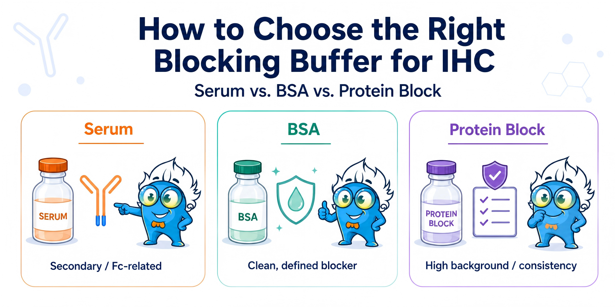
A practical rule: use serum when secondary antibody or Fc-related background is likely, BSA when you need a cleaner defined blocker, and commercial protein block when background control and workflow consistency matter most.
The real question is not “Which blocker is best?” It is “What kind of background am I trying to reduce?”
Do not change the blocker until you have looked at the shape of the background.
A diffuse haze across the whole tissue may suggest non-specific protein binding, excessive antibody concentration, or insufficient washing. Background concentrated in immune-rich tissues such as spleen, tonsil, lymph node, or inflamed tissue may point to Fc-mediated binding. A positive secondary-only control suggests the problem is coming from the secondary antibody or detection system, not the primary antibody. Edge staining or signal around damaged areas may reflect tissue drying, over-retrieval, section damage, or uneven washing.
This matters because blocking mainly addresses non-specific protein interactions. It will not correct endogenous peroxidase activity, endogenous biotin, tissue autofluorescence, over-retrieval damage, poor washing, or an antibody used too concentrated. Similar principles are also important in Western blots, where inappropriate blocking conditions may increase background noise and reduce assay specificity.
If the background pattern is unclear, separate true non-specific staining from detection or tissue-related artifacts before changing every condition at once. Boster’s guides to non-specific staining in IHC and high background in DAB staining are useful follow-up references.
Normal serum contains a broad mix of proteins that can occupy non-specific binding sites and reduce unwanted secondary antibody interactions. It is often a good starting point for indirect IHC workflows, especially in immune-rich tissues or samples with Fc receptor activity.
In many workflows, the blocking serum is selected from the same species as the secondary antibody host. For example, if the secondary antibody is raised in goat, normal goat serum is commonly used to reduce non-specific secondary binding.
The trade-off is variability. Serum contains immunoglobulins and other undefined components, so it can sometimes introduce cross-reactivity or lot-to-lot inconsistency. This becomes more important in multiplex IF, sensitive fluorescence workflows, or assays where serum components interfere with the antibody system.
Use serum when you need broad blocking against secondary or Fc-related background. Avoid treating it as a universal blocker.
Bovine serum albumin is often the cleaner choice when you want to reduce general non-specific binding without adding the complexity of serum. It is more defined, easier to standardize, and commonly used in antibody diluents to improve consistency.
BSA is a good starting point when background is mild to moderate, when serum may create extra variables, or when the workflow requires a cleaner blocking environment. It can also be useful in IF or multiplex staining, where avoiding serum-related cross-reactivity may matter. In fluorescence-based applications, optimized blockers can help improve signal-to-noise ratios during fluorescent detection.
The limitation is that BSA is not always strong enough for difficult tissue. If background is driven by Fc receptor binding, damaged tissue, sticky extracellular matrix, or high endogenous activity, BSA alone may not solve the problem.
Use BSA for a clean baseline. Do not expect it to fix every high-background slide.
Commercial protein blocks are useful when the background is difficult to control or when the staining workflow needs to stay consistent across many slides, operators, tissue types, or validation runs.
They are often helpful during assay optimization, repeated staining, high-throughput workflows, and tissue panels where serum or BSA gives variable results. The main advantage is not that they are “magic.” It is that they reduce one source of workflow variability.
A commercial protein block can be a better fit when tissue background remains high after basic blocking, when serum introduces extra background, or when the lab needs a more standardized starting condition. Some manufacturers also offer specialized reagents such as fluorescent blot blocking buffer formulations designed for fluorescence imaging compatibility and reduced interaction with fluorescent dyes.
For broader optimization around blocking, antibody dilution, retrieval, and detection conditions, Boster’s IHC optimization guide can help place blocking in the full workflow.
| Situation | Better starting choice | Why |
|---|---|---|
| Secondary antibody-related background | Normal serum | Reduces non-specific secondary binding |
| Fc receptor-rich tissue | Serum or Fc-focused blocking | BSA alone may be insufficient |
| Need a clean, defined blocker | BSA | Fewer undefined serum components |
| Mild non-specific haze | BSA or protein block | Depends on tissue and detection system |
| High tissue background | Commercial protein block | Broader and more standardized blocking |
| Multiplex IF workflow | BSA or validated protein block | Helps reduce serum-related cross-reactivity |
| Repeated staining across many samples | Commercial protein block | Improves run-to-run consistency |
| Serum adds background | BSA or protein block | Avoids serum-derived variability |
A simple rule: serum is best for secondary or Fc-related background, BSA is best for cleaner baseline blocking, and commercial protein block is best when consistency and stronger background control matter.
High background is not always a blocking problem. Before reaching for a stronger blocker, audit the rest of the workflow.
If the primary antibody is too concentrated, a stronger block may reduce haze but will not restore specificity. If washing is too short or too weak, an unbound antibody can remain trapped in the tissue. If antigen retrieval is too harsh, damaged tissue may mechanically trap antibodies and detection reagents. If the secondary-only control is positive, the problem is likely the secondary antibody, detection system, or endogenous activity.
Blocking also cannot replace specific steps for endogenous peroxidase, endogenous biotin, or autofluorescence. These require their own controls and blocking steps. In DAB-based IHC, for example, endogenous enzyme activity can look like non-specific staining even when the protein block is working.
If background appears together with weak specific signal, antibody dilution and incubation conditions should also be reviewed. Boster’s guide on optimizing primary antibody incubation in IHC is a useful next step. Similar optimization principles apply in chemiluminescent detection, alkaline phosphatase detection, and biotin-streptavidin detection workflows used across immunodetection platforms.
Serum should be selected around the antibody system, matching the secondary antibody host and avoiding serum from the same species as the primary antibody. Poor species matching can add background instead of reducing it.
Longer blocking time or higher protein concentration does not always improve staining. It may reduce specific signals or make results less consistent.
Endogenous peroxidase, endogenous biotin, and autofluorescence are not solved by serum, BSA, or general protein block alone.
High background often comes from overly concentrated primary or secondary antibody. Dilution optimization may solve the problem more directly than switching blockers. This is particularly important when working with sensitive secondary antibody conjugate systems or phospho-specific antibodies targeting low-abundance phosphorylated targets.
There is no universal best blocker. Serum is useful for secondary antibody or Fc-related background, BSA is useful for cleaner defined blocking, and commercial protein block is useful for high background or standardized workflows.
Use serum when secondary antibody binding or Fc receptor interactions are likely. Use BSA when you want a simpler, more defined blocker and serum may introduce variability or cross-reactivity.
Sometimes, but it should be tested. IF workflows can be more sensitive to autofluorescence and serum-related background. BSA or a validated protein block is often a cleaner starting point.
The background may not come from non-specific protein binding. Check antibody concentration, washing, antigen retrieval, endogenous peroxidase, endogenous biotin, autofluorescence, and secondary-only controls. For a wider workflow review, Boster’s IHC troubleshooting guide can help organize the next checks.
In some laboratories, related optimization workflows may also involve Western blot membranes such as Nitrocellulose membrane or PVDF membrane platforms, particularly when validating antibodies across IHC and Western Blotting applications. Buffer systems including Tris-buffered saline and phosphate-buffered saline are commonly used during wash and incubation steps to maintain staining consistency.
Choosing a blocking buffer in IHC is not about finding the strongest blocker. It is about matching the blocker to the likely source of background.
Use serum when secondary antibody binding or Fc-related background is likely. Use BSA when you need a cleaner, more defined condition. Use commercial protein block when tissue background is high or workflow consistency matters.
The best blocking strategy lowers background without weakening the specific signal you need to interpret. Effective blocking can also improve consistency across downstream applications, including fluorescent western blotting, protein quantitation, and broader proteomic analysis workflows. These considerations are especially relevant when working with complex mammalian proteins in cultured cells, including models such as HEK293 cells and Jurkat cells, or when analyzing frozen tissue sections for research or diagnostic or therapeutic purpose studies.
If you are still selecting reagents for the workflow, Boster’s IHC reagents page can help you review available options.






