This website uses cookies to ensure you get the best experience on our website.
- Table of Contents

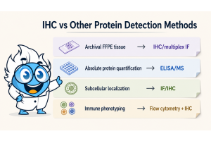
Choosing the wrong secondary antibody can quickly turn an IHC stain into a high-background result. This article explains how to reduce non-specific staining by looking beyond host-species matching and checking the factors that matter most: primary isotype, tissue species, cross-reactivity, blocking strategy, detection system, and no-primary controls. For the full staining workflow, review Boster’s IHC protocol before optimizing the detection step.
In a simplified workflow, the secondary antibody binds the primary antibody and carries the detection label. In real tissue, it also encounters endogenous immunoglobulins, Fc receptor-positive cells, blood components, extracellular matrix, endogenous enzymes, and autofluorescent structures. If it binds an unintended target, the detection system can amplify that weak interaction into visible background.
Secondary antibody background in IHC is context-dependent: the same reagent may be clean in one tissue but produce diffuse haze in another. If the pattern is difficult to interpret, compare it with common patterns of non-specific staining in IHC. Often, the antibody is not inherently unsuitable; it is too broad, insufficiently adsorbed, or poorly matched to the tissue or detection system. Similar issues may also appear during Western blot optimization and flow cytometry panel validation when secondary specificity is not carefully controlled.
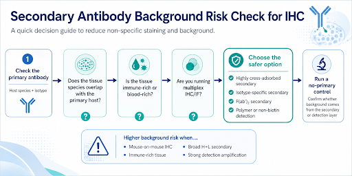
Secondary antibody selection starts with the primary antibody, but it should not stop at host-species matching. For rabbit primaries, Anti-Rabbit IgG detection is often straightforward. Mouse monoclonals need more caution because many are IgG1, IgG2a, IgG2b, IgG3, or IgM. For mouse IgG primaries, a broad anti-mouse IgG H+L secondary is convenient, but its breadth can increase background in sensitive IHC.
If the primary antibody is a mouse IgG1 monoclonal, an anti-mouse IgG1-specific secondary may be cleaner than a broad anti-mouse IgG H+L secondary. This matters even more in multiplex IHC, mouse-on-mouse staining, or immune-rich tissue, where broad recognition can blur the difference between true signal and endogenous Ig, Fc-related binding, or cross-reactivity.
For low-background IHC, the best secondary is not necessarily the broadest one. It detects the intended primary reliably without adding unnecessary tissue signals. If you are still selecting the upstream reagent, start with an IHC-validated primary antibody that matches your target antigens, sample species, and application. Careful review of the species of primary antibody and class and subclass of primary antibody is important during assay planning, especially when working with monoclonal antibodies, polyclonal antibodies, or newer formats such as VHH antibodies.
One common IHC mistake is choosing the secondary based only on the primary antibody while ignoring tissue species. In mouse-on-mouse IHC, an anti-mouse secondary may bind endogenous mouse immunoglobulins in the tissue, not just the mouse primary antibody. The result may look like diffuse haze, tissue-associated staining, or apparent positivity in negative areas.
Extra caution is needed for spleen, lymph node, bone marrow, inflamed tissue, blood-rich samples, and multiplex IHC or IF. A highly cross-adsorbed secondary, isotype-specific secondary, F(ab’)2 secondary, polymer-based detection system, or dedicated mouse-on-mouse strategy may be cleaner than a standard secondary.
This becomes particularly important when staining formalin fixed paraffin embedded samples, or frozen tissue sections, since tissue processing conditions can affect accessibility and background during IHC preparation and antigen retrieval workflows.
Cross-adsorbed, or pre-adsorbed, secondary antibodies are processed to reduce binding to immunoglobulins from non-target species. In IHC, they are useful when cross-reactivity would be hard to interpret later.
| IHC Setup | Main Background Risk | Recommended Strategy |
|---|---|---|
| Primary host matches tissue species | Endogenous Ig binding | Highly cross-adsorbed secondary or species-specific strategy |
| Mouse primary on mouse tissue | Mouse-on-mouse background | Mouse-on-mouse reagent, polymer-based detection, or highly cross-adsorbed secondary |
| Immune-rich tissue | Fc receptor or endogenous immunoglobulin background | F(ab’)2, Fc-specific, or highly cross-adsorbed secondary |
| Multiplex IHC / IF | Secondary cross-reacts with another primary | Highly cross-adsorbed species-specific secondary |
| No-primary control is positive | Secondary or detection-layer background | Change secondary, blocking, washing, or detection system |
Cross-adsorption will not fix poor washing, overdeveloped DAB, or unblocked endogenous enzymes. But when the problem is secondary cross-reactivity, it can be one of the fastest fixes. In many protocols, an adsorbed secondary antibody or pre-adsorbed secondary antibody is selected to reduce unwanted species reactivity and improve interpretation accuracy.
Secondary specificity is not only about species. It is also about which region or class of immunoglobulin the secondary recognizes.
H+L secondary antibodies recognize heavy and light chains. They work for routine IHC, but may be too broad for sensitive tissue or multiplex staining.
Fc-specific secondary antibodies recognize the Fc region of the primary antibody and may reduce recognition of light chains or fragments.
Isotype-specific secondary antibodies help with mouse monoclonals because they can distinguish IgG subclasses.
F(ab')2 fragment secondary antibodies lack the Fc region and can help in Fc receptor-rich tissues such as spleen, lymph node, bone marrow, or inflamed samples.
For clean IHC, broader is not always better. After narrowing the host species, isotype, adsorption level, and detection format, choose a secondary antibody that fits your IHC setup rather than the broadest reagent available. The species of secondary antibody and whether the reagent is a labeled or conjugated secondary antibody should also align with the selected detection chemistry and imaging platform.
Blocking should be planned with secondary antibody selection. For serum blocking, the blocking serum is commonly selected from the same species as the secondary antibody host, not the primary antibody host. If the secondary is goat anti-rabbit IgG, normal goat serum is often used, though protocols vary.
The detection system can amplify both real signal and background. In HRP-DAB IHC, unblocked endogenous peroxidase, non-specific secondary binding, or overdeveloped DAB can make low-level background look real. For brown chromogenic background, review causes of high background in DAB staining.
For biotin-streptavidin detection, endogenous biotin can create unwanted staining, so a biotin block or non-biotin polymer system may be cleaner. For fluorescent IHC, do not choose the secondary only by color; species cross-reactivity, tissue autofluorescence, and bleed-through controls also matter.
Common systems include Horseradish peroxidase, alkaline phosphatase, avidin-biotin complex, and enzyme polymer detection system approaches, each using a different reporter enzyme strategy for visualization. Some laboratories may also use a biotinylated secondary antibody to improve signal amplification in low-expression targets or during chromogenic IHC workflows. In fluorescence applications, optimized secondary antibodies can improve fluorescent detection and overall fluorescent readout quality when viewed under fluorescence or confocal microscopy.
When pairing a new primary with a secondary, Boster’s Buy Primary, Get Secondary promotion may help reduce setup cost. Check the promotion details to confirm eligible secondary antibody formats.
A no-primary control is one of the most useful ways to identify secondary antibody background. The primary antibody is omitted, but the secondary and detection reagents are still applied. If staining appears, the signal is not coming from specific primary binding.
A positive no-primary control points to secondary binding, endogenous Ig, Fc-mediated binding, detection reagent background, endogenous enzyme or biotin activity, or insufficient blocking and washing. It helps avoid blaming the primary antibody when the secondary layer is responsible. For broader control setup, see 6 IHC controls you should know.
Including both a negative control and positive control slide during experimental design can help separate true staining from detection-related artifacts, particularly in translational research and development studies or biomarker validation workflows such as HER2 amplification analysis.
If the no-primary control is clean but the test slide has a high background, check primary concentration, antigen retrieval, tissue preparation, blocking, and washing.
Common mistakes include:
Treat secondary selection as part of the full IHC detection setup, not a simple matching step. Validation using orthogonal methods such as CRISPR knockouts, siRNA knockdown, or comparison with flow cytometry datasets may further strengthen staining confidence in difficult targets.
Yes. It can bind endogenous immunoglobulins, Fc receptor-rich tissue areas, tissue components, or the wrong primary antibody in a multiplex experiment.
Use one when cross-reactivity is likely, such as mouse-on-mouse IHC, multiplex IHC/IF, immune-rich tissue, or species-overlap experiments.
No. H+L secondaries are useful for routine IHC, but an isotype-specific, Fc-specific, F(ab’)2 fragment, or highly cross-adsorbed secondary may be cleaner for sensitive staining.
It means staining occurs without the primary antibody, pointing to the secondary antibody, detection reagent, endogenous enzyme or biotin activity, blocking, or washing as possible sources.
Secondary antibody selection is a small step with a large effect on IHC background. Choose the secondary based on the full context: primary host and isotype, tissue species, cross-adsorption, blocking strategy, detection system, and no-primary control results. The best secondary detects the primary clearly while adding as little tissue background as possible.
Additional workflow optimization using advanced imaging tools, multiplex assays, or specialized reagents such as IHC detection reagent products may further improve sensitivity and reproducibility across different tissue sections and staining platforms.