ABCB7 / Iron-sulfur clusters transporter ABCB7, mitochondrial · IHC design guide

Design Immunohistochemistry for ABCB7

Plan chromogenic IHC for ABCB7 in paraffin sections using the observed cytoplasmic and membranous staining in endothelium and smooth muscle (HPA tissue IHC). The catalog antibody has an IHC dilution range of 1:50–1:200 (datasheet); ABCB7 localizes molecularly to the mitochondrial inner membrane (UniProt).

Evidence assembled Oct 2026 · For research use; verify linked source records and product datasheet before use
Immunohistochemistry protocol sheet for ABCB7 (IHC for ABCB7): expected localisation Cytoplasmic/membranous in tissue (HPA tissue IHC); mitochondrial inner membrane (UniProt), antibody A06088-2, validated IHC image, and IHC protocol steps
Printable ABCB7 IHC protocol sheet — expected localisation Cytoplasmic/membranous in tissue (HPA tissue IHC); mitochondrial inner membrane (UniProt), antibody A06088-2, controls and protocol steps. Open the full ABCB7 IHC guide →

ABCB7 Immunohistochemistry Experimental Design Guide

Expected localisation, validated protocols, controls and antibodies — the at-a-glance facts below, then the full design guide.

Must know before staining
Expected localisation Cytoplasmic/membranous in tissue (HPA tissue IHC); mitochondrial inner membrane (UniProt)
Staining pattern Endothelium and smooth muscle: cytoplasmic/membranous (HPA tissue IHC)
Antigen retrieval Citrate pH 6.0 HIER, 95–98 °C, 20 min (rule: cytoplasmic / membrane antigen)
Positive control ⓘ Lung+4 more · see all
Negative control ⓘ Skeletal muscle+1 more · see all
Important caveats
Reasons your staining may differ from the expected pattern.
Fixation Keep fixation consistent across paraffin sections. (standard IHC practice; not target-specific)
Caveat Staining and RNA show medium consistency (HPA tissue IHC)
Regulation Low tissue specificity at RNA level (HPA tissue RNA)
Isoform / epitope 3 isoforms; epitope coverage is unspecified (UniProt)
Section 1

Recommended ABCB7 IHC & IF Protocols

The catalog antibody’s IHC-P protocol is accompanied by published ABCB7 staining methods for K562 cell slides and paraffin heart sections (PMC3794445; PMC6739357).

Recommended immunohistochemistry (IHC-P) protocol parameters
SampleParaffin-embedded rat heart tissue; fixative not specified (datasheet A06088-2)
FixationImage fixative and duration unreported (datasheet A06088-2); verify before use.
Sectioning4–5 µm sections on charged slides (standard)
DeparaffinisationXylene, graded ethanol series to water (standard)
Antigen retrievalHeat-induced epitope retrieval in citrate buffer, pH 6.0, 20 min at 95–98 °C (standard rule: cytoplasmic / membrane antigen)
Peroxidase block3% H2O2, 10 min, room temperature (standard)
Blocking10% normal serum of the secondary host, 30 min, room temperature (standard)
Primary antibodyRabbit anti-ABCB7, 1:50-1:200 (datasheet A06088-2)
Primary incubationOvernight at 4 °C (standard)
DetectionHRP-polymer secondary, DAB chromogen 5–10 min (standard)
CounterstainHematoxylin, blue, dehydrate and mount (standard)
Expected resultABCB7-positive staining in alveolar cells of lung (HPA tissue IHC: High). HPA tissue profile: Distinct cytoplasmic and membranous expression in endothelium and smooth muscle. No signal in the no-primary control.
💡Decision noteStart with citrate pH 6.0 HIER at 95–98 °C for 20 min (page antigen-retrieval setting); the heart-section study used citrate pH 6.8 (PMC6739357).
Section 2

What Is the Expected ABCB7 Staining Pattern?

ABCB7 is an inner mitochondrial membrane transporter with six transmembrane segments (UniProt O75027 topology). In paraffin IHC, expect cytoplasmic and membranous staining, especially in endothelium and smooth muscle (HPA tissue IHC). HPA reports high staining in alveolar, smooth muscle, and testicular peritubular cells (HPA tissue IHC). Its IHC reliability is Approved, with medium consistency between staining and RNA data (HPA tissue IHC).

What am I looking at on my slide?
Distinct cytoplasmic staining, sometimes appearing granular, in the expected cells; some membranous staining may accompany it.This fits the reported tissue pattern (HPA tissue IHC) and mitochondrial localization (UniProt O75027; HPA ICC-IF). At light-microscope resolution, the IHC pattern alone cannot identify individual mitochondrial membranes (general IHC interpretation).
Strong staining in alveolar, smooth muscle, or testicular peritubular cells.These are useful positive comparators because HPA scores each cell population High (HPA tissue IHC). Judge the named cell population, since a whole tissue section can contain several cell types with different staining levels (general IHC interpretation).
Predominantly nuclear staining, with little corresponding cytoplasmic signal.Nuclear localization conflicts with the mitochondrial assignment (UniProt O75027; HPA ICC-IF). Treat it as a localization warning and review staining controls and morphology before scoring ABCB7 positivity (general IHC practice).
Prominent staining of skeletal myocytes or bladder urothelial cells.HPA reports ABCB7 as Not detected in those specific cell populations (HPA tissue IHC). Check cell identity, cross-reactivity, and endogenous chromogenic activity; a positive result there requires independent validation (general IHC practice).
Diffuse section-wide color, or no signal in an expected positive cell population.Neither pattern supports a confident cell-specific call (general IHC interpretation). Diffuse color warrants background controls; absent signal in HPA High cells warrants a run-performance check before interpreting other cells as negative (HPA tissue IHC; general IHC practice).
💡Expected ABCB7 appearanceCall a section positive when identifiable alveolar, smooth muscle, or peritubular cells show distinct, potentially strong cytoplasmic staining with possible membranous staining (HPA tissue IHC); isolated nuclear color or uniform section-wide deposit is suspect (UniProt O75027; general IHC interpretation).
How each factor affects the staining
Subcellular context (UniProt O75027 topology; HPA ICC-IF)ABCB7 resides in the inner mitochondrial membrane, and HPA ICC-IF assigns its main location to mitochondria (UniProt O75027; HPA ICC-IF). A chromogenic cytoplasmic pattern is compatible with that location, but IHC cannot establish membrane-level localization by appearance alone (general IHC interpretation).
Cell choice and staining range (HPA tissue IHC)HPA scores alveolar, smooth muscle, and testicular peritubular cells High; adipocytes and several glandular populations are Medium; skeletal myocytes and bladder urothelial cells are Not detected (HPA tissue IHC). Compare the same named cell type when assessing intensity, since tissue-wide averages can obscure cell-specific patterns (general IHC interpretation).
IHC evidence strength (HPA tissue IHC; HPA antibody validation)The tissue profile has Approved reliability but only medium staining–RNA consistency (HPA tissue IHC). Antibody HPA034982 is Approved for IHC and ICC, without an Enhanced IHC designation in the supplied antibody record (HPA antibody validation). Treat unexpected patterns as requiring confirmation, rather than extending the reference profile automatically (general IHC practice).
Topology and epitope limits (UniProt O75027 topology)ABCB7 has six membrane-spanning segments and matrix-facing and intermembrane-space regions (UniProt O75027 topology). The supplied record gives no antibody epitope or target-specific fixation response, so it cannot predict which retrieval condition will work best (supplied HPA and UniProt records). Optimize retrieval using the IHC-validated antibody's documented conditions and appropriate controls (general IHC practice).
Why is my staining missing, weak or wrong?
SituationLikely causeNext action
No staining in an expected positive cell population (HPA tissue IHC).The IHC run may have failed, or the sampled area may not contain enough of the named positive cells (general IHC practice).Confirm cell identity and section quality; check the catalog antibody's IHC-P conditions, retrieval, detection reagents, and a positive control in the same run (general IHC practice).
Color spreads across tissue without clear cell boundaries (general IHC interpretation).Nonspecific binding, excess antibody, or chromogen background can obscure localization (general IHC practice).Review blocking, antibody dilution, washing, and a no-primary control; score only resolved cell-associated signal (general IHC practice).
Strong color in skeletal myocytes or bladder urothelial cells (HPA tissue IHC).Those cells are listed as Not detected, so cross-reactivity or endogenous detection activity is possible (HPA tissue IHC; general IHC practice).Verify the cell type; use a no-primary control and an appropriate endogenous-enzyme control for the chromogen, then seek independent confirmation (general IHC practice).
Nuclear staining dominates the section (general IHC interpretation).A predominantly nuclear pattern conflicts with reported mitochondrial localization (UniProt O75027; HPA ICC-IF).Inspect counterstain and no-primary controls, lower nonspecific background, and reassess whether cytoplasmic signal remains in HPA positive cells (general IHC practice; HPA tissue IHC).
Membranous staining seems stronger than cytoplasmic staining (HPA tissue IHC).HPA describes both cytoplasmic and membranous expression, while UniProt places ABCB7 in the inner mitochondrial membrane (HPA tissue IHC; UniProt O75027).Do not reject membranous color alone; compare its cell distribution with HPA and seek independent localization evidence if the pattern is exclusively cell-surface-like (general IHC interpretation; HPA tissue IHC).
What should IF/ICC show for ABCB7?HPA assigns the main ICC-IF location to mitochondria and lists A-431 and U2OS images (HPA ICC-IF).Expect a mitochondrial cytoplasmic pattern and assess localization separately from the paraffin IHC result (HPA ICC-IF; general IF interpretation).

Sample controls for ABCB7 IHC & IF

🧪Run lung first: alveolar cells should stain (HPA: High in alveolar cells). Use skeletal muscle myocytes as the negative tissue (HPA: Not detected in myocytes); on the lung slide, cells lacking specific staining provide a background reference, but the supplied HPA rows do not establish a particular lung cell type as negative.
Positive control tissue: Lung (Alveolar cells, HPA High)
Negative control tissue: Skeletal muscle (HPA Not detected)
ICC-IF cell lines (HPA subcellular resource): HPA ICC-IF images show ABCB7 in A-431, U2OS, with annotated localisation: Mitochondria (approved) (HPA subcellular).
Technical controls: Include a no-primary (secondary-only) control, an isotype control matched to the primary antibody’s host species and clonality, and ABCB7 knockout tissue or a blocking peptide control if a suitable peptide is available (standard IHC specificity controls). Quench endogenous peroxidase for chromogenic staining and assess pigment in lung separately from reaction product (standard lung IHC practice).
⚠️Feasibility: A target-specific fixation window and fixation effect are unreported in the supplied evidence; the selected A06088-2 rat-heart paraffin-section caption does not state a fixative (selected-SKU tissue-IHC caption: fixative unreported). That caption uses microwave retrieval in 10 mM PBS, pH 7.2, but does not establish that retrieval is required for other tissues (selected-SKU tissue-IHC caption). IF may help resolve the expected mitochondrial pattern (HPA: mitochondria, approved; UniProt O75027: inner mitochondrial membrane); the supplied evidence does not establish that frozen sections are easier.

HPA tissue IHC evidence for ABCB7

Comprehensive Human Protein Atlas IHC scoring per tissue (reliability: Approved — Medium consistency between antibody staining and RNA expression data.). Rows are taken directly from the HPA tissue chart — click any row's HPA link to view the source.

Positive expression · recommended positive controls

TissueCell typeLevelEvidenceSource
Lung Alveolar cells High Protein (IHC) HPA →
Smooth muscle Smooth muscle cells High Protein (IHC) HPA →
Testis Peritubular cells High Protein (IHC) HPA →
Adipose tissue Adipocytes Medium Protein (IHC) HPA →
Adrenal gland Glandular cells Medium Protein (IHC) HPA →

Undetected expression · recommended negative controls

TissueCell typeLevelEvidenceSource
Skeletal muscle Myocytes Not detected Protein (IHC) HPA →
Urinary bladder Urothelial cells Not detected Protein (IHC) HPA →
Section 3

Advanced ABCB7 IHC Tips

Troubleshoot ABCB7 chromogenic IHC in paraffin sections by checking retrieval, staining controls, cellular pattern and compartment before comparing signal across samples.

Which retrieval should I start with for weak ABCB7 staining in paraffin sections?
Start with citrate buffer at pH 6.0, heated to 95–98 °C for 20 min (page retrieval setting). Allow sections to cool in buffer before washing, and keep retrieval conditions identical across comparison slides (standard IHC practice). If staining remains weak, a published image for catalog antibody A06088-2 used microwave retrieval in 10 mM PBS, pH 7.2, on paraffin-embedded rat heart at 1:100; evaluate that as a separate condition for that antibody (selected A06088-2 IHC caption). Compare specific cellular staining and background on matched sections, since ABCB7 is an inner mitochondrial membrane protein and stronger overall color alone does not establish specificity (UniProt O75027 localisation; standard IHC practice).
How should I investigate ABCB7 staining differences between fixation batches?
ABCB7-specific sensitivity to fixation is unknown from the supplied evidence; the A06088-2 rat-heart image states that sections were paraffin embedded but does not state their fixative (selected A06088-2 IHC caption). Record fixative, fixation duration, processing batch and section age for every specimen before interpreting intensity differences (standard IHC practice). Run sections from the batches together using citrate pH 6.0 retrieval at 95–98 °C for 20 min, with the same antibody and detection conditions (page retrieval setting; standard IHC practice). If a difference persists, compare an internal positive cell population and a no-primary control in each batch before assigning it to ABCB7 abundance (standard IHC practice).
What staining pattern is plausible for ABCB7 in tissue sections?
ABCB7 resides in the mitochondrial inner membrane, with six transmembrane segments and a large matrix-facing region spanning residues 431–752 (UniProt O75027 localisation and topology). In chromogenic sections, expect cellular cytoplasmic staining rather than a resolvable inner-membrane outline; the HPA tissue profile describes cytoplasmic and membranous staining in endothelium and smooth muscle (UniProt O75027 localisation; HPA tissue IHC profile; standard light-microscopy practice). Compare those cell types with adjacent structures and check whether signal follows cell boundaries or instead pools over debris (HPA tissue IHC profile; standard IHC practice). Predominantly nuclear staining needs independent validation, because the supplied subcellular evidence places ABCB7 in mitochondria (HPA subcellular; UniProt O75027 localisation).
Could isoforms or epitope access explain inconsistent ABCB7 IHC?
ABCB7 has 3 annotated isoforms, but the supplied antibody evidence does not map the recognized epitope or establish isoform coverage (UniProt O75027 isoforms; supplied A06088-2 caption). Record the antibody identifier and any verified immunogen region before comparing results across reagents (standard IHC practice). Epitope position matters because ABCB7 contains six membrane spans at residues 141–430 and a matrix-facing region at 431–752 (UniProt O75027 topology). If two antibodies disagree, compare their mapped epitopes and staining patterns on serial sections under matched retrieval conditions, then seek an independent specificity control before attributing the difference to an isoform (standard IHC practice).
How can IF help assess an ambiguous ABCB7 IHC pattern?
Use IF as a separate localisation check: ABCB7 has approved mitochondrial localisation in cell imaging, while this page’s curated antibody protocol is for IHC in paraffin sections (HPA subcellular; supplied application scope). In multiplex tissue imaging, pair ABCB7 with a marker for the cell population being assessed, such as an appropriate endothelial or smooth-muscle marker when evaluating those HPA-described populations (HPA tissue IHC profile; standard IF practice). Select fluorophores after measuring tissue autofluorescence and include single-color controls for bleed-through (standard IF practice). If the antibody epitope is mapped, optimize permeabilisation for access to its membrane-facing side; ABCB7 exposes regions to both mitochondrial matrix and intermembrane space (UniProt O75027 topology; standard IF practice).
How do I reduce diffuse brown background without losing ABCB7 signal?
Check a no-primary control and inspect whether color appears across multiple compartments rather than in the cells being scored (standard IHC practice). For peroxidase-based chromogenic detection, block endogenous peroxidase before DAB development and use the detection system’s recommended blocking and wash steps (standard IHC practice). Titrate the IHC-validated antibody against a matched positive section; the A06088-2 rat-heart image documents 1:100 for that image, not a universal dilution (selected A06088-2 IHC caption). Shorten development or adjust antibody concentration only after confirming that the control background is reduced while the expected cellular pattern remains visible (standard IHC practice; HPA tissue IHC profile).
How should I score ABCB7 staining across paraffin sections? ⚠ ANSWER MARKED FOR VERIFICATION
Define the cell population and compartment before scoring, because HPA reports cytoplasmic and membranous staining in endothelium and smooth muscle (HPA tissue IHC profile). For a defined population, record % positive cells and staining intensity, or calculate an H-score using the same thresholds on every slide (standard IHC practice). For spatial comparisons, report positive-cell density per mm² of viable tissue or of the relevant compartment, with area boundaries set before review (standard IHC practice). Normalize comparisons to the number of eligible cells or viable compartment area, and process reference sections together to monitor batch effects (standard IHC practice).
When is an ABCB7-positive IHC result convincing rather than artefactual?
A convincing result has reproducible cellular staining in a plausible cytoplasmic distribution, consistent with ABCB7’s mitochondrial inner-membrane location (UniProt O75027 localisation; standard IHC practice). HPA reports distinct staining in endothelium and smooth muscle and high staining in lung alveolar cells, smooth-muscle cells and testis peritubular cells; use cell identity when interpreting a section (HPA tissue IHC profile). Treat isolated nuclear color, stained tissue edges, necrotic areas or color present in a no-primary control as reasons to investigate artefact (UniProt O75027 localisation; standard IHC practice). Check endogenous peroxidase contribution in DAB assays and seek independent corroboration before treating intensity alone as proof of ABCB7 expression (standard IHC practice).
Boster reagents

Best ABCB7 / Iron-sulfur clusters transporter ABCB7, mitochondrial IHC Antibodies

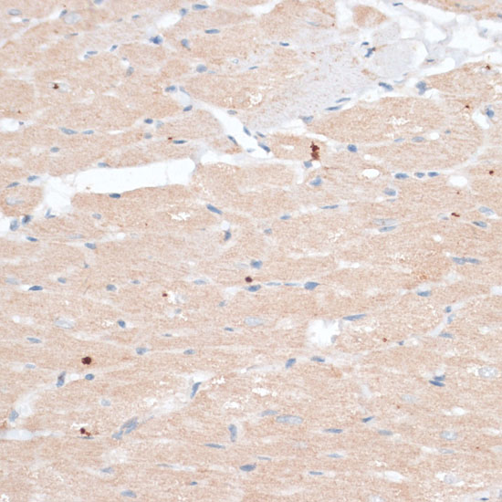
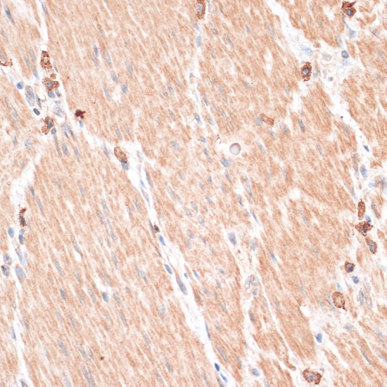
A06088-2 has real IHC images from paraffin-embedded rat heart and human gastric cancer (IHC image captions); IF/ICC use is listed, with no IF image supplied (catalog applications; IF image alts).

Real IHC data Immunohistochemistry of paraffin-embedded rat heart using ABCB7 antibody at dilution of 1:100 .Perform microwave antigen retrieval with 10 mM PBS buffer pH 7.2 before commencing with IHC staining protocol.
Anti-ABCB7 Antibody
Cat # A06088-2

A06088-2 will render with its rat-heart IHC figure; the catalog also supplies a human gastric-cancer IHC image, both from paraffin-embedded tissue at 1:100 (IHC image captions). The antibody lists IHC and IF/ICC applications and Human, Mouse, and Rat reactivity; no IF image is supplied (catalog applications; catalog reactivity; IF image alts).

Which to pick: For tissue IHC, choose A06088-2: its own captions document paraffin-embedded rat heart and human gastric cancer, with microwave retrieval in 10 mM PBS, pH 7.2; the fixative is unreported (IHC image captions). For IF/ICC, A06088-2 lists those applications at 1:50–1:200, although no IF image is supplied (catalog applications; catalog IF dilution; IF image alts). For Human, Mouse, or Rat samples, A06088-2 lists reactivity across all three species and has a Rabbit host; the supplied IHC images document Human and Rat samples only (catalog reactivity; catalog host; IHC image captions).

Each figure is that product's own IHC / IF validation image from its datasheet.

References

  1. UniProt Consortium. UniProt entry O75027 (ABCB7_HUMAN, Iron-sulfur clusters transporter ABCB7, mitochondrial).
  2. Human Protein Atlas. ABCB7 tissue IHC expression (reliability: Approved).
  3. Human Protein Atlas. ABCB7 subcellular location (ICC-IF): Localized to the mitochondria..
  4. Human Protein Atlas. ABCB7 antibody validation summary (1 antibodies).
  5. The transporter ABCB7 is a mediator of the phenotype of acquired refractory anemia with ring sideroblasts. Leukemia 2013 — PMC3794445.
  6. Chronic Pressure Overload Results in Deficiency of Mitochondrial Membrane Transporter ABCB7 Which Contributes to Iron Overload, Mitochondrial Dysfunction, Metabolic Shift and Worsens Cardiac Function. Scientific reports 2019 — PMC6739357.
  7. Role of transferrin receptor and the ABC transporters ABCB6 and ABCB7 for resistance and differentiation of tumor cells towards artesunate. PloS one 2007 — PMC1949049.
  8. PubMed PMID:9621516 — UniProt-cited evidence.
  9. PubMed PMID:10196363 — UniProt-cited evidence.
  10. PubMed PMID:11050011 — UniProt-cited evidence.