This website uses cookies to ensure you get the best experience on our website.


- Table of Contents
Plan chromogenic IHC of ACADSB in paraffin sections using hepatocytes as a high-staining reference (HPA tissue IHC). The catalog antibody is listed at 2–5 μg/mL for human IHC-P (datasheet A08652-1), and granular cytoplasmic staining is reported in most tissues (HPA tissue IHC).

Expected localisation, validated protocols, controls and antibodies — the at-a-glance facts below, then the full design guide.
| Expected localisation | Granular cytoplasm (HPA tissue IHC); mitochondrial matrix (UniProt) | |
| Staining pattern | Granular cytoplasmic staining in most tissues (HPA tissue IHC) | |
| Antigen retrieval | EDTA pH 8.0 HIER, heat-mediated (datasheet A08652-1) | |
| Positive control | Breast+4 more · see all | |
| Negative control | Adipose tissue+4 more · see all |
| Fixation | Matched tissue-IHC evidence does not establish the fixation claim. Validate the specimen-specific method before use. (selected-SKU IHC image A08652-1) | |
| Caveat | Tissue staining has medium agreement with RNA data (HPA tissue IHC) | |
| Regulation | Liver-enhanced RNA expression (HPA tissue RNA) | |
| Isoform / epitope | 2 isoforms; mature chain aa 34–432 (UniProt) |
The catalog antibody’s IHC protocol (datasheet A08652-1) is accompanied by three published ACADSB IHC methods (PMC6506390; PMC8791850; PMC12585056).
| Sample | Paraffin-embedded human liver cancer tissue; fixative not specified (datasheet A08652-1) |
| Fixation | Image fixative and duration unreported (datasheet A08652-1); verify before use. |
| Sectioning | 4–5 µm sections on charged slides (standard) |
| Deparaffinisation | Xylene, graded ethanol series to water (standard) |
| Antigen retrieval | Heat retrieval: EDTA pH 8.0 (datasheet A08652-1); 20 min, 95–100 °C (standard) |
| Peroxidase block | 3% H2O2, 10 min, room temperature (standard) |
| Blocking | 10% goat serum (datasheet A08652-1) |
| Primary antibody | Rabbit anti-ACADSB, 2-5μg/ml (datasheet A08652-1) |
| Primary incubation | Overnight at 4 °C (datasheet A08652-1) |
| Detection | HRP-conjugated secondary, DAB chromogen (datasheet A08652-1) |
| Counterstain | Hematoxylin, blue, dehydrate and mount (standard) |
| Expected result | ACADSB-positive staining in glandular cells of breast (HPA tissue IHC: High). HPA tissue profile: Granular cytoplasmic expression in most tissues. No signal in the no-primary control. |
ACADSB is a mitochondrial matrix protein with no transmembrane segment (UniProt P45954 topology). In paraffin sections, expect granular cytoplasmic staining, particularly in hepatocytes, kidney distal tubules, and several glandular cell populations (HPA: tissue IHC). HPA rates the tissue profile Enhanced, with medium agreement between staining and RNA data; external verification remains pending (HPA: reliability).
| Granular cytoplasmic staining in hepatocytes, kidney distal tubules, or listed glandular cells (HPA: High). | This fits the reported tissue pattern and mitochondrial matrix location (HPA: tissue IHC; UniProt P45954). Judge intensity within the identified cell population: HPA reports high staining for these cells, while its broader description is granular cytoplasmic expression in most tissues (HPA: tissue IHC). |
| Predominantly nuclear, membranous, or extracellular staining, without convincing cytoplasmic granules. | Treat this as a localization mismatch that needs investigation, rather than assigning it to ACADSB: the protein is assigned to the mitochondrial matrix and has no transmembrane segment (UniProt P45954 topology). Compare morphology and controls before interpreting an unusual compartment as specific staining (general IHC practice). |
| Strong staining in adipocytes, lung alveolar cells, or esophageal squamous epithelial cells (HPA: Not detected). | These cell populations are reported as not detected, so strong signal warrants a check for cross-reactivity or endogenous chromogen activity (HPA: tissue IHC; general IHC practice). A mismatch alone does not identify the cause; verify the cell type and inspect detection controls (general IHC practice). |
| Uniform haze across cells and surrounding tissue, with little cell-specific granular detail. | Diffuse background obscures the expected granular cytoplasmic pattern (HPA: tissue IHC). Check the no-primary control, blocking, washing, and chromogen development as general IHC troubleshooting steps; this appearance does not establish an ACADSB-specific fixation or epitope effect (general IHC practice). |
| No convincing signal in hepatocytes or kidney distal tubules despite intact tissue morphology. | Both are reported as high-staining populations, making them useful checks for an apparently negative run (HPA: tissue IHC). Review antibody identity and the recorded retrieval and detection steps before calling the sample negative; this record supplies no target-specific fixation sensitivity or retrieval condition (general IHC practice). |
| Compartment and cellular distribution | ACADSB is assigned to the mitochondrial matrix, while tissue IHC describes granular cytoplasmic expression (UniProt P45954; HPA: tissue IHC). Read chromogenic granules in their tissue context; light microscopy does not by itself prove that each granule is mitochondrial (general IHC practice). |
| Choice of comparison tissue | HPA reports high staining in hepatocytes, kidney distal tubules, and specified glandular cells, but not-detected staining in several other named populations (HPA: tissue IHC). Compare the same cell type when judging a result; a whole-section average can conceal cell-specific differences (general IHC practice). |
| Strength of the reference pattern | HPA lists the tissue profile as Enhanced and both listed antibodies as IHC Enhanced, while describing only medium staining–RNA consistency and pending external verification (HPA: reliability; HPA: antibodies). Use the profile as a reference pattern, with its stated verification limit. |
| Protein processing and isoforms | UniProt lists a processed chain spanning residues 34–432 and two isoforms (UniProt P45954). The supplied record gives no antibody epitope or isoform-specific IHC pattern, so it cannot predict which form produces staining or explain a particular negative section. |
| IF/ICC Q: Where should signal localize? | A: HPA reports supported mitochondrial localization in ICC-IF and lists HPA041458 as ICC Supported (HPA: subcellular; HPA: antibodies). This is a localization reference for the separate IF/ICC guide, with no IF/ICC protocol specified here. |
| Situation | Likely cause | Next action |
|---|---|---|
| A hepatocyte or kidney distal-tubule control is blank. | The result conflicts with HPA's high staining in those cell populations; the record does not establish why (HPA: tissue IHC). | Confirm the cell type and antibody identity, then check the documented retrieval, primary-antibody, detection, and chromogen steps against the run record (general IHC practice). Do not infer ACADSB-specific fixation sensitivity. |
| The slide has widespread brown haze. | Background may arise from nonspecific binding, endogenous detection activity, or overdevelopment (general IHC practice). | Inspect a no-primary control, blocking and wash steps, and chromogen development; repeat with corrected general IHC conditions if needed (general IHC practice). Look for recovery of the granular cell pattern (HPA: tissue IHC). |
| Signal is mainly nuclear or outlines cell membranes. | That distribution conflicts with the mitochondrial matrix assignment and granular cytoplasmic tissue profile (UniProt P45954; HPA: tissue IHC). | Check tissue morphology and detection controls, and compare the suspect cells with a high-staining reference cell population (HPA: High; general IHC practice). Treat the compartment mismatch as unresolved until corroborated. |
| A reported not-detected cell population stains strongly. | Cross-reactivity or endogenous chromogen activity is possible; staining alone cannot distinguish them (HPA: tissue IHC; general IHC practice). | Verify the cell identity and inspect no-primary and detection controls (general IHC practice). Compare with a reported high-staining population on a suitable section before interpreting the unexpected signal (HPA: tissue IHC). |
| A section looks weak overall despite some positive cells. | Cell populations differ in the HPA profile: some are high, some low, and some not detected (HPA: tissue IHC). | Score identified cell populations separately, then compare like cells across sections (general IHC practice). Use the HPA levels as a reference and avoid treating low staining as proof of a failed run (HPA: tissue IHC). |
| Two antibodies give different tissue patterns. | Both listed antibodies have IHC Enhanced status, but the tissue profile still has medium staining–RNA consistency and awaits external verification (HPA: antibodies; HPA: reliability). | Document each antibody and compare the same cell populations and controls across sections (general IHC practice). Report the disagreement rather than assigning an unsupported epitope, isoform, or fixation mechanism. |
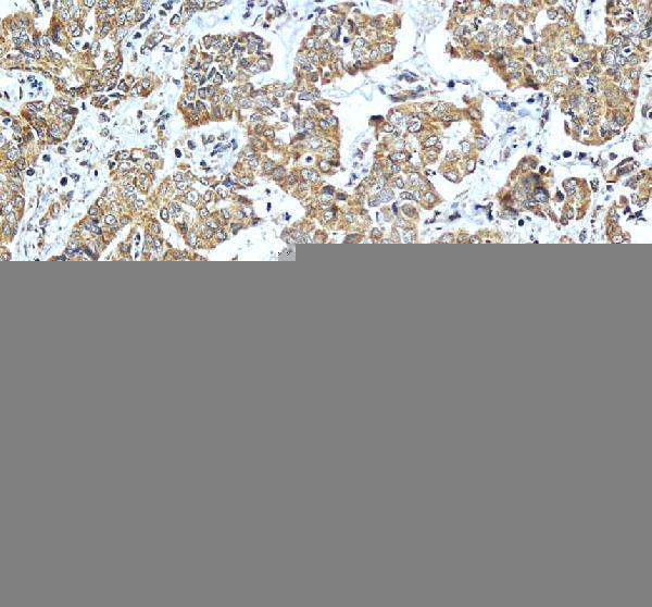
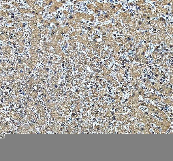
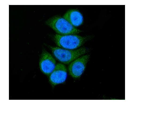


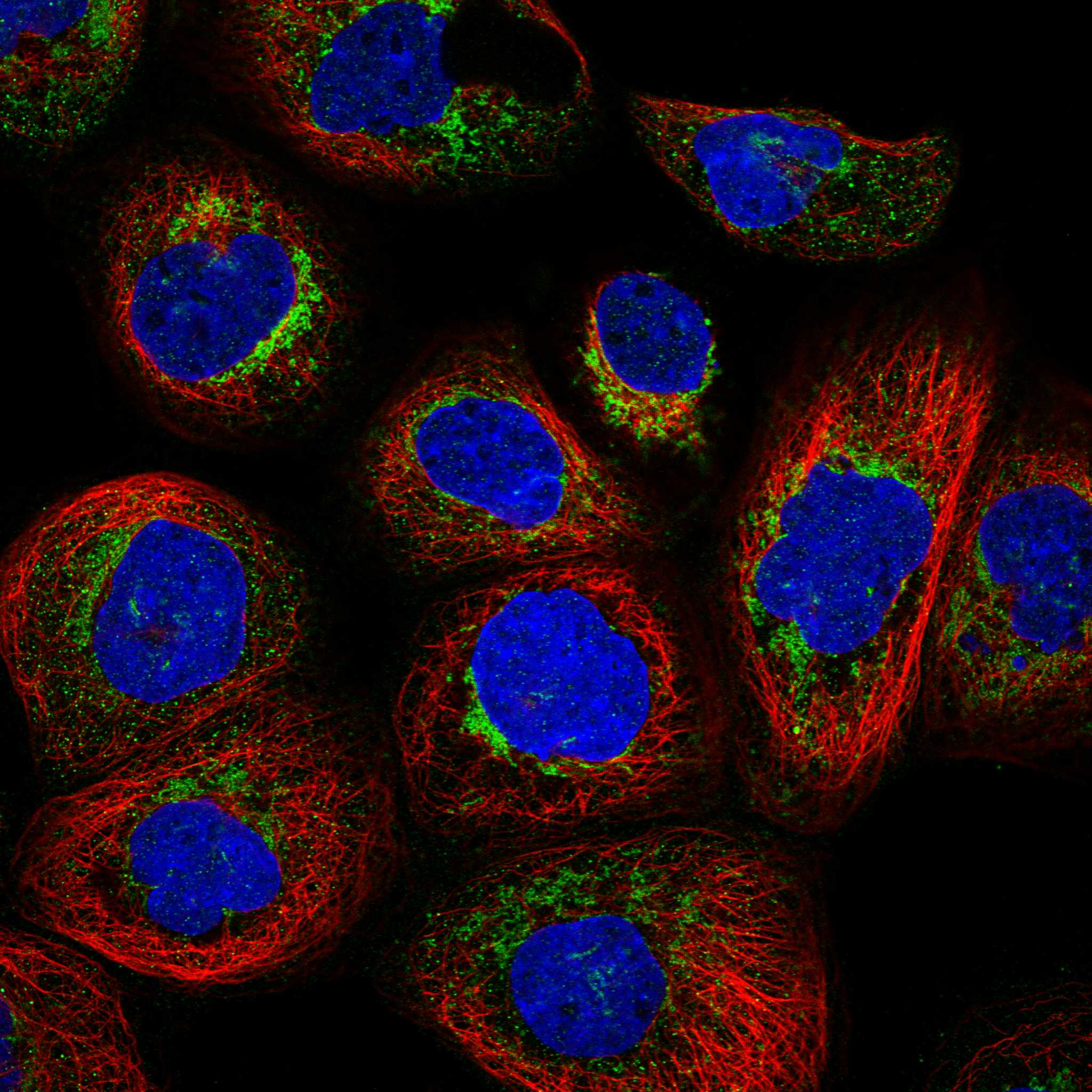
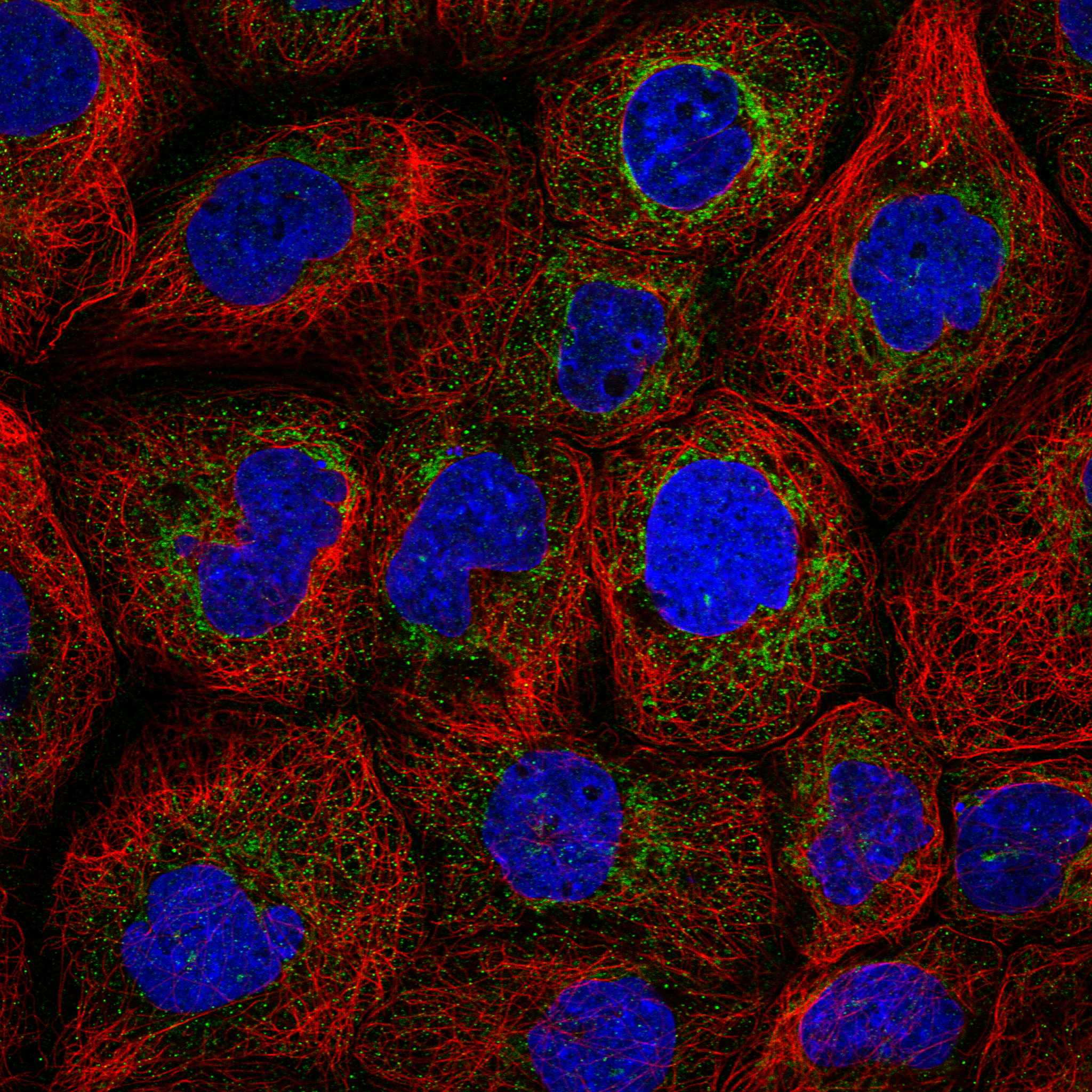
Comprehensive Human Protein Atlas IHC scoring per tissue (reliability: Enhanced — Medium consistency between antibody staining and RNA expression data. Pending external verification.). Rows are taken directly from the HPA tissue chart — click any row's HPA link to view the source.
| Tissue | Cell type | Level | Evidence | Source |
|---|---|---|---|---|
| Breast | Glandular cells | High | Protein (IHC) | HPA → |
| Colon | Glandular cells | High | Protein (IHC) | HPA → |
| Duodenum | Glandular cells | High | Protein (IHC) | HPA → |
| Gallbladder | Glandular cells | High | Protein (IHC) | HPA → |
| Kidney | Distal tubules | High | Protein (IHC) | HPA → |
| Tissue | Cell type | Level | Evidence | Source |
|---|---|---|---|---|
| Adipose tissue | Adipocytes | Not detected | Protein (IHC) | HPA → |
| Bone marrow | Hematopoietic cells | Not detected | Protein (IHC) | HPA → |
| Caudate | Glial cells | Not detected | Protein (IHC) | HPA → |
| Cerebellum | Cells in granular layer | Not detected | Protein (IHC) | HPA → |
| Epididymis | Glandular cells | Not detected | Protein (IHC) | HPA → |
Troubleshoot ACADSB staining in paraffin sections using the catalog antibody’s tissue IHC conditions, mitochondrial localisation, and cell specific reference patterns.
A08652-1 has IHC images from human paraffin liver and liver cancer sections and an IF image from Mcf-7 cells (A08652-1 image captions).
A08652-1 is listed for IHC and IF/ICC (A08652-1 catalog applications). Its IHC images show human paraffin liver and liver cancer sections, while its IF image shows Mcf-7 cells (A08652-1 image captions).
Which to pick: For tissue IHC, choose A08652-1: its own images show staining in human paraffin sections, with 2 μg/ml primary antibody after heat retrieval in EDTA at pH 8.0 (A08652-1 IHC image captions). For IF/ICC, the same SKU has an image from Mcf-7 cells; its listed IHC/IF reactivity includes human, mouse, rat and monkey, though the supplied IHC images demonstrate human tissue only (A08652-1 IF image caption; A08652-1 catalog reactivity; A08652-1 IHC image captions). The IHC captions identify paraffin sections but do not report the fixative (A08652-1 IHC image captions).