This website uses cookies to ensure you get the best experience on our website.


- Table of Contents
Plan ACO2 paraffin-section IHC around granular cytoplasmic staining (HPA tissue IHC) and mitochondrial localisation (UniProt). This guide covers fixation, retrieval, a catalog antibody concentration of 2–5 μg/ml (datasheet M03096-3), chromogenic detection, and scoring.

Expected localisation, validated protocols, controls and antibodies — the at-a-glance facts below, then the full design guide.
| Expected localisation | Granular cytoplasm (HPA tissue IHC); mitochondrion (UniProt) | |
| Staining pattern | Widespread granular cytoplasmic staining (HPA tissue IHC) | |
| Antigen retrieval | EDTA pH 8.0 HIER, heat-mediated (datasheet M03096-3) | |
| Positive control | Adrenal gland+4 more · see all | |
| Negative control | Adipose tissue |
| Fixation | Keep fixation consistent across sections. (standard IHC practice; not target-specific) | |
| Caveat | Adipocytes may be unstained (HPA tissue IHC) | |
| Regulation | RNA enhanced: heart/skeletal/tongue (HPA tissue RNA) | |
| Isoform / epitope | No isoforms annotated; mature chain spans residues 28–780 (UniProt) |
The catalog antibody’s IHC-P protocol is paired with one published ACO2 IHC protocol using osteosarcoma and paracancerous tissues (PMC9534693 methods).
| Sample | Paraffin-embedded human breast cancer tissue; fixative not specified (datasheet M03096-3) |
| Fixation | Image fixative and duration unreported (datasheet M03096-3); verify before use. |
| Sectioning | 4–5 µm sections on charged slides (standard) |
| Deparaffinisation | Xylene, graded ethanol series to water (standard) |
| Antigen retrieval | Heat retrieval: EDTA pH 8.0 (datasheet M03096-3); 20 min, 95–100 °C (standard) |
| Peroxidase block | 3% H2O2, 10 min, room temperature (standard) |
| Blocking | 10% goat serum (datasheet M03096-3) |
| Primary antibody | Mouse monoclonal (clone 4C12D1) anti-ACO2, 2-5 μg/ml (datasheet M03096-3) |
| Primary incubation | Overnight at 4 °C (datasheet M03096-3) |
| Detection | HRP-conjugated secondary, DAB chromogen (datasheet M03096-3) |
| Counterstain | Hematoxylin, blue, dehydrate and mount (standard) |
| Expected result | ACO2-positive staining in glandular cells of adrenal gland (HPA tissue IHC: High). HPA tissue profile: Ubiquitous cytoplasmic expression in a granular pattern. No signal in the no-primary control. |
ACO2 localizes to mitochondria (UniProt Q99798; HPA subcellular ICC-IF). In paraffin-section IHC, expect granular cytoplasmic staining across many tissues, including glandular cells and glia reported as high (HPA tissue IHC). HPA describes the tissue pattern as ubiquitous and rates its IHC evidence Enhanced, citing agreement between staining and RNA expression (HPA tissue IHC). ACO2 has no transmembrane segment (UniProt Q99798 topology).
| Granular cytoplasmic signal in adrenal or appendix glandular cells (HPA tissue IHC: High). | This fits the reported high staining in those cells and ACO2's mitochondrial location (HPA tissue IHC; UniProt Q99798). Judge the granules against tissue structure and controls; a positive control alone does not establish that every stained structure is specific (general IHC practice). |
| Predominantly nuclear staining or a sharp cell-surface rim. | Neither pattern matches mitochondrial localization or the reported granular cytoplasmic IHC profile (UniProt Q99798; HPA tissue IHC). Check section morphology, counterstain and control slides before interpreting a compartment mismatch as ACO2 (general IHC practice). |
| Strong signal restricted to adipocytes, with little signal in adjacent cell types. | HPA reports ACO2 as not detected in adipocytes, while describing a broad granular cytoplasmic tissue pattern (HPA tissue IHC). Treat this discordance as possible cross-reactivity or endogenous chromogen activity; compare negative controls before assigning it to ACO2 (general IHC practice). |
| Uniform color across cells, extracellular areas and section edges. | A uniform deposit obscures the granular cytoplasmic pattern reported by HPA (HPA tissue IHC). Assess background with a primary-antibody omission control, then review blocking, washing and detection conditions (general IHC practice). |
| No convincing signal in adrenal glandular cells or bone-marrow hematopoietic cells. | Both are reported as high-staining cell populations (HPA tissue IHC). First confirm that the relevant cells are present and preserved; then compare a suitable positive control and review the antibody, retrieval and detection workflow (general IHC practice). |
| Compartment and topology | Mitochondrial localization supports granular cytoplasmic interpretation (UniProt Q99798; HPA subcellular ICC-IF). With no transmembrane segment, a crisp cell-surface outline lacks support as the expected ACO2 pattern (UniProt Q99798 topology). |
| Cell population | HPA reports high staining in several glandular populations, hematopoietic cells and glia, but no detected signal in adipocytes (HPA tissue IHC). Score the named cell population rather than treating a whole tissue as uniformly positive or negative (general IHC practice). |
| Evidence strength | The reported tissue pattern has Enhanced reliability, and HPA001097 has Enhanced IHC validation (HPA tissue IHC; HPA antibody record). These ratings support the reference pattern; they do not validate an arbitrary reagent or every signal on an individual slide (general IHC practice). |
| Processing and epitope limits | UniProt annotates the mature ACO2 chain as residues 28–780 and lists no isoforms (UniProt Q99798). The supplied record gives no antibody epitope or target-specific fixation response, so neither differential epitope staining nor fixation sensitivity can be inferred. |
| Situation | Likely cause | Next action |
|---|---|---|
| Weak or absent staining in an HPA high-staining cell population (HPA tissue IHC). | The slide may lack an interpretable positive cell population, or a general IHC workflow step may have underperformed (general IHC practice). | Verify cell identity and section integrity, then check the positive control, antigen-retrieval run, antibody dilution and detection reagents (general IHC practice). |
| Nuclear or surface-rim signal dominates the slide. | The compartment conflicts with mitochondrial localization and granular cytoplasmic IHC staining (UniProt Q99798; HPA tissue IHC). | Review morphology and counterstain, compare control slides, and adjust staining or detection conditions if the misplaced signal persists (general IHC practice). |
| Adipocytes stain strongly despite an otherwise weak section. | HPA reports adipocytes as not detected (HPA tissue IHC). Cross-reactivity or endogenous chromogen activity is possible (general IHC practice). | Check a primary-antibody omission control and the applicable endogenous-activity block; interpret staining by cell type (general IHC practice). |
| Diffuse deposit hides cytoplasmic granules. | Background from the staining or detection workflow can prevent assessment of the reported granular pattern (general IHC practice; HPA tissue IHC). | Compare omission and positive controls, then review blocking, antibody concentration, washes and chromogen development (general IHC practice). |
| Two sections show different apparent staining intensity. | The proportion of HPA high-staining cell populations may differ between sections (HPA tissue IHC); staining-run variation is also possible (general IHC practice). | Compare like cell populations and morphology, then evaluate both slides against the same suitable controls before scoring intensity (general IHC practice). |
| IF/ICC Q&A: What localization should be expected? | HPA approves a mainly mitochondrial ICC-IF location and lists images from A-431, U-251MG and U2OS (HPA subcellular ICC-IF). | Expect mitochondrial localization in that separate application; assess IF/ICC conditions in its own guide, without treating this IHC pattern as an IF/ICC protocol (HPA subcellular ICC-IF). |
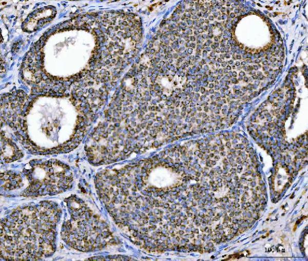
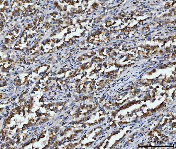


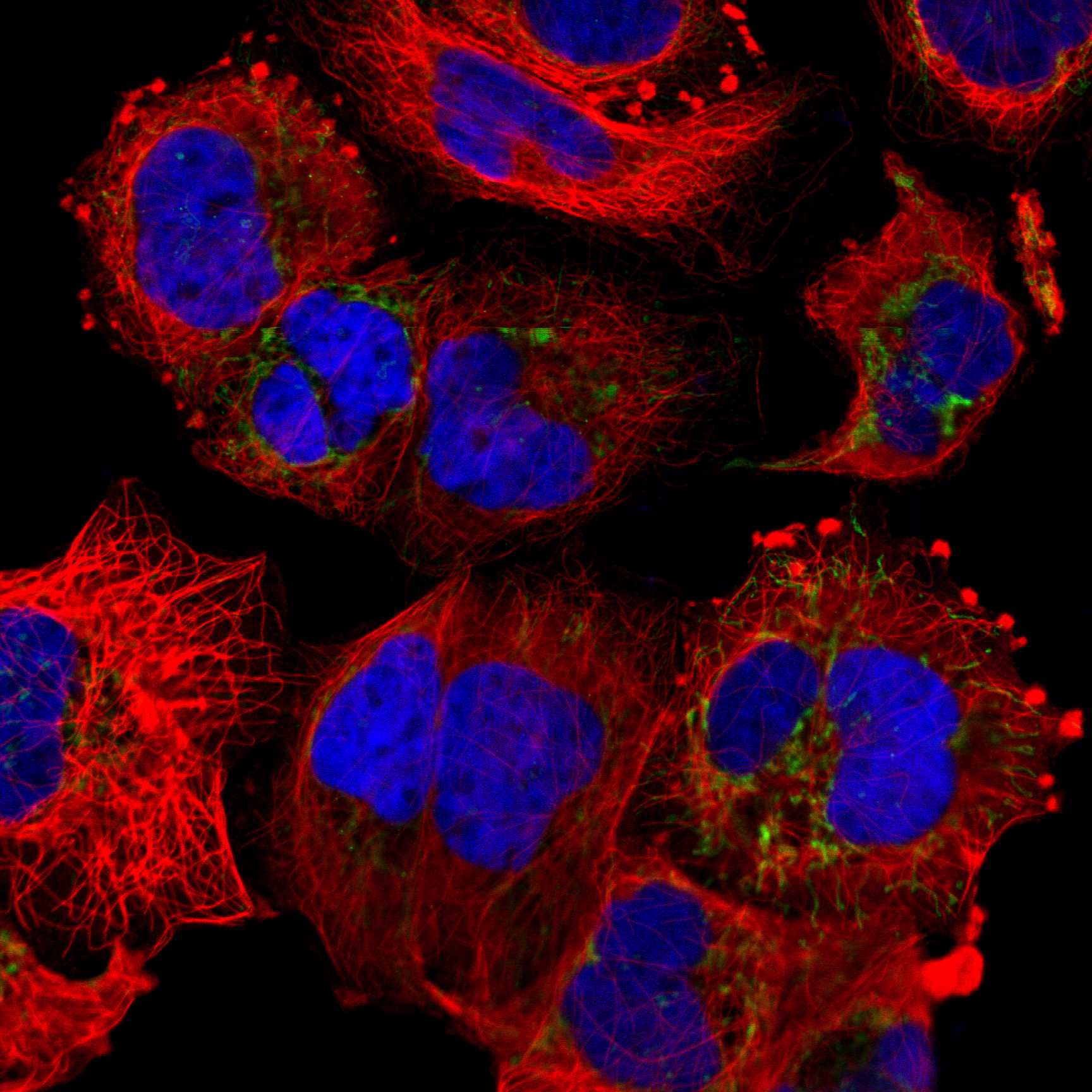
Comprehensive Human Protein Atlas IHC scoring per tissue (reliability: Enhanced — High consistency between antibody staining and RNA expression data.). Rows are taken directly from the HPA tissue chart — click any row's HPA link to view the source.
| Tissue | Cell type | Level | Evidence | Source |
|---|---|---|---|---|
| Adrenal gland | Glandular cells | High | Protein (IHC) | HPA → |
| Appendix | Glandular cells | High | Protein (IHC) | HPA → |
| Bone marrow | Hematopoietic cells | High | Protein (IHC) | HPA → |
| Breast | Glandular cells | High | Protein (IHC) | HPA → |
| Caudate | Glial cells | High | Protein (IHC) | HPA → |
| Tissue | Cell type | Level | Evidence | Source |
|---|---|---|---|---|
| Adipose tissue | Adipocytes | Not detected | Protein (IHC) | HPA → |
Troubleshoot ACO2 staining in paraffin sections by checking retrieval, cellular pattern, controls and scoring against its expected mitochondrial localisation (UniProt Q99798; HPA subcellular).
M03096-3 has a real ACO2 IHC figure from a human paraffin section (M03096-3 image caption). PB9973 is listed for human ICC/IF (PB9973 catalog).
The rendered M03096-3 card documents IHC on paraffin-embedded human breast cancer; its other captions show human hepatocellular carcinoma, laryngeal squamous cell carcinoma and lung adenocarcinoma (M03096-3 image captions). The catalog lists M03096-3 for IHC with human, mouse and rat reactivity (M03096-3 catalog).
Which to pick: For tissue IHC, choose mouse monoclonal M03096-3: its own figure shows a human paraffin section, and the fixative is unreported (M03096-3 image caption; M03096-3 catalog). For IF/ICC, rabbit polyclonal PB9973 is listed for human ICC/IF at 5 μg/ml (PB9973 catalog). For cross-species planning, rabbit monoclonal M03096-2 lists IHC and ICC/IF applications and human, mouse and rat reactivity, but has no image caption documenting tissue processing (M03096-2 catalog).