ACRBP / Acrosin-binding protein · IHC design guide

Design Immunohistochemistry for ACRBP

Plan chromogenic IHC on paraffin sections using testis, where elongated or late spermatids show high staining (HPA tissue IHC). The guide covers fixation consistency, antibody titration, detection controls, and interpretation of processed ACRBP (UniProt).

Evidence assembled Oct 2026 · For research use; verify linked source records and product datasheet before use
Immunohistochemistry protocol sheet for ACRBP (IHC for ACRBP): expected localisation Acrosomal vesicles expected; tissue compartment unverified (UniProt), antibody A12806-1, validated IHC image, and IHC protocol steps
Printable ACRBP IHC protocol sheet — expected localisation Acrosomal vesicles expected; tissue compartment unverified (UniProt), antibody A12806-1, controls and protocol steps. Open the full ACRBP IHC guide →

ACRBP Immunohistochemistry Experimental Design Guide

Expected localisation, validated protocols, controls and antibodies — the at-a-glance facts below, then the full design guide.

Must know before staining
Expected localisation Acrosomal vesicles expected; tissue compartment unverified (UniProt)
Staining pattern Selective staining of elongated or late spermatids; compartment unspecified (HPA tissue IHC)
Antigen retrieval Tris-EDTA pH 9.0 HIER, heat-mediated (datasheet A12806-1)
Positive control ⓘ Testis
Negative control ⓘ Adipose tissue+4 more · see all
Important caveats
Reasons your staining may differ from the expected pattern.
Fixation Keep fixation consistent across sections. (standard IHC practice; not target-specific)
Caveat Presumed off-target staining may complicate interpretation (HPA tissue IHC)
Regulation Expression is testis-restricted in normal tissue (UniProt)
Isoform / epitope No isoforms; epitopes in aa 26–273 may miss the mature form (UniProt)
Section 1

Recommended ACRBP IHC & IF Protocols

The catalog antibody’s IHC-P protocol is paired with published ACRBP staining methods for ovarian tissue and liposarcoma (PMC8589352; PMC4287229).

Recommended immunohistochemistry (IHC-P) protocol parameters
SampleParaffin-embedded human tonsil tissue; fixative not specified (datasheet A12806-1)
FixationImage fixative and duration unreported (datasheet A12806-1); verify before use.
Sectioning4–5 µm sections on charged slides (standard)
DeparaffinisationXylene, graded ethanol series to water (standard)
Antigen retrievalHeat retrieval: Tris-EDTA pH 9.0 (datasheet A12806-1); 20 min, 95–100 °C (standard)
Peroxidase block3% H2O2, 10 min, room temperature (standard)
Blocking10% normal serum of the secondary host, 30 min, room temperature (standard)
Primary antibodyRabbit anti-ACRBP, 1:100-1:300 (datasheet A12806-1)
Primary incubationOvernight at 4 °C (standard)
DetectionHRP-polymer secondary, DAB chromogen 5–10 min (standard)
CounterstainHematoxylin, blue, dehydrate and mount (standard)
Expected resultACRBP-positive staining in elongated or late spermatids of testis (HPA tissue IHC: High). HPA tissue profile: Selective expression in subsets of cells in seminiferous ducts. No signal in the no-primary control.
💡Decision noteStart with pH 9.0 Tris-EDTA heat retrieval for the catalog antibody (datasheet A12806-1). Both published methods used pH 6.0 retrieval with their own antibodies (PMC8589352; PMC4287229).
Section 2

What Is the Expected ACRBP Staining Pattern?

ACRBP is an acrosomal secretory-vesicle protein with no transmembrane segment (UniProt Q8NEB7: localization, topology). In normal tissue, expect selective staining of elongated or late spermatids in seminiferous ducts, where HPA reports high staining (HPA: testis IHC). HPA rates the tissue profile Enhanced, while noting medium consistency with RNA data and disregarded presumed off-target binding (HPA: tissue IHC reliability).

What am I looking at on my slide?
Strong staining in a subset of elongated or late spermatids, concentrated toward their heads.This fits the high, selective testis IHC pattern (HPA: testis IHC) and acrosomal localization (UniProt Q8NEB7: localization). Score the relevant spermatids rather than the whole duct: other cells can make the section look mostly unstained despite a valid positive result.
Predominantly nuclear staining in testis, without a convincing spermatid-head pattern.Treat this as a compartment mismatch requiring artifact review against the expected acrosomal pattern (UniProt Q8NEB7: localization). Do not dismiss all nuclear signal categorically: HPA reports approved nucleoplasmic staining in cultured-cell ICC-IF, which does not establish the expected testis IHC pattern (HPA: ICC-IF; testis IHC).
Staining mainly in unrelated cell types, with little signal in late spermatids.Consider cross-reactivity or endogenous chromogenic activity before calling the section ACRBP-positive (standard IHC practice). HPA reports presumed off-target binding in its tissue assessment; its supported positive cell population is elongated or late spermatids (HPA: tissue IHC reliability; testis IHC).
Diffuse chromogen across the section, including cells expected to be negative.This obscures the selective pattern reported in testis (HPA: testis IHC). Review background controls, blocking, detection chemistry and wash conditions (standard IHC practice). ACRBP is annotated as secreted, so extracellular signal alone should not be accepted as proof of the expected cell-specific pattern (UniProt Q8NEB7: localization).
No signal in a testis section containing elongated or late spermatids.The known-positive cell population is present, so review assay performance before interpreting the result as absent ACRBP (HPA: testis IHC; standard IHC practice). Check control staining, antibody and detection steps, and whether the scored ducts contain the relevant spermatid stage.
💡Expected ACRBP appearanceCall testis IHC positive when high, selective staining marks elongated or late spermatids with an acrosome-consistent head pattern (HPA: testis IHC; UniProt Q8NEB7: localization); diffuse staining or staining centered on unrelated cells warrants a false-positive review (standard IHC practice).
How each factor affects the staining
Cell stage and scoring regionHPA places the high signal in elongated or late spermatids, within subsets of cells in seminiferous ducts (HPA: testis IHC). A duct without the relevant stage is a weak basis for judging assay sensitivity.
Secretory-vesicle location and topologyACRBP is annotated in the acrosome and as secreted, with no transmembrane segment (UniProt Q8NEB7: localization, topology). Use cell identity and compartment together when interpreting chromogen; a membrane outline alone is not the predicted pattern.
Protein processing and epitope coverageUniProt annotates a signal peptide at residues 1–25, a propeptide at 26–273, and a mature chain at 274–543 (UniProt Q8NEB7: processing). Antibody epitope location is not supplied, so the record cannot predict which processed form a given stain detects.
Antibody validation and tissue caveatHPA marks HPA039082 IHC Enhanced and HPA039081 IHC Supported (HPA: antibody validation). The tissue profile is Enhanced but has medium RNA–staining consistency and presumed off-target binding that was disregarded (HPA: tissue IHC reliability); investigate unexpected staining independently.
Normal versus cancer tissueUniProt describes normal-tissue expression as restricted to testis and reports expression in a spectrum of cancers (UniProt Q8NEB7: tissue specificity). The supplied HPA IHC observations establish a testis pattern; they do not establish an expected staining pattern for any particular tumor.
Why is my staining missing, weak or wrong?
SituationLikely causeNext action
Testis positive control is blank.Relevant late spermatids may be absent from the scored ducts, or an assay step may have failed (HPA: testis IHC; standard IHC practice).Confirm the cell stage on the counterstained section, then check reagent application, detection and a working positive control (standard IHC practice). Do not infer ACRBP absence from a section lacking the expected cells.
Signal is weak but confined to the expected spermatids.This is below the high staining level described for elongated or late spermatids, although slide and assay conditions can affect visible chromogen (HPA: testis IHC; standard IHC practice).Check the positive control, detection and counterstain, then optimize the validated IHC procedure within its documented conditions (standard IHC practice). No ACRBP-specific retrieval or dilution requirement is supplied here.
Many cell types show brown signal.Nonspecific antibody staining or endogenous detection activity can mimic a broad positive result (standard IHC practice); HPA also notes presumed off-target binding (HPA: tissue IHC reliability).Compare a negative reagent control and detection-only control, assess endogenous activity, and require the selective spermatid pattern before scoring positive (standard IHC practice; HPA: testis IHC).
A crisp nuclear pattern dominates testis IHC.It conflicts with the acrosomal localization annotation (UniProt Q8NEB7: localization), although HPA reports nucleoplasmic signal in ICC-IF (HPA: ICC-IF).Check morphology, compartment assignment and controls; interpret the testis section against its selective late-spermatid IHC reference (HPA: testis IHC; standard IHC practice).
Background masks the spermatid pattern.Inadequate blocking, washing or detection control can produce diffuse chromogen (standard IHC practice). The supplied sources do not identify a target-specific cause.Review blocking and wash steps and inspect negative controls before changing the interpretation threshold (standard IHC practice). Reassess whether individual elongated or late spermatids can be distinguished (HPA: testis IHC).
Does an ICC-IF nuclear or vesicular image validate this IHC result?HPA approves nucleoplasm and vesicles, with additional cytosol, in ICC-IF images from HEL, U-251MG and U2OS (HPA: ICC-IF). Those cultured-cell observations address a different application.Use the testis cell-stage and acrosome-consistent pattern to judge paraffin IHC (HPA: testis IHC; UniProt Q8NEB7: localization). Consult the separate IF/ICC guide for that application's design.

Sample controls for ACRBP IHC & IF

🧪Run testis first and score elongated or late spermatids as the expected positive cells (HPA: High in elongated or late spermatids). Run adipose tissue as a negative comparator (HPA: Not detected in adipocytes), and use adjacent non-spermatid cells on the testis slide as internal background comparators rather than assuming they are validated negatives (HPA: positive call is specific to elongated or late spermatids).
Positive control tissue: Testis (Elongated or late spermatids, HPA High)
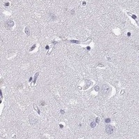
Negative control tissue: Adipose tissue (HPA Not detected)
ICC-IF cell lines (HPA subcellular resource): HPA ICC-IF images show ACRBP in HEL, U-251MG, U2OS, with annotated localisation: Nucleoplasm (approved), Vesicles (approved) (HPA subcellular).
Technical controls: Include a no-primary, secondary-only control; an isotype control matched to the primary antibody’s host species and clonality; and ACRBP knockout tissue or peptide competition, if available (standard IHC practice). For chromogenic testis IHC, quench endogenous peroxidase and check for residual background before scoring spermatids (standard IHC practice).
⚠️Feasibility: A target-specific fixation window or fixation effect is unreported, and the selected A12806-1 paraffin-section caption does not state its fixative (selected-SKU tissue-IHC caption: fixative unreported). The caption reports Tris-EDTA retrieval at pH 9.0 and primary antibody dilution of 1:200, but does not establish whether retrieval is required; the supplied evidence also does not establish whether frozen sections or IF are easier (selected-SKU tissue-IHC caption). Score staining in the HPA-defined spermatid population carefully, since the expected acrosomal localization can be obscured by diffuse background (HPA: High in elongated or late spermatids; UniProt Q8NEB7: acrosome; standard IHC practice).

HPA tissue IHC evidence for ACRBP

Comprehensive Human Protein Atlas IHC scoring per tissue (reliability: Enhanced — Medium consistency between antibody staining and RNA expression data. Secreted protein, tissue location of RNA and protein is expected to differ. Presumed off target binding observed and disregarded.). Rows are taken directly from the HPA tissue chart — click any row's HPA link to view the source.

Positive expression · recommended positive controls

TissueCell typeLevelEvidenceSource
Testis Elongated or late spermatids High Protein (IHC) HPA →

Undetected expression · recommended negative controls

TissueCell typeLevelEvidenceSource
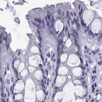
Adipose tissue Adipocytes Not detected Protein (IHC) HPA →
Adrenal gland Glandular cells Not detected Protein (IHC) HPA →
Appendix Glandular cells Not detected Protein (IHC) HPA →
Bone marrow Hematopoietic cells Not detected Protein (IHC) HPA →
Breast Adipocytes Not detected Protein (IHC) HPA →
Section 3

Advanced ACRBP IHC Tips

Troubleshoot ACRBP staining in paraffin sections by checking retrieval, expected spermatid staining, compartment, and controls before scoring (datasheet A12806-1; HPA tissue IHC; UniProt Q8NEB7).

Which retrieval conditions should I start with for ACRBP in paraffin sections?
Use heat-mediated retrieval in Tris-EDTA at pH 9.0 for ACRBP paraffin sections (datasheet A12806-1). The selected tonsil image used that retrieval, primary antibody at 1:200 overnight at 4 °C, and secondary antibody at 1:200 for 30 minutes at room temperature (datasheet A12806-1). Run testis containing late spermatids alongside the study sections, because high staining in those cells provides a useful biological reference (HPA tissue IHC). If staining is weak, compare retrieval duration on adjacent sections while keeping the antibody incubation and detection conditions fixed; assess tissue damage and background as well as signal (standard IHC practice).
How should I troubleshoot fixation when ACRBP staining is weak?
The selected paraffin-section caption does not state a fixative, so ACRBP sensitivity to a particular fixative or fixation duration is unknown (datasheet A12806-1). Record the fixative and processing history for each specimen, and compare sections processed alike before attributing weak staining to fixation (standard IHC practice). Include a testis reference with elongated or late spermatids and evaluate its staining in the same run (HPA tissue IHC; standard IHC practice). If reference staining is weak too, repeat the documented Tris-EDTA retrieval at pH 9.0 and the 1:200 primary incubation overnight at 4 °C before testing a fixation explanation (datasheet A12806-1; standard IHC practice).
Where should convincing ACRBP staining appear in tissue?
In normal testis, expect selective staining of elongated or late spermatids within seminiferous ducts (HPA tissue IHC). ACRBP is associated with the acrosome, a secretory vesicle, and is also annotated as secreted; it has no transmembrane segment (UniProt Q8NEB7). Score the relevant cells and their intracellular staining pattern separately from diffuse extracellular colour, because a secreted protein can complicate assignment of tissue signal to its source cell (UniProt Q8NEB7; HPA tissue IHC). HPA also reports approved nucleoplasmic, vesicular, and cytosolic locations from subcellular imaging, so investigate an unexpected compartment with controls before calling it specific tissue staining (HPA subcellular; standard IHC practice).
Could ACRBP processing change which cells stain with this antibody?
ACRBP has a 543-amino-acid precursor with a signal peptide at residues 1–25, a propeptide at 26–273, and a mature chain at 274–543 (UniProt Q8NEB7). No isoforms, glycosylation sites, or other modified residues are annotated in the supplied record (UniProt Q8NEB7). The selected image caption gives incubation and retrieval conditions but no epitope position, so it cannot establish whether this antibody detects precursor, mature ACRBP, or both (datasheet A12806-1; UniProt Q8NEB7). If staining differs across spermatid stages, interpret that pattern against the epitope location when available and compare matched sections under identical retrieval conditions (HPA tissue IHC; standard IHC practice).
How can I check ACRBP localisation by multiplex immunofluorescence?
For a complementary IF experiment, pair ACRBP with a validated marker identifying elongated or late spermatids, the high-staining population in tissue IHC (HPA tissue IHC; standard IF practice). Choose spectrally separated fluorophores and inspect an unstained section first, then place the weaker signal in a channel with less tissue autofluorescence (standard IF practice). ACRBP has no transmembrane segment and is associated with the acrosomal secretory vesicle, so optimise permeabilisation for access to the antibody’s epitope and check whether vesicular structure remains intact (UniProt Q8NEB7; standard IF practice). Do not treat the paraffin-section 1:200 dilution or pH 9.0 retrieval as IF validation; they belong to the selected IHC image (datasheet A12806-1).
What should I check when ACRBP IHC shows widespread brown staining?
First compare the pattern with testis late spermatids, where staining is high, and with an omitted-primary section processed through the same detection steps (HPA tissue IHC; standard IHC practice). HPA reports presumed off-target binding in its tissue assessment, so widespread colour alone is insufficient evidence of ACRBP expression (HPA tissue IHC). Check endogenous peroxidase blocking, secondary-antibody background, and whether DAB development has obscured cell boundaries; these are general chromogenic IHC controls (standard IHC practice). If background persists, titrate the catalog antibody around the caption’s 1:200 dilution while holding Tris-EDTA retrieval at pH 9.0 constant, and compare signal against background on adjacent sections (datasheet A12806-1; standard IHC practice).
How should I score ACRBP staining across paraffin sections? ⚠ ANSWER MARKED FOR VERIFICATION
Define the scoring compartment and cell population before reviewing study groups, using elongated or late spermatids as the normal-tissue reference population (HPA tissue IHC; standard IHC practice). Report the percentage of positive target cells and an H-score if staining intensity varies, or positive-cell density per mm² when counting discrete cells (standard IHC practice). Normalise cell counts to the number of evaluable target cells or their measured tissue area, and apply one threshold and counterstain criterion across slides (standard IHC practice). Record retrieval and antibody conditions with each run, particularly Tris-EDTA at pH 9.0 and the caption’s 1:200 primary dilution, so scores remain comparable (datasheet A12806-1; standard IHC practice).
How can I distinguish true ACRBP staining from artefact?
In normal tissue, give greatest weight to staining in elongated or late spermatids within seminiferous ducts, the high-staining population reported by HPA (HPA tissue IHC). Check whether the pattern is consistent with acrosomal or vesicular ACRBP, while recognising that UniProt also annotates secretion and HPA subcellular imaging reports nucleoplasmic and cytosolic locations (UniProt Q8NEB7; HPA subcellular). Investigate staining confined to section edges, necrotic areas, unexpected cells, or an implausible compartment before assigning it to ACRBP (HPA tissue IHC; standard IHC practice). Compare an omitted-primary control for endogenous enzyme or detection background, and seek independent support for unexpected staining because HPA reports presumed off-target binding (HPA tissue IHC; standard IHC practice).
Boster reagents

Best ACRBP / Acrosin-binding protein IHC Antibodies

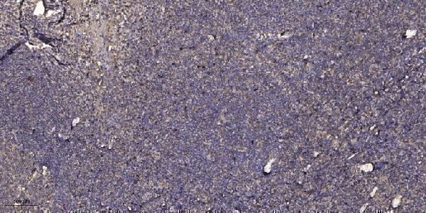
A12806-1 has a human tonsil paraffin-section IHC image (catalog IHC caption). IF is listed for human, mouse, and rat (catalog applications/reactivity), but no IF image is supplied (catalog IF images).

Real IHC data Immunohistochemical analysis of paraffin-embedded human tonsil. 1, Antibody was diluted at 1:200 (4° overnight). 2, Tris-EDTA, pH9.0 was used for antigen retrieval. 3, Secondary antibody was diluted at 1:200 (room temperature, 30min).
Anti-OY-TES-1 ACRBP Antibody
Cat # A12806-1

A12806-1 is listed for IHC and IF in human, mouse, and rat (catalog applications/reactivity). Its IHC image shows a human tonsil paraffin section (catalog IHC caption); no IF image is supplied (catalog IF images).

Which to pick: For tissue IHC, choose A12806-1: its image documents Tris-EDTA retrieval at pH 9.0 and primary antibody at 1:200 overnight at 4 °C (catalog IHC caption); the fixative is unreported (catalog IHC caption). For IF/ICC, A12806-1 lists IF, but ICC and an IF image are unreported; its host is rabbit and clonality is unreported (catalog applications/IF images/host/clone). For cross-species work, A12806-1 lists mouse and rat reactivity, while the supplied IHC image documents human tissue only (catalog reactivity/IHC caption).

Each figure is that product's own IHC / IF validation image from its datasheet.

References

  1. UniProt Consortium. UniProt entry Q8NEB7 (ACRBP_HUMAN, Acrosin-binding protein).
  2. Human Protein Atlas. ACRBP tissue IHC expression (reliability: Enhanced).
  3. Human Protein Atlas. ACRBP subcellular location (ICC-IF): Mainly localized to the nucleoplasm and vesicles. In addition localized to the cytosol..
  4. Human Protein Atlas. ACRBP antibody validation summary (2 antibodies).
  5. Cancer-testis antigen ACRBP expression and serum immunoreactivity in ovarian cancer: Its association with prognosis. Immunity, inflammation and disease 2021 — PMC8589352.
  6. Expression of cancer-testis antigens MAGEA1, MAGEA3, ACRBP, PRAME, SSX2, and CTAG2 in myxoid and round cell liposarcoma. Modern pathology : an official journal of the United States and Canadian Academy of Pathology, Inc 2014 — PMC4287229.
  7. In vitro transplantation of spermatogonial stem cells isolated from human frozen-thawed testis tissue can induce spermatogenesis under 3-dimensional tissue culture conditions. Biological research 2019 — PMC6438003.
  8. Mini Bioreactor Can Support In Vitro Spermatogenesis of Mouse Testicular Tissue. Cell journal 2022 — PMC9445517.
  9. PubMed PMID:11248070 — UniProt-cited evidence.
  10. PubMed PMID:15489334 — UniProt-cited evidence.