ADCY3 / Adenylate cyclase type 3 · IHC design guide

Design Immunohistochemistry for ADCY3

Plan ADCY3 chromogenic IHC on paraffin sections around the reported cytoplasmic tissue signal (HPA tissue IHC). Use the catalog antibody’s 1:200 tissue example (datasheet A04795) and annotated positive and negative cells as controls (HPA tissue IHC).

Evidence assembled Oct 2026 · For research use; verify linked source records and product datasheet before use
Immunohistochemistry protocol sheet for ADCY3 (IHC for ADCY3): expected localisation Cytoplasmic tissue staining (HPA tissue IHC); ciliary membrane location annotated (UniProt), antibody A04795, validated IHC image, and IHC protocol steps
Printable ADCY3 IHC protocol sheet — expected localisation Cytoplasmic tissue staining (HPA tissue IHC); ciliary membrane location annotated (UniProt), antibody A04795, controls and protocol steps. Open the full ADCY3 IHC guide →

ADCY3 Immunohistochemistry Experimental Design Guide

Expected localisation, validated protocols, controls and antibodies — the at-a-glance facts below, then the full design guide.

Must know before staining
Expected localisation Cytoplasmic tissue staining (HPA tissue IHC); ciliary membrane location annotated (UniProt)
Staining pattern Cytoplasmic signal in many tissues, including glandular cells (HPA tissue IHC)
Antigen retrieval EDTA pH 8.0 HIER, heat-mediated (datasheet A04795)
Positive control ⓘ Adrenal gland+4 more · see all
Negative control ⓘ Adipose tissue+4 more · see all
Important caveats
Reasons your staining may differ from the expected pattern.
Fixation Keep fixation consistent between sections. (standard IHC practice; not target-specific)
Caveat Antibody staining has low consistency with RNA expression (HPA tissue IHC)
Regulation No specific staining regulation documented (UniProt)
Isoform / epitope 2 isoforms; cytoplasmic versus external epitope may matter (UniProt)
Section 1

Recommended ADCY3 IHC & IF Protocols

The catalog antibody’s IHC-P protocol (datasheet A04795) is followed by published ADCY3 staining protocols for gastric tissue (PMC3858564) and kidney (PMC9204326).

Recommended immunohistochemistry (IHC-P) protocol parameters
SampleParaffin-embedded human endometrial cancer tissue; fixative not specified (datasheet A04795)
FixationImage fixative and duration unreported (datasheet A04795); verify before use.
Sectioning4–5 µm sections on charged slides (standard)
DeparaffinisationXylene, graded ethanol series to water (standard)
Antigen retrievalHeat retrieval: EDTA pH 8.0 (datasheet A04795); 20 min, 95–100 °C (standard)
Peroxidase block3% H2O2, 10 min, room temperature (standard)
Blocking10% goat serum (datasheet A04795)
Primary antibodyRabbit anti-ADCY3, 1:50 recommended; image 1:200 (datasheet A04795)
Primary incubationOvernight at 4 °C (datasheet A04795)
DetectionHRP-conjugated secondary, DAB chromogen (datasheet A04795)
CounterstainHematoxylin, blue, dehydrate and mount (standard)
Expected resultADCY3-positive staining in glandular cells of adrenal gland (HPA tissue IHC: Medium). HPA tissue profile: Cytoplasmic expression in most tissues. No signal in the no-primary control.
💡Decision noteStart with heat-mediated EDTA, pH 8.0, for the catalog antibody (datasheet A04795); neither published excerpt specifies antigen retrieval (PMC3858564; PMC9204326).
Section 2

What Is the Expected ADCY3 Staining Pattern?

ADCY3 is a 12-pass membrane protein assigned to the cell membrane and primary cilia, with cytoplasmic and Golgi localization also reported (UniProt O60266 topology and subcellular location). In tissue IHC, expect chiefly cytoplasmic staining in selected epithelial, glandular, and muscle cells (HPA: tissue IHC profile). HPA rates its tissue staining Approved but reports low consistency with RNA expression, so interpret intensity and distribution cautiously (HPA: tissue IHC reliability).

What am I looking at on my slide?
Cytoplasmic staining in adrenal glandular cells, respiratory epithelium of bronchus, or heart cardiomyocytes.These cell types show Medium staining in HPA tissue IHC (HPA: adrenal gland, bronchus, heart muscle). A predominantly cytoplasmic pattern fits HPA’s overall tissue profile (HPA: cytoplasmic expression in most tissues). Compare cells within the section; Medium is an HPA category, not a fixed chromogen intensity for every run (general IHC practice).
A narrow apical or ciliary signal accompanies cytoplasmic staining in an appropriate cell.Membrane or ciliary localization is biologically plausible because ADCY3 has 12 transmembrane segments and is assigned to the cell and cilium membranes (UniProt O60266 topology and subcellular location). HPA’s tissue IHC summary chiefly describes cytoplasmic staining, however, so do not require a visible cilium in paraffin sections (HPA: tissue IHC profile; general IHC practice).
Strong, widespread nuclear-only staining appears, without the expected cytoplasmic pattern.Treat this as a discordant IHC result and check controls and detection background before calling it ADCY3 (HPA: cytoplasmic tissue IHC profile; general IHC practice). HPA lists nucleoplasm as an uncertain ICC-IF location; that separate assay does not establish nuclear-only paraffin IHC staining (HPA: subcellular localization).
Strong staining appears mainly in adipocytes, bone-marrow hematopoietic cells, or lymph-node germinal-center cells.Those specified cell populations were Not detected in HPA tissue IHC (HPA: adipose tissue, bone marrow, lymph node). Unexpected signal may reflect antibody cross-reactivity or endogenous detection activity; inspect a matched negative control and the distribution of chromogen before interpreting it as ADCY3 (general IHC practice).
Fine, diffuse color covers both tissue and empty areas, or an expected positive population has no detectable signal.Uniform haze favors background from detection or washing rather than a cell-specific pattern (general IHC practice). Conversely, absent staining in HPA Medium adrenal glandular cells may indicate a failed IHC run, although one negative section cannot establish absent ADCY3 protein (HPA: adrenal gland; general IHC practice).
💡Expected ADCY3 appearanceCall a convincing positive when selected glandular or epithelial cells, or cardiomyocytes, show predominantly cytoplasmic staining of interpretable intensity (HPA: Medium examples and tissue IHC profile); isolated pan-tissue haze or nuclear-only color is discordant and needs control review (HPA: tissue IHC profile; general IHC practice).
How each factor affects the staining
Subcellular distribution and topologyADCY3 spans membranes 12 times, while UniProt also assigns cytoplasmic and Golgi locations (UniProt O60266 topology and subcellular location). In paraffin IHC, assess the whole cellular pattern rather than demanding a sharply resolved membrane rim (general IHC practice).
Tissue and cell selectionHPA reports Medium staining in adrenal glandular cells, bronchial respiratory epithelial cells, and cardiomyocytes, but Not detected in selected adipocytes and hematopoietic cells (HPA: tissue IHC). Use these named populations as comparisons, without treating their categories as universal assay controls (general IHC practice).
Evidence strengthThe tissue profile is Approved despite low consistency between antibody staining and RNA expression; the listed tissue-IHC antibody is Approved rather than Enhanced (HPA: tissue IHC reliability; HPA: CAB010223 IHC validation). A plausible pattern therefore still needs appropriate assay controls (general IHC practice).
Isoforms and epitope assignmentUniProt lists two ADCY3 isoforms and one full-length chain; this payload gives no antibody epitope or isoform-specific IHC result (UniProt O60266 isoforms and processing). Do not infer which isoform produces a stained compartment or a negative result from the available record.
Why is my staining missing, weak or wrong?
SituationLikely causeNext action
Expected adrenal glandular cells show no stain (HPA: adrenal gland, Medium).The run may have lost target detection through a general staining-workflow problem; a negative section alone does not identify which step failed (general IHC practice).Check that the positive-control section stained in the same run, then review antigen retrieval, primary-antibody incubation, detection reagents, and chromogen development against the validated IHC procedure (general IHC practice).
The whole section has pale, diffuse chromogen (general IHC practice).Nonspecific detection, inadequate washing, or endogenous enzyme activity can create background that lacks the cell distribution described by HPA (general IHC practice; HPA: tissue IHC profile).Inspect a no-primary control and compare tissue with blank areas; review blocking, washes, and the enzyme-blocking step appropriate to the detection system (general IHC practice).
Unexpected cells stain more strongly than the named HPA positive populations.Cross-reactivity or endogenous detection activity is possible, especially if HPA records the specific unexpected cell population as Not detected (HPA: named negative populations; general IHC practice).Compare matched negative and no-primary controls, identify the stained cell type precisely, and avoid extending HPA’s cell-specific negatives to every cell in an entire organ (general IHC practice; HPA: tissue IHC).
Nuclear-only staining dominates the slide (HPA: cytoplasmic tissue IHC profile).The pattern conflicts with the tissue IHC profile; nucleoplasm is an uncertain finding from a separate ICC-IF assay (HPA: tissue IHC and subcellular localization).Check counterstain versus chromogen, assess the no-primary control, and score any cytoplasmic staining independently before assigning ADCY3 positivity (general IHC practice).
A ciliary or membrane rim is hard to see in an otherwise plausible positive section.UniProt assigns ADCY3 to cilium and cell membranes, but HPA describes chiefly cytoplasmic tissue IHC; a cilium may be difficult to resolve in a section (UniProt O60266 subcellular location; HPA: tissue IHC; general IHC practice).Judge the named cell population and cytoplasmic pattern first, then inspect suitable high-magnification fields for localized signal without making visible cilia a pass criterion (HPA: tissue IHC; general IHC practice).
Q: Does an IF/ICC ciliary or nucleoplasmic image settle an ambiguous paraffin IHC result?HPA calls both primary-cilium and nucleoplasm ICC-IF locations uncertain; ICC-IF and tissue IHC are separate observations (HPA: subcellular localization and tissue IHC).A: No. Interpret paraffin IHC against its own cell-type pattern and controls; use IF/ICC localization only as contextual evidence, with its stated uncertainty (HPA: tissue IHC and subcellular localization; general IHC practice).

Sample controls for ADCY3 IHC & IF

🧪Run adrenal gland first: glandular cells should stain at the HPA Medium level (HPA: adrenal gland glandular cells, Medium; UniProt O60266: adrenal detection). Use adipose tissue as the negative comparator (HPA: adipocytes, Not detected); on the adrenal slide, adjacent nonglandular cells should lack specific staining when compared with a no-primary section, but confirm their status rather than assuming they are ADCY3-negative.
Positive control tissue: Adrenal gland (Glandular cells, HPA Medium)
Negative control tissue: Adipose tissue (HPA Not detected)
ICC-IF cell lines (HPA subcellular resource): HPA ICC-IF images show ADCY3 in HEK293, Rh30, U2OS, ASC52telo, RPTEC/TERT1, hTERT-RPE1 (serum starved), KOLF2.1J, with annotated localisation: Nucleoplasm (uncertain), Primary cilium (uncertain) (HPA subcellular).
Technical controls: Include no-primary (secondary-only) and concentration-matched rabbit IgG isotype controls alongside an ADCY3 knockout sample or validated immunizing-peptide block (caption: rabbit anti-ADCY3 antibody; standard IHC controls). Quench endogenous peroxidase for HRP–DAB detection, and check the adrenal no-primary section for pigment that could resemble DAB (caption: HRP–DAB detection; standard IHC practice).
⚠️Feasibility: A target-specific fixation window or fixation effect is unreported, and the selected A04795 paraffin-section caption does not state the fixative (caption: fixative unreported). The reported IHC result used heat retrieval in EDTA at pH 8.0, making that a documented starting condition rather than proof that retrieval is required (caption: EDTA retrieval, pH 8.0). The supplied evidence does not establish that frozen sections or IF are easier; for adrenal IHC, compare suspected brown pigment with the no-primary section before scoring DAB staining (HPA: adrenal gland glandular cells, Medium; standard IHC practice).

HPA tissue IHC evidence for ADCY3

Comprehensive Human Protein Atlas IHC scoring per tissue (reliability: Approved — Low consistency between antibody staining and RNA expression data.). Rows are taken directly from the HPA tissue chart — click any row's HPA link to view the source.

Positive expression · recommended positive controls

TissueCell typeLevelEvidenceSource
Adrenal gland Glandular cells Medium Protein (IHC) HPA →
Bronchus Respiratory epithelial cells Medium Protein (IHC) HPA →
Cervix Squamous epithelial cells Medium Protein (IHC) HPA →
Endometrium Glandular cells Low Protein (IHC) HPA →
Epididymis Glandular cells Medium Protein (IHC) HPA →

Undetected expression · recommended negative controls

TissueCell typeLevelEvidenceSource
Adipose tissue Adipocytes Not detected Protein (IHC) HPA →
Bone marrow Hematopoietic cells Not detected Protein (IHC) HPA →
Hippocampus Glial cells Not detected Protein (IHC) HPA →
Lymph node Germinal center cells Not detected Protein (IHC) HPA →
Ovary Ovarian stroma cells Not detected Protein (IHC) HPA →
Section 3

Advanced ADCY3 IHC Tips

Troubleshoot ADCY3 chromogenic IHC in paraffin sections using the catalog antibody’s tissue image, ADCY3 topology, and independent localisation evidence.

How should I adjust antigen retrieval when ADCY3 staining is weak or uneven?
Start with heat mediated retrieval in EDTA pH 8.0 for paraffin sections (datasheet A04795). The catalog antibody produced a tissue IHC image after this retrieval, followed by 1:200 primary antibody overnight at 4°C (datasheet A04795). If signal is weak, confirm complete deparaffinisation and consistent heating and cooling across slides before changing retrieval conditions (standard IHC practice). Include a matched positive control such as adrenal glandular cells, and assess whether staining appears in the expected cell population (HPA: Medium in adrenal glandular cells). Record retrieval time and temperature for each run so changes in intensity can be traced to processing rather than biology (standard IHC practice).
Could fixation explain variable ADCY3 staining between paraffin blocks?
Target specific fixation sensitivity is unknown because the selected tissue IHC caption does not state its fixative (datasheet A04795). Compare blocks with documented fixation histories using identical section thickness, EDTA pH 8.0 retrieval, and antibody incubation to isolate processing differences (datasheet A04795; standard IHC practice). If histories differ, interpret weaker staining cautiously and test a control block in every run before assigning a biological difference (standard IHC practice). Check section morphology and tissue preservation alongside the stain, because damaged or detached areas can make cell level comparisons unreliable (standard IHC practice). Do not use staining intensity alone to infer how a block was fixed (standard IHC practice).
Where should ADCY3 staining appear, and how should diffuse cytoplasm be handled?
ADCY3 is annotated at the cell membrane, cilium, cytoplasm, and Golgi apparatus, including cytoplasm near lipid droplets (UniProt O60266 localisation). HPA describes predominantly cytoplasmic tissue IHC staining but reports low consistency between antibody staining and RNA expression (HPA: tissue IHC profile and reliability). Score membrane or cilium associated signal separately from diffuse cytoplasmic signal when resolution permits, and retain cell type annotations (UniProt O60266 localisation; standard IHC practice). A thin cilium may be difficult to resolve in chromogenic sections, so absence of a visible ciliary structure alone cannot exclude ADCY3 (standard IHC practice). Confirm disputed patterns with an independent control or orthogonal method (standard IHC practice).
How can epitope position affect ADCY3 staining in paraffin sections?
ADCY3 has 2 annotated isoforms and 12 transmembrane segments, with cytoplasmic regions at residues 1–79, 402–632, and 854–1144 (UniProt O60266 isoforms and topology). The supplied antibody caption does not identify its epitope, so staining cannot establish which isoform or membrane facing region the antibody recognises (datasheet A04795). Obtain the immunogen or epitope documentation before attributing differences in signal to isoform expression or epitope accessibility (standard IHC practice). Keep retrieval at EDTA pH 8.0 while assessing such differences, and compare sections with matched processing (datasheet A04795; standard IHC practice). Interpret a changed pattern as provisional until specificity is independently supported (standard IHC practice).
How should I investigate an ADCY3 pattern with multiplex immunofluorescence?
For an IF follow up, multiplex ADCY3 with a marker identifying the cell population being scored, such as glandular cells in an adrenal section (HPA: Medium in adrenal glandular cells; standard IF practice). Choose fluorophores in channels with low measured tissue autofluorescence, and include single stain and no primary controls to assess bleed through and background (standard IF practice). ADCY3 spans the membrane 12 times, but the supplied antibody epitope is unspecified; choose permeabilisation only after determining whether that epitope faces the cytoplasm or extracellular space (UniProt O60266 topology; datasheet A04795). Assess ciliary, membrane, Golgi, and cytoplasmic signals as distinct patterns rather than pooling them (UniProt O60266 localisation; standard IF practice).
What should I check when ADCY3 DAB staining is widespread?
Compare the stained section with a no primary control to assess secondary reagent binding and endogenous chromogenic signal (standard IHC practice). For the catalog antibody, the demonstrated workflow used 10% goat serum blocking, 1:200 primary antibody overnight at 4°C, and peroxidase linked detection with DAB (datasheet A04795). Apply a peroxidase block and check wash stringency and DAB development time when diffuse colour persists; these are general chromogenic IHC controls (standard IHC practice). ADCY3 can show cytoplasmic staining, so diffuse colour alone is insufficient to call background (HPA: tissue IHC profile). Examine whether signal follows intact cells and disappears appropriately in controls before scoring it (standard IHC practice).
How should I quantify ADCY3 IHC across samples with different cell composition? ⚠ ANSWER MARKED FOR VERIFICATION
Define the compartment and cell population before scoring: for example, score glandular cells separately from surrounding tissue where that population is present (HPA: Medium in adrenal glandular cells; standard IHC practice). Use an H-score or percent positive cells with fixed intensity thresholds, and report the number of evaluable cells or tissue area examined (standard IHC practice). Normalise positive counts to the eligible cell population, or report positive cell density per mm² of evaluable tissue (standard IHC practice). Exclude folds, damaged edges, and necrotic regions by a prespecified rule, then apply identical acquisition and scoring settings across groups (standard IHC practice). Record cytoplasmic and membrane associated signal separately when resolvable (UniProt O60266 localisation; standard IHC practice).
How can I distinguish convincing ADCY3 staining from artefact?
Prioritise signal in intact cells and assess whether its cytoplasmic, membrane, or cilium associated distribution is plausible for ADCY3 (UniProt O60266 localisation; standard IHC practice). Compare the implicated cell type with an appropriate reference: HPA reports Medium staining in adrenal glandular cells and no detection in adipocytes of adipose tissue (HPA: tissue IHC). Treat isolated nuclear staining cautiously because HPA marks nucleoplasmic localisation uncertain, while UniProt lists membrane, cilium, cytoplasm, and Golgi locations (HPA: subcellular; UniProt O60266 localisation). Reject edge accentuation, necrotic deposits, or colour persisting in no primary and peroxidase controls as evidence of specific ADCY3 staining (standard IHC practice). HPA also reports low agreement between tissue staining and RNA, so confirm consequential findings independently (HPA: reliability).
Boster reagents

Best ADCY3 / Adenylate cyclase type 3 IHC Antibodies

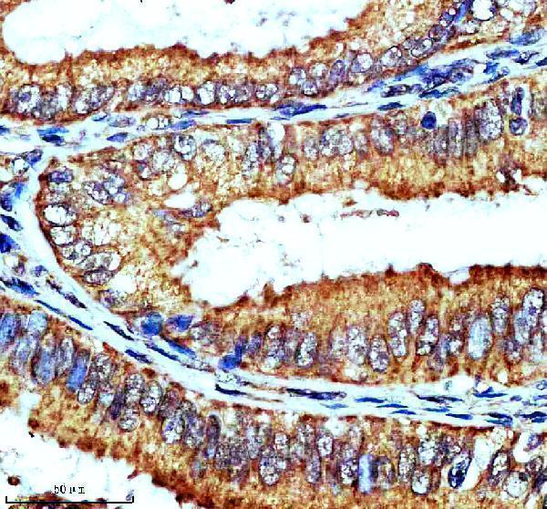
A04795 has real IHC data from a paraffin-embedded human endometrial cancer section (A04795 image caption). IF/ICC is listed without an IF figure (catalog applications; catalog IF images).

Real IHC data IHC analysis of ADCY3 using anti-ADCY3 antibody (A04795). ADCY3 was detected in a paraffin-embedded section of human endometrial cancer tissue. Heat mediated antigen retrieval was performed in EDTA buffer (pH 8.0, epitope retrieval solution). The tissue section was blocked with 10% goat serum. The tissue section was then incubated with 1:200 rabbit anti-ADCY3 Antibody (A04795) overnight at 4°C. Peroxidase Conjugated Goat Anti-rabbit IgG was used as secondary antibody and incubated for 30 minutes at 37°C. The tissue section was developed using HRP Conjugated Rabbit IgG Super Vision Assay Kit (Catalog # SV0002) with DAB as the chromogen.
Anti-ADCY3 antibody
Cat # A04795

A04795 will render with an IHC image from a paraffin-embedded human endometrial cancer section (A04795 image caption). The catalog lists IF/ICC applications and human, mouse, and rat reactivity for A04795, but provides no IF image (catalog applications; catalog reactivity; catalog IF images).

Which to pick: For tissue IHC, choose A04795: its image documents EDTA retrieval at pH 8.0 and a 1:200 primary dilution in a paraffin-embedded human section; the fixative is unreported (A04795 image caption). For IF/ICC, A04795 is the listed option, with a catalog IF dilution of 1:50, although no IF image is provided (catalog applications; catalog IF dilution; catalog IF images). For mouse or rat samples, A04795 lists reactivity with both species, while its displayed IHC example is human (catalog reactivity; A04795 image caption).

Each figure is that product's own IHC / IF validation image from its datasheet.

References

  1. UniProt Consortium. UniProt entry O60266 (ADCY3_HUMAN, Adenylate cyclase type 3).
  2. Human Protein Atlas. ADCY3 tissue IHC expression (reliability: Approved).
  3. Human Protein Atlas. ADCY3 subcellular location (ICC-IF): Mainly localized to the nucleoplasm and primary cilium. In addition localized to the nucleoli, plasma membrane, the Golgi apparatus, primary cilium transition zone and basal body..
  4. Human Protein Atlas. ADCY3 antibody validation summary (2 antibodies).
  5. Upregulation of adenylate cyclase 3 (ADCY3) increases the tumorigenic potential of cells by activating the CREB pathway. Oncotarget 2013 — PMC3858564.
  6. Expression Regulation of Water Reabsorption Genes and Transcription Factors in the Kidneys of Lepus yarkandensis. Frontiers in physiology 2022 — PMC9204326.
  7. Muscle-derived Mimecan regulates hypothalamus-brown adipose tissue communication and promotes health and lifespan in mice. Cell metabolism 2026 — PMC13180260.
  8. Regulation of Brain Primary Cilia Length by MCH Signaling: Evidence from Pharmacological, Genetic, Optogenetic, and Chemogenic Manipulations. Molecular neurobiology 2022 — PMC9083846.
  9. PubMed PMID:9920776 — UniProt-cited evidence.
  10. PubMed PMID:14702039 — UniProt-cited evidence.
  11. PubMed PMID:15815621 — UniProt-cited evidence.