This website uses cookies to ensure you get the best experience on our website.


- Table of Contents
Use kidney tubules and pancreatic exocrine cells as high staining reference tissues for ADCY7 IHC (HPA tissue IHC). Start the IHC-validated antibody at 1:100–1:300 (datasheet), and interpret staining cautiously because HPA rates tissue IHC reliability uncertain (HPA tissue IHC).

Expected localisation, validated protocols, controls and antibodies — the at-a-glance facts below, then the full design guide.
| Expected localisation | Cytoplasmic tissue staining (HPA tissue IHC); membrane protein (UniProt) | |
| Staining pattern | Cytoplasmic staining in kidney tubules and pancreatic exocrine cells (HPA tissue IHC) | |
| Antigen retrieval | Citrate pH 6.0 HIER, 95–98 °C, 20 min (rule: cytoplasmic / membrane antigen) | |
| Positive control | Kidney+4 more · see all | |
| Negative control | Adipose tissue+4 more · see all |
| Fixation | Keep fixation consistent across paraffin sections. (standard IHC practice; not target-specific) | |
| Caveat | Antibody staining and RNA expression show low consistency (HPA tissue IHC) | |
| Regulation | Expression regulation not specified (UniProt) | |
| Isoform / epitope | No isoforms annotated; epitope side matters across 12 transmembrane spans (UniProt) |
The catalog antibody’s IHC-P protocol is accompanied by one published ADCY7 IHC workflow using paraffin sections (PMC8919236).
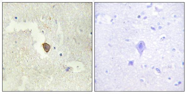
| Sample | Paraffin-embedded human brain tissue; fixative not specified (datasheet A11384) |
| Fixation | Image fixative and duration unreported (datasheet A11384); verify before use. |
| Sectioning | 4–5 µm sections on charged slides (standard) |
| Deparaffinisation | Xylene, graded ethanol series to water (standard) |
| Antigen retrieval | Heat-induced epitope retrieval in citrate buffer, pH 6.0, 20 min at 95–98 °C (standard rule: cytoplasmic / membrane antigen) |
| Peroxidase block | 3% H2O2, 10 min, room temperature (standard) |
| Blocking | 10% normal serum of the secondary host, 30 min, room temperature (standard) |
| Primary antibody | Rabbit anti-ADCY7, 1:100-1:300 (datasheet A11384) |
| Primary incubation | Overnight at 4 °C (standard) |
| Detection | HRP-polymer secondary, DAB chromogen 5–10 min (standard) |
| Counterstain | Hematoxylin, blue, dehydrate and mount (standard) |
| Expected result | ADCY7-positive staining in cells in tubules of kidney (HPA tissue IHC: High). HPA tissue profile: Cytoplasmic expression in selected tissues. No signal in the no-primary control. |
ADCY7 is a 12-pass membrane protein with substantial cytoplasmic regions (UniProt P51828 topology). In paraffin-section IHC, HPA reports cytoplasmic staining, high in kidney tubule cells and pancreatic exocrine glandular cells, and medium in bone-marrow hematopoietic cells (HPA: tissue IHC). Treat these as comparison patterns rather than definitive controls: tissue IHC reliability is Uncertain because antibody staining and RNA expression have low consistency (HPA: tissue IHC reliability).
| Strong cytoplasmic staining in kidney tubule cells or pancreatic exocrine glandular cells, with identifiable cell boundaries. | This resembles the reported high IHC signal in those specific cell populations (HPA: kidney tubule cells High; pancreas exocrine glandular cells High). Record the proportion of stained cells and where stain lies within them. The match supports interpretation but does not establish specificity, because HPA rates the tissue IHC profile Uncertain (HPA: tissue IHC reliability). |
| A dominant nuclear-only pattern, or staining without a discernible cytoplasmic or membrane-associated component. | This does not match the reported cytoplasmic tissue pattern (HPA: tissue IHC profile) or membrane localization (UniProt P51828 topology). Check the negative reagent control and reassess whether the visible signal belongs to the intended cells before scoring ADCY7. Nuclear-only staining alone is insufficient evidence for a new ADCY7 location. |
| Strong staining in adipocytes or alveolar cells while the expected cell populations remain faint. | HPA reports ADCY7 as Not detected in adipocytes and alveolar cells (HPA: adipose tissue adipocytes Not detected; lung alveolar cells Not detected). Such a distribution raises concern for cross-reactivity or detection-related signal; it does not prove either cause. Compare cell identity, morphology and reagent controls before assigning the stain to ADCY7. |
| Brown signal spreads across cells, connective tissue and empty spaces, obscuring individual cell outlines. | Diffuse deposit cannot be scored reliably as cellular ADCY7 staining. Examine a negative reagent control and the detection chemistry for nonspecific background (general IHC practice). HPA describes staining in selected cells, not a uniform tissue-wide deposit (HPA: tissue IHC profile). |
| No visible signal in kidney tubule cells and pancreatic exocrine glandular cells. | The absence conflicts with HPA's reported high staining in those cells (HPA: kidney tubule cells High; pancreas exocrine glandular cells High). Review the section, retrieval, antibody application and detection controls as general IHC checks. Because HPA calls its tissue profile Uncertain, a negative result cannot by itself establish assay failure or absence of ADCY7. |
| Topology and tissue compartment | Twelve transmembrane segments anchor ADCY7, while two long regions are cytoplasmic (UniProt P51828 topology); HPA describes cytoplasmic tissue IHC staining (HPA: tissue IHC profile). A cytoplasmic-looking chromogenic result can therefore be assessed alongside membrane biology without requiring a sharply outlined cell border. |
| Strength of tissue evidence | Kidney tubule and pancreatic exocrine glandular cells are reported High, whereas bone-marrow hematopoietic and cerebellar Purkinje cells are Medium (HPA: tissue IHC). These are observed staining levels, not guaranteed intensities in a new section; HPA rates the overall tissue IHC profile Uncertain (HPA: tissue IHC reliability). |
| Antibody validation | The listed tissue-IHC antibody HPA048241 has IHC status Uncertain; HPA041218 has ICC status Approved but no listed IHC status (HPA: antibody validation). Do not transfer ICC approval to paraffin-section IHC or treat agreement with one stained tissue as independent validation. |
| Epitope and retrieval | ADCY7 has cytoplasmic and transmembrane regions (UniProt P51828 topology), but these sources give no epitope position or target-specific retrieval or fixation response. Select and document retrieval conditions using the chosen antibody's IHC instructions, then interpret any change as an assay observation, not an established ADCY7 property (general IHC practice). |
| Situation | Likely cause | Next action |
|---|---|---|
| Kidney tubules or pancreatic exocrine cells are unstained. | The assay may have weak detection, or the observed tissue pattern may not reproduce; HPA's tissue IHC rating is Uncertain (HPA: tissue IHC reliability). | Check section quality, antibody application, retrieval and chromogenic detection with appropriate controls; document which cell population was assessed (general IHC practice; HPA: kidney tubule and pancreatic exocrine cells High). |
| Nuclei dominate the chromogenic signal. | A nuclear-only result conflicts with the reported cytoplasmic IHC profile and membrane topology (HPA: tissue IHC profile; UniProt P51828 topology). | Inspect cell boundaries and a negative reagent control, then withhold an ADCY7-positive score until the cellular location is resolved (general IHC practice). |
| Adipocytes or lung alveolar cells stain strongly. | These named populations are reported Not detected; cross-reactivity or detection background is possible, not established (HPA: adipocytes Not detected; alveolar cells Not detected). | Confirm the stained cell type and compare negative reagent controls and a reported positive cell population on the same staining run (general IHC practice; HPA: tissue IHC). |
| Brown precipitate is widespread or obscures cell detail. | Diffuse background can reflect nonspecific reagent binding or, with peroxidase-based detection, endogenous activity (general IHC practice). | Review blocking and the negative reagent control; if using peroxidase detection, check its endogenous-activity control before adjusting detection conditions (general IHC practice). |
| A membrane rim is absent despite cytoplasmic staining. | HPA reports cytoplasmic tissue IHC staining, while UniProt describes a membrane protein; a crisp rim is not a stated tissue-IHC requirement (HPA: tissue IHC profile; UniProt P51828 topology). | Score identifiable cells and cytoplasmic distribution, then compare the reported positive cell types; do not reject a result solely for lacking a rim (HPA: tissue IHC). |
| Q: Can IF/ICC localization settle an ambiguous IHC result? | HPA approves plasma-membrane localization in ICC-IF, with additional vesicles and rods & rings, but the listed IHC antibody has Uncertain IHC status (HPA: subcellular ICC-IF; HPA: antibody validation). | A: Use IF/ICC localization as context only. Resolve paraffin-section IHC through its own cell pattern and controls; ICC approval does not validate an IHC stain (HPA: antibody validation; general IHC practice). |


Comprehensive Human Protein Atlas IHC scoring per tissue (reliability: Uncertain — Low consistency between antibody staining and RNA expression data. Pending external verification.). Rows are taken directly from the HPA tissue chart — click any row's HPA link to view the source.
| Tissue | Cell type | Level | Evidence | Source |
|---|---|---|---|---|
| Kidney | Cells in tubules | High | Protein (IHC) | HPA → |
| Pancreas | Exocrine glandular cells | High | Protein (IHC) | HPA → |
| Bone marrow | Hematopoietic cells | Medium | Protein (IHC) | HPA → |
| Cerebellum | Purkinje cells | Medium | Protein (IHC) | HPA → |
| Cervix | Squamous epithelial cells | Medium | Protein (IHC) | HPA → |
| Tissue | Cell type | Level | Evidence | Source |
|---|---|---|---|---|
| Adipose tissue | Adipocytes | Not detected | Protein (IHC) | HPA → |
| Appendix | Glandular cells | Not detected | Protein (IHC) | HPA → |
| Caudate | Glial cells | Not detected | Protein (IHC) | HPA → |
| Colon | Endothelial cells | Not detected | Protein (IHC) | HPA → |
| Fallopian tube | Glandular cells | Not detected | Protein (IHC) | HPA → |
Use membrane aware controls and cell resolved scoring when interpreting ADCY7 staining in paraffin sections (UniProt P51828 topology; HPA tissue IHC reliability: Uncertain).
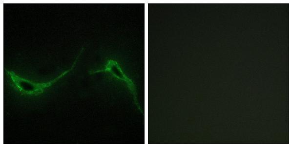
A11384 has IHC imagery from paraffin-embedded human brain tissue and IF imagery from NIH/3T3 cells (image captions); the catalog lists Human and Mouse reactivity (catalog: A11384).
A11384 is listed for IHC and IF, with Human and Mouse reactivity (catalog: A11384). Its IHC image shows paraffin-embedded human brain tissue, while its IF image shows NIH/3T3 cells (image captions: A11384).
Which to pick: For tissue IHC, choose A11384: its own image documents staining in paraffin-embedded human brain tissue, and the listed IHC dilution is 1:100–1:300 (IHC image caption and datasheet: A11384). For cell IF, choose A11384 based on its NIH/3T3 image and listed IF dilution of 1:200–1:1000; its polyclonal format and Human/Mouse reactivity also make it the catalog choice for evaluating both species (IF image caption and catalog: A11384). The IHC caption does not report a fixative or document mouse tissue staining (IHC image caption: A11384).