This website uses cookies to ensure you get the best experience on our website.


- Table of Contents
Plan ADGRG6 staining in paraffin sections using endothelial cells as a tissue-level reference (HPA tissue IHC). This guide covers the expected membrane location and receptor cleavage (UniProt), plus the catalog antibody’s 1:100–1:300 IHC dilution (datasheet A30803).

Expected localisation, validated protocols, controls and antibodies — the at-a-glance facts below, then the full design guide.
| Expected localisation | Cell membrane expected (UniProt); endothelial cells stain (HPA tissue IHC) | |
| Staining pattern | Endothelial cells stain; compartment unresolved (HPA tissue IHC) | |
| Antigen retrieval | Tris-EDTA pH 9.0 HIER, heat-mediated (datasheet A30803) | |
| Positive control | Skeletal muscle+4 more · see all | |
| Negative control | Adipose tissue+4 more · see all |
| Fixation | Keep fixation consistent across sections. (standard IHC practice; not target-specific) | |
| Caveat | Staining and RNA show low consistency (HPA tissue IHC) | |
| Regulation | Surface detection in activated cells (UniProt) | |
| Isoform / epitope | 4 isoforms; cleavage makes N- vs C-terminal epitope key (UniProt) |
The catalog antibody’s IHC-P protocol (datasheet: A30803) is accompanied by published ADGRG6 IHC protocols from bladder tumors (PMC6372626) and mouse spine sections (PMC8328515).
| Sample | Paraffin-embedded human liver cancer tissue; fixative not specified (datasheet A30803) |
| Fixation | Image fixative and duration unreported (datasheet A30803); verify before use. |
| Sectioning | 4–5 µm sections on charged slides (standard) |
| Deparaffinisation | Xylene, graded ethanol series to water (standard) |
| Antigen retrieval | Heat retrieval: Tris-EDTA pH 9.0 (datasheet A30803); 20 min, 95–100 °C (standard) |
| Peroxidase block | 3% H2O2, 10 min, room temperature (standard) |
| Blocking | 10% normal serum of the secondary host, 30 min, room temperature (standard) |
| Primary antibody | Rabbit anti-ADGRG6, 1:100 - 1:300 (datasheet A30803) |
| Primary incubation | Overnight at 4 °C (standard) |
| Detection | HRP-polymer secondary, DAB chromogen 5–10 min (standard) |
| Counterstain | Hematoxylin, blue, dehydrate and mount (standard) |
| Expected result | ADGRG6-positive staining in myocytes of skeletal muscle (HPA tissue IHC: High). HPA tissue profile: Highly expressed in endothelial cells. No signal in the no-primary control. |
ADGRG6 is a cell membrane receptor with 7 transmembrane segments (UniProt Q86SQ4 topology). In paraffin section IHC, assess staining in myocytes and smooth muscle cells, where HPA reports high signal, and in endothelial cells, where its tissue profile emphasizes expression (HPA: tissue IHC). HPA rates the IHC evidence Approved but reports low consistency between antibody staining and RNA expression (HPA: reliability description).
| Defined staining at cell borders in skeletal muscle myocytes or smooth muscle cells. | This fits the expected membrane compartment (UniProt Q86SQ4: cell membrane) and HPA's High staining in both cell types (HPA: skeletal muscle myocytes; smooth muscle cells). Assess cell borders against the counterstain and surrounding tissue; a uniformly colored section cannot establish membrane localisation (general IHC practice). |
| Endothelial staining is visible, while adjacent cells stain differently or remain pale. | An endothelial pattern is plausible (HPA: highly expressed in endothelial cells); cerebral cortex endothelial cells are Medium and colon endothelial cells Low in the supplied observations (HPA: tissue IHC). Do not require every vessel to match: UniProt reports ADGRG6 in aortic but not skin microvascular endothelial cells and on activated but not resting umbilical vein cell surfaces (UniProt Q86SQ4: tissue specificity; subcellular location). |
| Strong nuclear staining dominates, without convincing cell border staining. | A nuclear dominant result conflicts with the annotated membrane location (UniProt Q86SQ4: cell membrane; topology). Treat it as suspect rather than as proof of a new ADGRG6 compartment; compare the positive control and a detection control before assigning the signal to the target (general IHC practice). |
| Cells annotated as negative stain as strongly as the expected positive cells. | For example, HPA reports adipocytes and adrenal gland glandular cells as Not detected (HPA: tissue IHC). Signal in those specific cells warrants a specificity check; antibody cross reactivity or endogenous detection activity are possible causes, rather than established explanations for an individual slide (general IHC practice). |
| Diffuse color obscures cell boundaries, or the known positive cell population has no signal. | Diffuse color prevents a defensible localisation call (general IHC practice). An absent result in skeletal muscle myocytes or smooth muscle cells conflicts with HPA's High observations (HPA: tissue IHC), but does not alone prove biological absence or a particular preparation failure. |
| Compartment and epitope position | UniProt places residues 38–862 extracellularly and describes 7 transmembrane segments with cytoplasmic regions (UniProt Q86SQ4: topology). The supplied record gives no antibody epitope, so topology supports a membrane expectation but cannot predict which receptor segment the IHC antibody detects. |
| Proteolytic processing | UniProt describes extracellular N terminal and membrane bound C terminal fragments that predominantly remain associated (UniProt Q86SQ4: processing; subunit, by similarity). Interpret border staining with this architecture in mind; the record does not establish that a specific IHC pattern reflects fragment separation or shedding. |
| Cell type and endothelial state | HPA grades skeletal and smooth muscle cells High, cerebral cortex endothelium Medium, and colon endothelium Low (HPA: tissue IHC). UniProt's endothelial observations differ by vessel source and activation state (UniProt Q86SQ4: tissue specificity; subcellular location); do not treat every unstained vessel as assay failure. |
| Isoforms and antibody evidence | UniProt lists 4 isoforms (UniProt Q86SQ4: isoforms). The supplied HPA antibody record lists HPA017346 as IHC Approved and gives no ICC validation or isoform specific epitope information (HPA: antibody record). Neither isoform recognition nor an IHC Enhanced validation claim follows from these data. |
| Agreement with RNA and tissue controls | HPA calls the IHC reliability Approved while noting low consistency with RNA and staining in unannotated cells or structures (HPA: reliability description). Interpret the pictured cell type directly; an RNA enriched tissue designation for liver or placenta is not a substitute for a cell resolved IHC positive control (HPA: RNA specificity; tissue IHC). |
| IF/ICC Q: what location should be expected? | A: Membrane is the supported location (HPA: subcellular summary; UniProt Q86SQ4: cell membrane). HPA supplies no ICC IF images or ICC antibody validation in this payload (HPA: subcellular record; antibody record), so it does not establish an IF staining pattern. Use the separate IF/ICC guide for that application. |
| Situation | Likely cause | Next action |
|---|---|---|
| No signal in a section expected to contain skeletal muscle myocytes or smooth muscle cells. | HPA reports High staining in those cells (HPA: tissue IHC); absent color may reflect a failed stain or a sampling or cell identification issue (general IHC practice). | Confirm the expected cells are present on the counterstained section, then review the IHC antibody and detection controls and the run's retrieval and detection steps (general IHC practice). No ADGRG6 specific retrieval condition is supplied. |
| Endothelial cells are pale or negative while a muscle control stains. | The endothelial result is context dependent: cortex endothelial cells are Medium and colon endothelial cells Low (HPA: tissue IHC); UniProt also reports vessel source and activation differences (UniProt Q86SQ4). | Identify the vessel and cell population before calling failure; compare with the run's positive control and HPA's matched tissue observation, where available (HPA: tissue IHC; general IHC practice). |
| Nuclei are strongly colored but cell borders are not. | Predominant nuclear staining is inconsistent with the membrane annotation (UniProt Q86SQ4: subcellular location). The cause cannot be assigned from compartment alone. | Check a detection control and the positive tissue for the same nuclear pattern, then reassess antibody specificity and interpretation (general IHC practice). Do not score nuclear color alone as ADGRG6 positive. |
| Adipocytes or adrenal gland glandular cells show convincing color. | Those cell populations are Not detected in the supplied HPA observations (HPA: tissue IHC). Cross reactivity or endogenous detection activity is possible (general IHC practice). | Compare a reagent omission control and the expected positive cells on the same run; review endogenous activity blocking as appropriate for chromogenic detection (general IHC practice). |
| Color spreads broadly across tissue and masks cell boundaries. | Nonspecific background or detection activity can obscure compartment assignment (general IHC practice); HPA's membrane summary cannot validate diffuse color (HPA: subcellular summary). | Inspect reagent omission and detection controls, blocking, washing, and development time; score only signal that can be assigned to cells and location (general IHC practice). |
| Two specimens show different distributions or staining strengths. | HPA reports uneven cell type levels and low antibody staining to RNA consistency (HPA: tissue IHC; reliability description). UniProt also lists 4 isoforms, without an epitope map for this antibody (UniProt Q86SQ4: isoforms; HPA: antibody record). | Compare the same cell types, compartments, and run controls before calling a biological difference; avoid attributing discordance to an isoform or fixation effect without separate evidence (general IHC practice). |



Comprehensive Human Protein Atlas IHC scoring per tissue (reliability: Approved — Low consistency between antibody staining and RNA expression data. Antibody staining in cells/structures not annotated, view images.). Rows are taken directly from the HPA tissue chart — click any row's HPA link to view the source.
| Tissue | Cell type | Level | Evidence | Source |
|---|---|---|---|---|
| Skeletal muscle | Myocytes | High | Protein (IHC) | HPA → |
| Smooth muscle | Smooth muscle cells | High | Protein (IHC) | HPA → |
| Breast | Myoepithelial cells | Medium | Protein (IHC) | HPA → |
| Cerebral cortex | Endothelial cells | Medium | Protein (IHC) | HPA → |
| Heart muscle | Cardiomyocytes | Medium | Protein (IHC) | HPA → |
| Tissue | Cell type | Level | Evidence | Source |
|---|---|---|---|---|
| Adipose tissue | Adipocytes | Not detected | Protein (IHC) | HPA → |
| Adrenal gland | Glandular cells | Not detected | Protein (IHC) | HPA → |
| Appendix | Glandular cells | Not detected | Protein (IHC) | HPA → |
| Bronchus | Respiratory epithelial cells | Not detected | Protein (IHC) | HPA → |
| Caudate | Glial cells | Not detected | Protein (IHC) | HPA → |
Troubleshoot ADGRG6 staining in paraffin section IHC using the catalog antibody’s tissue image, receptor topology, and cell specific reference staining.
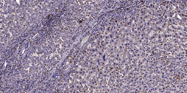
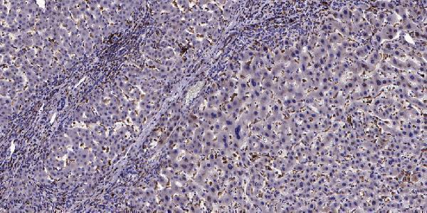
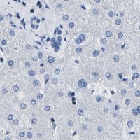
Both anti-ADGRG6 antibodies have IHC images from paraffin-embedded human liver cancer (catalog image captions). Both list Human and Mouse reactivity; A30803 also lists Rat (catalog reactivity).
A30803 will render with its paraffin-embedded human liver cancer IHC image; it lists IHC, IF and ICC applications and Human, Mouse and Rat reactivity (catalog image caption; applications/reactivity). A32344-1 will render with its own paraffin-embedded human liver cancer IHC image; it lists IHC and IF applications and Human and Mouse reactivity (catalog image caption; applications/reactivity).
Which to pick: For tissue IHC, either A30803 or A32344-1 has its own paraffin-section image using 1:200 primary antibody and Tris-EDTA retrieval at pH 9.0; neither caption reports the fixative (each SKU’s IHC image caption). For IF/ICC, choose A30803 when ICC is needed because it lists both applications, while A32344-1 lists IF only; both are rabbit polyclonals (catalog applications; host; dilution_raw). For Rat samples, choose A30803, which lists Rat reactivity; A32344-1 lists Human and Mouse (catalog reactivity).