This website uses cookies to ensure you get the best experience on our website.


- Table of Contents
Plan ADGRL1 chromogenic IHC in paraffin sections using the catalog antibody’s 1:50–1:300 IHC dilution range (datasheet A30832). This guide covers tissue staining patterns and control selection while accounting for the uncertain reliability of HPA tissue staining (HPA tissue IHC).

Expected localisation, validated protocols, controls and antibodies — the at-a-glance facts below, then the full design guide.
| Expected localisation | Membranous and cytoplasmic tissue staining (HPA tissue IHC) | |
| Staining pattern | Glandular cells in several tissues: membranous and cytoplasmic (HPA tissue IHC) | |
| Antigen retrieval | Tris-EDTA pH 9.0 HIER, heat-mediated (datasheet A30832) | |
| Positive control | Breast+4 more · see all | |
| Negative control | Adipose tissue+4 more · see all |
| Fixation | Keep fixation consistent across sections (standard IHC practice; not target-specific) | |
| Caveat | Staining has low consistency with RNA expression (HPA tissue IHC) | |
| Regulation | No target-specific regulator reported (UniProt; HPA tissue IHC) | |
| Isoform / epitope | 2 isoforms; check whether the epitope is extracellular or cytoplasmic (UniProt) |
The catalog antibody has an IHC-P protocol (datasheet A30832); the published protocol below stains rat brain sections with chromogenic immunoperoxidase (PMC13038874 methods).
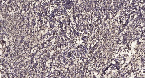
| Sample | Paraffin-embedded human meningioma tissue; fixative not specified (datasheet A30832) |
| Fixation | Image fixative and duration unreported (datasheet A30832); verify before use. |
| Sectioning | 4–5 µm sections on charged slides (standard) |
| Deparaffinisation | Xylene, graded ethanol series to water (standard) |
| Antigen retrieval | Heat retrieval: Tris-EDTA pH 9.0 (datasheet A30832); 20 min, 95–100 °C (standard) |
| Peroxidase block | 3% H2O2, 10 min, room temperature (standard) |
| Blocking | 10% normal serum of the secondary host, 30 min, room temperature (standard) |
| Primary antibody | Rabbit anti-ADGRL1, 1:50 recommended; image 1:200 (datasheet A30832) |
| Primary incubation | Overnight at 4 °C (standard) |
| Detection | HRP-polymer secondary, DAB chromogen 5–10 min (standard) |
| Counterstain | Hematoxylin, blue, dehydrate and mount (standard) |
| Expected result | ADGRL1-positive staining in glandular cells of breast (HPA tissue IHC: High). HPA tissue profile: Membranous and cytoplasmic expression in several tissues, including cerebral cortex. No signal in the no-primary control. |
ADGRL1 is a seven-pass membrane receptor reported at the cell surface, axons, growth cones and presynaptic sites (UniProt O94910 localization and topology). In paraffin-section IHC, look for membranous staining, with possible cytoplasmic staining, in HPA-reported positive cell populations (HPA: tissue IHC profile). Treat tissue predictions cautiously: HPA rates the IHC pattern Uncertain because antibody staining and RNA expression have low consistency (HPA: reliability).
| Membranous staining, with some cytoplasmic staining, in breast or duodenal glandular cells. | This matches the reported compartment pattern and cells scored High in those tissues (HPA: tissue IHC). Membrane localization is biologically plausible for a seven-pass receptor (UniProt O94910 topology). A matching image supports interpretation but is not definitive validation because HPA rates the IHC evidence Uncertain (HPA: reliability). |
| Low staining in cerebral cortex neuropil or cerebellar Purkinje cells. | A weak result can fit the reported IHC pattern: both populations are scored Low (HPA: tissue IHC). Neuropil staining is compatible with the reported axonal and synaptic localization (UniProt O94910 localization). Avoid requiring strong neuronal staining solely because brain RNA is tissue enhanced; HPA reports low consistency between staining and RNA (HPA: RNA specificity and reliability). |
| Predominantly nuclear staining, without a discernible membrane-associated pattern. | Nuclear localization is outside the supplied ADGRL1 localization record (UniProt O94910 localization; HPA: tissue IHC profile). Consider nonspecific staining or detection artefact, especially if the pattern persists in controls (general IHC practice). Cytoplasmic staining alone is less decisive because HPA also reports a cytoplasmic component (HPA: tissue IHC profile). |
| Strong signal in adipocytes or bone-marrow hematopoietic cells. | These populations are scored Not detected (HPA: tissue IHC). Check cross-reactivity and endogenous detection activity with appropriate controls (general IHC practice). Their HPA-negative status is a warning sign, not proof that every stained cell is false positive, given the Uncertain antibody-level IHC assessment (HPA: HPA037974 IHC; HPA: reliability). |
| Diffuse chromogen across cells and tissue spaces, or no signal in a section expected to stain. | Diffuse background obscures the reported membranous and cytoplasmic distribution (HPA: tissue IHC profile). An absent signal in an HPA High population warrants a technical check, but one negative section cannot establish absent ADGRL1: the tissue calls have Uncertain reliability (HPA: positive tissue list and reliability). |
| Membrane topology and compartment | Seven transmembrane segments support membrane-associated interpretation (UniProt O94910 topology). HPA also reports cytoplasmic staining, so score membrane and cytoplasmic components separately rather than treating every cytoplasmic signal as an artefact (HPA: tissue IHC profile). |
| Extracellular processing and unknown antibody epitope | ADGRL1 forms a heterodimer with a large extracellular region non-covalently linked to a seven-transmembrane moiety (UniProt O94910 subunit). The supplied record does not locate the IHC antibody epitope; do not infer which moiety a stained section detects or interpret a negative result as loss of one moiety. |
| Tissue selection and evidence strength | Breast glandular cells and kidney tubule cells are scored High, whereas adipocytes and bone-marrow hematopoietic cells are Not detected (HPA: tissue IHC). These provide comparison regions, subject to HPA's Uncertain reliability and low staining–RNA consistency (HPA: reliability). |
| Antigen retrieval | Use a controlled retrieval condition and compare it across sections as general paraffin-IHC practice. No ADGRL1-specific retrieval requirement or fixation sensitivity is established by the supplied UniProt and HPA records; do not assign a tissue result to retrieval alone. |
| Situation | Likely cause | Next action |
|---|---|---|
| No signal in breast or duodenal glandular cells. | These cells are scored High, so failed staining or detection is possible; HPA's IHC reliability is Uncertain (HPA: tissue IHC and reliability). | Check section integrity, retrieval, primary incubation and detection with a run control; compare another HPA High population before calling ADGRL1 absent (general IHC practice; HPA: positive tissue list). |
| Only diffuse background is visible. | Nonlocalized chromogen may reflect incomplete blocking, washing or endogenous detection activity (general IHC practice). | Review blocking and washes, and run a no-primary control; judge any remaining signal against the reported membrane and cytoplasmic profile (general IHC practice; HPA: tissue IHC profile). |
| Staining is mainly nuclear. | The supplied localization places ADGRL1 at membranes, projections and synaptic sites, not nuclei (UniProt O94910 localization). | Compare a no-primary control and examine whether membrane-associated staining survives background reduction before scoring the result as ADGRL1 (general IHC practice; UniProt O94910 localization). |
| Strong signal appears in an HPA Not detected cell population. | Cross-reactivity or endogenous detection activity is possible; the reference tissue calls are themselves uncertain (general IHC practice; HPA: tissue IHC and reliability). | Check controls and compare cell types within the same section; report the discrepancy rather than treating the HPA negative call as absolute (general IHC practice; HPA: reliability). |
| Cerebral cortex neuropil looks weaker than a glandular comparator. | Neuropil is scored Low, while several glandular populations are scored High (HPA: tissue IHC). | Score each cell population against its own reported level; do not raise chromogen development solely to force equally strong staining (general IHC practice; HPA: tissue IHC). |
| Can an ICC/IF membrane image confirm this paraffin-IHC result? | HPA lists a membrane summary but no available main-location assignment or ICC/IF cell-line images (HPA: subcellular record). | Use membrane localization as a biological expectation (UniProt O94910 localization); assess ICC/IF on its separate guide page and do not treat the supplied HPA record as image-based ICC/IF validation. |


Comprehensive Human Protein Atlas IHC scoring per tissue (reliability: Uncertain — Low consistency between antibody staining and RNA expression data.). Rows are taken directly from the HPA tissue chart — click any row's HPA link to view the source.
| Tissue | Cell type | Level | Evidence | Source |
|---|---|---|---|---|
| Breast | Glandular cells | High | Protein (IHC) | HPA → |
| Duodenum | Glandular cells | High | Protein (IHC) | HPA → |
| Endometrium | Glandular cells | High | Protein (IHC) | HPA → |
| Fallopian tube | Glandular cells | High | Protein (IHC) | HPA → |
| Kidney | Cells in tubules | High | Protein (IHC) | HPA → |
| Tissue | Cell type | Level | Evidence | Source |
|---|---|---|---|---|
| Adipose tissue | Adipocytes | Not detected | Protein (IHC) | HPA → |
| Bone marrow | Hematopoietic cells | Not detected | Protein (IHC) | HPA → |
| Caudate | Glial cells | Not detected | Protein (IHC) | HPA → |
| Esophagus | Squamous epithelial cells | Not detected | Protein (IHC) | HPA → |
| Heart muscle | Cardiomyocytes | Not detected | Protein (IHC) | HPA → |
Troubleshoot ADGRL1 staining in paraffin sections by checking retrieval, cellular pattern, controls and scoring against the documented IHC conditions (caption A30832).
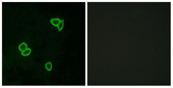
A30832 has an IHC image from paraffin-embedded human meningioma and an IF image from LOVO cells (catalog image captions); its listed reactivity covers human, mouse and rat (catalog reactivity).
A30832 lists IHC and shows staining of paraffin-embedded human meningioma (catalog applications; IHC image caption). A30832 also has an IF image of LOVO cells, although IF is absent from its listed applications (IF image caption; catalog applications).
Which to pick: Choose A30832 for paraffin-section IHC because its own caption documents that preparation; the fixative is unreported (A30832 IHC image caption). For IF/ICC, A30832 offers an IF image in LOVO cells, but IF/ICC is not a listed application (A30832 IF image caption; catalog applications). A30832 is the only cross-species candidate here: this rabbit polyclonal lists human, mouse and rat reactivity, while its IHC image documents a human sample only (catalog host, clonality and reactivity; A30832 IHC image caption).