AIFM2 / Ferroptosis suppressor protein 1 · IHC design guide

Design Immunohistochemistry for AIFM2

Plan AIFM2 staining in paraffin sections using cytoplasmic expression and high staining in spleen red-pulp cells as tissue references (HPA tissue IHC). Start the IHC-validated antibody A06541-2 at 1:50 (datasheet: A06541-2), and include a no-primary control (standard IHC practice).

Evidence assembled Oct 2026 · For research use; verify linked source records and product datasheet before use
Immunohistochemistry protocol sheet for AIFM2 (IHC for AIFM2): expected localisation Observed cytoplasmic staining (HPA tissue IHC); membrane and nuclear locations are annotated (UniProt), antibody A06541-2, validated IHC image, and IHC protocol steps
Printable AIFM2 IHC protocol sheet — expected localisation Observed cytoplasmic staining (HPA tissue IHC); membrane and nuclear locations are annotated (UniProt), antibody A06541-2, controls and protocol steps. Open the full AIFM2 IHC guide →

AIFM2 Immunohistochemistry Experimental Design Guide

Expected localisation, validated protocols, controls and antibodies — the at-a-glance facts below, then the full design guide.

Must know before staining
Expected localisation Observed cytoplasmic staining (HPA tissue IHC); membrane and nuclear locations are annotated (UniProt)
Staining pattern Cytoplasmic staining in several tissues, including red-pulp cells (HPA tissue IHC)
Antigen retrieval EDTA pH 8.0 HIER, heat-mediated (datasheet A06541-2)
Positive control ⓘ Spleen+4 more · see all
Negative control ⓘ Adipose tissue+4 more · see all
Important caveats
Reasons your staining may differ from the expected pattern.
Fixation Keep paraffin-section fixation consistent (standard IHC practice; not target-specific)
Caveat Antibody staining has low consistency with RNA expression (HPA tissue IHC)
Regulation TP53 induces AIFM2 transcripts (UniProt)
Isoform / epitope 2 isoforms; confirm antibody epitope coverage (UniProt)
Section 1

Recommended AIFM2 IHC & IF Protocols

The catalog antibody’s IHC-P protocol is paired with published AIFM2 IHC methods for cervical tumor samples (PMC10195139) and mouse kidney (PMC7893796).

Recommended immunohistochemistry (IHC-P) protocol parameters
SampleParaffin-embedded human liver cancer tissue; fixative not specified (datasheet A06541-2)
FixationImage fixative and duration unreported (datasheet A06541-2); verify before use.
Sectioning4–5 µm sections on charged slides (standard)
DeparaffinisationXylene, graded ethanol series to water (standard)
Antigen retrievalHeat retrieval: EDTA pH 8.0 (datasheet A06541-2); 20 min, 95–100 °C (standard)
Peroxidase block3% H2O2, 10 min, room temperature (standard)
Blocking10% goat serum (datasheet A06541-2)
Primary antibodyRabbit anti-AIFM2, 1:50 recommended; image 2 μg/ml (datasheet A06541-2)
Primary incubationOvernight at 4 °C (datasheet A06541-2)
DetectionHRP-conjugated secondary, DAB chromogen (datasheet A06541-2)
CounterstainHematoxylin, blue, dehydrate and mount (standard)
Expected resultAIFM2-positive staining in cells in red pulp of spleen (HPA tissue IHC: High). HPA tissue profile: Cytoplasmic expression in several tissues. No signal in the no-primary control.
💡Decision noteStart with heat-mediated EDTA at pH 8.0 for the catalog antibody (datasheet A06541-2); citrate at pH 6.0 is a published alternative (PMC7893796).
Section 2

What Is the Expected AIFM2 Staining Pattern?

AIFM2 is expected mainly in the cytoplasm on tissue IHC, with the strongest reported staining in spleen red pulp cells and medium staining in selected hematopoietic, alveolar, glandular and spermatogonial cells (HPA tissue IHC). Membrane association is biologically plausible given its N-terminal transmembrane segment at residues 7–27 (UniProt Q9BRQ8 topology). HPA rates the tissue IHC profile Approved but reports low consistency with RNA expression (HPA tissue IHC).

What am I looking at on my slide?
Distinct cytoplasmic staining in spleen red pulp cells; weaker staining in other reported positive cell populations (HPA tissue IHC).This best matches the observed tissue profile: red pulp cells are High; bone marrow hematopoietic cells, lung alveolar cells, small-intestinal glandular cells and testis spermatogonia are Medium (HPA tissue IHC). Compare intensity within the same staining run; these categories are observations, not universal scoring thresholds.
Predominantly nuclear staining in an otherwise routine tissue section.Recheck specificity and scoring before calling this the expected IHC pattern: HPA describes cytoplasmic tissue expression (HPA tissue IHC). UniProt lists nuclear localization and stress-associated translocation, so nuclear signal is not inherently impossible, but the supplied tissue IHC data do not establish it as the routine positive pattern (UniProt Q9BRQ8).
Strong signal in a cell population reported as Not detected, such as adipocytes in adipose tissue (HPA tissue IHC).Treat a discordant cell-type pattern as a specificity or detection-control question. Cross-reactivity or endogenous chromogenic activity are possible explanations under standard IHC practice; one negative HPA observation alone does not prove the cells lack AIFM2 in every specimen (HPA tissue IHC).
Diffuse chromogen across cells and extracellular areas, with little compartment or cell-type distinction.This does not reproduce the cytoplasmic, cell-selective HPA profile (HPA tissue IHC). Under standard chromogenic IHC practice, excess antibody, insufficient blocking or residual endogenous detection activity can obscure genuine staining; inspect control sections before scoring.
No detectable signal in spleen red pulp cells (HPA: High).A failed positive reference weakens interpretation of negative test sections. First assess tissue integrity and the IHC run, then review antibody dilution, antigen retrieval and detection using standard IHC controls; the supplied sources give no AIFM2-specific retrieval condition or working dilution.
💡Expected AIFM2 appearanceCall the run positive when spleen red pulp cells show distinct, relatively strong cytoplasmic chromogen, with an appropriate low-background control; diffuse staining or strong signal in HPA Not detected cell types is suspect (HPA tissue IHC; standard IHC practice).
How each factor affects the staining
Observed tissue distribution and evidence strengthHPA reports cytoplasmic staining in several tissues, including High red pulp cells, while rating the IHC profile Approved with low consistency between staining and RNA data (HPA tissue IHC). Use the listed cell populations as observed references, not an absolute expression map.
Membrane association and compartment scoringUniProt lists cell, mitochondrial and lipid-droplet membranes plus cytoplasm and nucleus, and annotates a transmembrane segment at residues 7–27 (UniProt Q9BRQ8). These locations support biological plausibility; HPA tissue IHC supplies the practical cytoplasmic expectation for paraffin sections.
Transcript and protein evidenceUniProt reports two transcripts and 2 isoforms (UniProt Q9BRQ8). HPA reports low tissue RNA specificity but cell-selective IHC staining and low RNA–staining consistency (HPA tissue IHC). Neither source supplies an isoform-specific tissue IHC pattern.
Stress-associated localizationUniProt describes nuclear translocation after oxidative stress and interaction with nuclear-import proteins (UniProt Q9BRQ8). Record experimental stress context before interpreting nuclear signal; the supplied HPA tissue IHC profile remains cytoplasmic, so nuclear staining needs independent validation.
Antibody validation scopeThe listed tissue-IHC antibody HPA042309 has Approved IHC status; HPA028961 and HPA075576 have Approved ICC status, with no IHC status supplied for them (HPA antibodies). Those labels do not establish interchangeability across IHC and IF/ICC.
Why is my staining missing, weak or wrong?
SituationLikely causeNext action
Spleen red pulp is blank despite an otherwise clean run (HPA: High).A technical failure is possible under standard IHC practice; the HPA reference alone cannot identify whether retrieval, antibody concentration or detection is responsible.Check a same-run positive reference and detection controls, then review retrieval and titrate the IHC-validated antibody according to its verified IHC instructions; no target-specific setting is supplied here.
The entire section is brown, including reported negative cells (HPA tissue IHC).Diffuse background can arise from excess primary antibody, inadequate blocking or endogenous enzyme activity in chromogenic IHC (standard IHC practice).Compare no-primary and detection-only controls; optimize blocking, washes and antibody concentration before assigning AIFM2-positive cells (standard IHC practice).
Adipocytes stain strongly while spleen red pulp cells do not (HPA tissue IHC).This reverses the supplied reference pattern: adipocytes are Not detected in adipose tissue, whereas red pulp cells are High (HPA tissue IHC). Cross-reactivity or a run problem is possible.Repeat with positive and negative reference sections in the same run, inspect localization, and require independent support before interpreting the adipocyte signal.
Signal appears mainly nuclear in an unstressed tissue section.HPA tissue IHC describes cytoplasmic expression; UniProt allows nuclear localization and describes stress-associated translocation, without establishing routine nuclear IHC staining (HPA tissue IHC; UniProt Q9BRQ8).Document the sample context, check background controls, and avoid scoring nuclei alone as a routine positive result without independent validation.
Lung or small intestine looks weaker than spleen.That ranking can fit the supplied observations: alveolar and small-intestinal glandular cells are Medium, while spleen red pulp cells are High (HPA tissue IHC).Compare the specified cell populations under the same run conditions. Score their compartment and local background before treating lower intensity as assay failure (HPA tissue IHC; standard IHC practice).
Can IF/ICC images guide the expected IHC pattern?HPA ICC-IF places AIFM2 mainly in vesicles, with additional plasma-membrane and cytosolic localization; HPA tissue IHC describes cytoplasmic expression (HPA subcellular; HPA tissue IHC).Use IF/ICC as a compartment comparison only. Interpret paraffin-section chromogen against the tissue-IHC pattern and IHC controls; the listed ICC approvals do not establish IHC performance (HPA antibodies; standard IHC practice).

Sample controls for AIFM2 IHC & IF

🧪Run spleen first: cells in the red pulp should stain (HPA: High in spleen red-pulp cells). Use adipose tissue as the negative tissue: adipocytes should remain unstained (HPA: Not detected in adipocytes); on the spleen slide, cells without specific signal should show only counterstain and no DAB deposit, but HPA does not identify a validated negative spleen cell type (HPA: High in red-pulp cells).
Positive control tissue: Spleen (Cells in red pulp, HPA High)
Negative control tissue: Adipose tissue (HPA Not detected)
ICC-IF cell lines (HPA subcellular resource): HPA ICC-IF images show AIFM2 in A-549, Hep-G2, U2OS, with annotated localisation: Vesicles (supported) (HPA subcellular).
Technical controls: Include a no-primary, secondary-only slide, a concentration-matched nonimmune rabbit IgG control, and an AIFM2 knockout sample if available (caption: rabbit primary; standard IHC control practice). Block endogenous peroxidase and assess pigment in spleen red pulp before interpreting DAB signal (standard chromogenic IHC practice).
⚠️Feasibility: A target-specific fixation window or fixation effect is unreported, and the selected A06541-2 tissue-IHC caption does not state a fixative (caption: fixative not stated). The caption uses heat retrieval in EDTA at pH 8.0, but does not establish that retrieval is required (caption: EDTA retrieval). Frozen sections are not established as easier; HPA provides ICC-IF images in A-549, Hep-G2 and U2OS cells, with mainly vesicular localization, but these do not establish an easier IF tissue workflow (HPA subcellular: ICC-IF images and supported vesicular localization).

HPA tissue IHC evidence for AIFM2

Comprehensive Human Protein Atlas IHC scoring per tissue (reliability: Approved — Low consistency between antibody staining and RNA expression data.). Rows are taken directly from the HPA tissue chart — click any row's HPA link to view the source.

Positive expression · recommended positive controls

TissueCell typeLevelEvidenceSource
Spleen Cells in red pulp High Protein (IHC) HPA →
Bone marrow Hematopoietic cells Medium Protein (IHC) HPA →
Lung Alveolar cells Medium Protein (IHC) HPA →
Small intestine Glandular cells Medium Protein (IHC) HPA →
Stomach Glandular cells Low Protein (IHC) HPA →

Undetected expression · recommended negative controls

TissueCell typeLevelEvidenceSource
Adipose tissue Adipocytes Not detected Protein (IHC) HPA →
Appendix Glandular cells Not detected Protein (IHC) HPA →
Breast Adipocytes Not detected Protein (IHC) HPA →
Bronchus Respiratory epithelial cells Not detected Protein (IHC) HPA →
Cerebellum Cells in granular layer Not detected Protein (IHC) HPA →
Section 3

Advanced AIFM2 IHC Tips

These questions address AIFM2 staining in paraffin sections by chromogenic IHC, with one note on the separate IF/ICC application.

What retrieval should I use when AIFM2 staining is weak in paraffin sections?
Start with heat-mediated retrieval in EDTA at pH 8.0 for this IHC-validated antibody (datasheet A06541-2). The selected paraffin-section image used that retrieval before staining human liver cancer tissue, but its caption does not state the fixative (datasheet A06541-2). Keep section thickness, heating, cooling, and detection conditions consistent while comparing retrieval runs (standard IHC practice). If staining remains weak, assess a small retrieval-condition series alongside a positive control; record any alternative buffer as a fallback condition, not the documented method (standard IHC practice; datasheet A06541-2). Judge improvement by cellular staining and tissue preservation together, since stronger DAB alone can reflect excess background (standard IHC practice).
Could fixation explain inconsistent AIFM2 staining between specimens?
Target-specific fixation sensitivity is unknown: the selected paraffin-section caption does not report its fixative (datasheet A06541-2). Record each specimen’s fixation and processing history, then compare sections processed alike before attributing differences to AIFM2 abundance (standard IHC practice). Use the documented EDTA pH 8.0 retrieval and keep antibody concentration and detection consistent during that comparison (datasheet A06541-2; standard IHC practice). The selected image used 2 μg/ml primary antibody overnight at 4°C, providing a documented staining condition, not a fixation study (datasheet A06541-2). Include a known staining control in each run and inspect morphology for processing damage (standard IHC practice).
How should I assess membrane, cytoplasmic, or nuclear AIFM2 staining?
Score the compartment visible in each cell rather than treating every DAB deposit as equivalent AIFM2 signal (standard IHC practice). UniProt lists lipid droplets, cell membrane, cytoplasm, mitochondrial membrane, and nucleus for AIFM2, while HPA tissue IHC describes cytoplasmic expression in several tissues (UniProt Q9BRQ8 subcellular; HPA tissue IHC profile). Its annotated transmembrane segment spans residues 7–27, but that annotation alone does not establish which compartment an IHC antibody will highlight (UniProt Q9BRQ8 topology; standard IHC interpretation). Review membrane-associated, granular cytoplasmic, and nuclear patterns separately against controls and intact morphology (standard IHC practice). Nuclear staining can be biologically plausible under oxidative stress, but staining alone cannot establish that mechanism (UniProt Q9BRQ8 subunit and function).
Can this antibody distinguish AIFM2 isoforms or reveal its membrane-facing epitope?
Do not assign isoform specificity from a chromogenic staining pattern: UniProt records 2 AIFM2 isoforms, and the supplied caption does not identify the antibody epitope (UniProt Q9BRQ8 isoforms; datasheet A06541-2). UniProt annotates a transmembrane segment at residues 7–27 and a chain spanning residues 2–373; neither locates this antibody’s binding site (UniProt Q9BRQ8 topology and processing). It also records an acetylation example at residue 168, without evidence that this modification changes staining with this antibody (UniProt Q9BRQ8 modified residues; datasheet A06541-2). Obtain an epitope map or isoform-specific validation before making either claim (standard antibody-validation practice). Until then, report the observed compartment and staining conditions without assigning an isoform or membrane-facing orientation (standard IHC practice).
How can I check an AIFM2 pattern by multiplex IF?
On the separate IF/ICC guide, pair AIFM2 with a validated marker for the cell population being examined; HPA reports staining in lung alveolar cells and small-intestinal glandular cells (HPA tissue IHC positive-cell entries; standard IF practice). Select fluorophores after checking autofluorescence in the actual tissue, and favor a spectrally separated, longer-wavelength channel when background is troublesome (standard IF practice). Plan permeabilisation around the antibody’s verified epitope and its side of the membrane; the epitope is not specified here despite AIFM2’s residues 7–27 transmembrane annotation (datasheet A06541-2; UniProt Q9BRQ8 topology). Include single-label and secondary-only controls to assess bleed-through and background (standard IF practice). Treat apparent colocalisation as an imaging observation, not proof of molecular interaction (standard IF interpretation).
What should I check when DAB background obscures AIFM2 staining?
First inspect a no-primary control and the distribution of DAB in intact tissue; widespread deposition can arise from detection chemistry or incomplete washing (standard chromogenic IHC practice). Include a peroxidase-blocking step when using HRP and DAB, and verify that the block itself does not damage morphology (standard chromogenic IHC practice). The selected paraffin-section image used 10% goat serum, 2 μg/ml rabbit primary antibody overnight at 4°C, and DAB development (datasheet A06541-2). If background persists, titrate the primary and detection conditions while holding retrieval at the documented EDTA pH 8.0 starting condition (standard IHC practice; datasheet A06541-2). Compare cellular signal with controls before accepting a darker stain as greater AIFM2 expression (standard IHC practice).
How should I quantify AIFM2 across paraffin sections? ⚠ ANSWER MARKED FOR VERIFICATION
Define the cell population and compartment before scoring, because HPA tissue IHC reports cytoplasmic expression across several tissues (HPA tissue IHC profile; standard IHC practice). For a cellular readout, report the percentage of positive cells and an H-score based on staining intensity; for spatial comparisons, report positive-cell density per mm² of viable tissue (standard IHC quantification practice). Normalize counts to the number of evaluable cells or viable tissue area, and use the same threshold, regions, and imaging settings across groups (standard IHC quantification practice). Exclude necrosis and section folds during annotation, and document how thresholds and exclusions were chosen (standard IHC quantification practice). Keep compartment-specific results separate if both cytoplasmic and nuclear staining are present (UniProt Q9BRQ8 subcellular; standard IHC practice).
How can I distinguish credible AIFM2 staining from artefact?
Look for reproducible cellular staining in intact regions that matches an annotated AIFM2 compartment, then compare it with no-primary and tissue controls (UniProt Q9BRQ8 subcellular; standard IHC practice). HPA reports high staining in spleen red-pulp cells and no detected staining in adipose-tissue adipocytes, although its tissue-IHC reliability note cites low consistency with RNA expression (HPA tissue IHC positive and negative entries; HPA reliability description). Be cautious with stain concentrated at section edges, necrotic areas, or sites of endogenous peroxidase activity (standard chromogenic IHC practice). Review unexpectedly exclusive staining in an unannotated compartment or cell population against morphology and controls before calling it positive (UniProt Q9BRQ8 subcellular; standard IHC interpretation). DAB localisation alone cannot establish AIFM2 enzymatic activity or ferroptosis suppression (UniProt Q9BRQ8 function; standard IHC interpretation).
Boster reagents

Best AIFM2 / Ferroptosis suppressor protein 1 IHC Antibodies

The catalog shows real IHC images for human paraffin sections with A06541 and A06541-2; it lists IF for A06541 and M06541 without IF images (catalog applications and image captions).

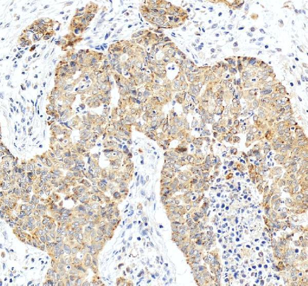
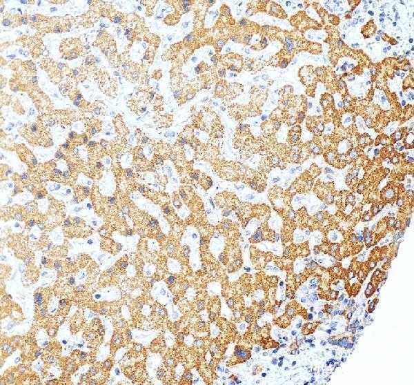
Real IHC data IHC analysis of FSP1/AIFM2 using anti-FSP1/AIFM2 antibody (A06541-2). FSP1/AIFM2 was detected in a paraffin-embedded section of human liver cancer tissue. Heat mediated antigen retrieval was performed in EDTA buffer (pH 8.0, epitope retrieval solution). The tissue section was blocked with 10% goat serum. The tissue section was then incubated with 2 μg/ml rabbit anti-FSP1/AIFM2 Antibody (A06541-2) overnight at 4°C. Peroxidase Conjugated Goat Anti-rabbit IgG was used as secondary antibody and incubated for 30 minutes at 37°C. The tissue section was developed using HRP Conjugated Rabbit IgG Super Vision Assay Kit (Catalog # SV0002) with DAB as the chromogen.
Anti-FSP1/AIFM2 Antibody
Cat # A06541-2
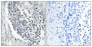
Real IHC data Immunohistochemical analysis of paraffin-embedded Human lung cancer. Antibody was diluted at 1:100 (4° overnight). High-pressure and temperature Tris-EDTA, pH8.0 was used for antigen retrieval. Negetive contrl (right) obtaned from antibody was pre-absorbed by immunogen peptide.
Anti-AMID AIFM2 Antibody
Cat # A06541

A06541 will render with an IHC image of paraffin-embedded human lung cancer (A06541 image caption). A06541-2 will render with an IHC image of a paraffin-embedded human liver cancer section (A06541-2 image caption).

Which to pick: For tissue IHC, choose A06541-2 for the human liver cancer example or A06541 for the human lung cancer example; both captions specify paraffin sections, but neither reports the fixative (A06541-2 and A06541 image captions). For IF/ICC, M06541 is a rabbit monoclonal listed for both applications, while A06541 is listed for IF; neither has an IF image in the payload (catalog applications and IF image fields). For cross-species work, A06541 lists human, monkey and mouse reactivity, while M06541 lists human, mouse and rat reactivity (catalog reactivity fields).

Each figure is that product's own IHC / IF validation image from its datasheet.

References

  1. UniProt Consortium. UniProt entry Q9BRQ8 (FSP1_HUMAN, Ferroptosis suppressor protein 1).
  2. Human Protein Atlas. AIFM2 tissue IHC expression (reliability: Approved).
  3. Human Protein Atlas. AIFM2 subcellular location (ICC-IF): Mainly localized to vesicles. In addition localized to the plasma membrane and cytosol..
  4. Human Protein Atlas. AIFM2 antibody validation summary (3 antibodies).
  5. IMPA2 blocks cervical cancer cell apoptosis and induces paclitaxel resistance through p53-mediated AIFM2 regulation. Acta biochimica et biophysica Sinica 2023 — PMC10195139.
  6. ATF6 aggravates acinar cell apoptosis and injury by regulating p53/AIFM2 transcription in Severe Acute Pancreatitis. Theranostics 2020 — PMC7381726.
  7. Metabolomic profiling identifies a novel mechanism for heat stroke‑related acute kidney injury. Molecular medicine reports 2021 — PMC7893796.
  8. Characterization of the proteome of stable and unstable carotid atherosclerotic plaques using data-independent acquisition mass spectrometry. Journal of translational medicine 2024 — PMC10921703.
  9. PubMed PMID:12135761 — UniProt-cited evidence.
  10. PubMed PMID:11980907 — UniProt-cited evidence.
  11. PubMed PMID:14702039 — UniProt-cited evidence.