This website uses cookies to ensure you get the best experience on our website.
- Table of Contents
3 Q&As


Facts about Autoimmune regulator.
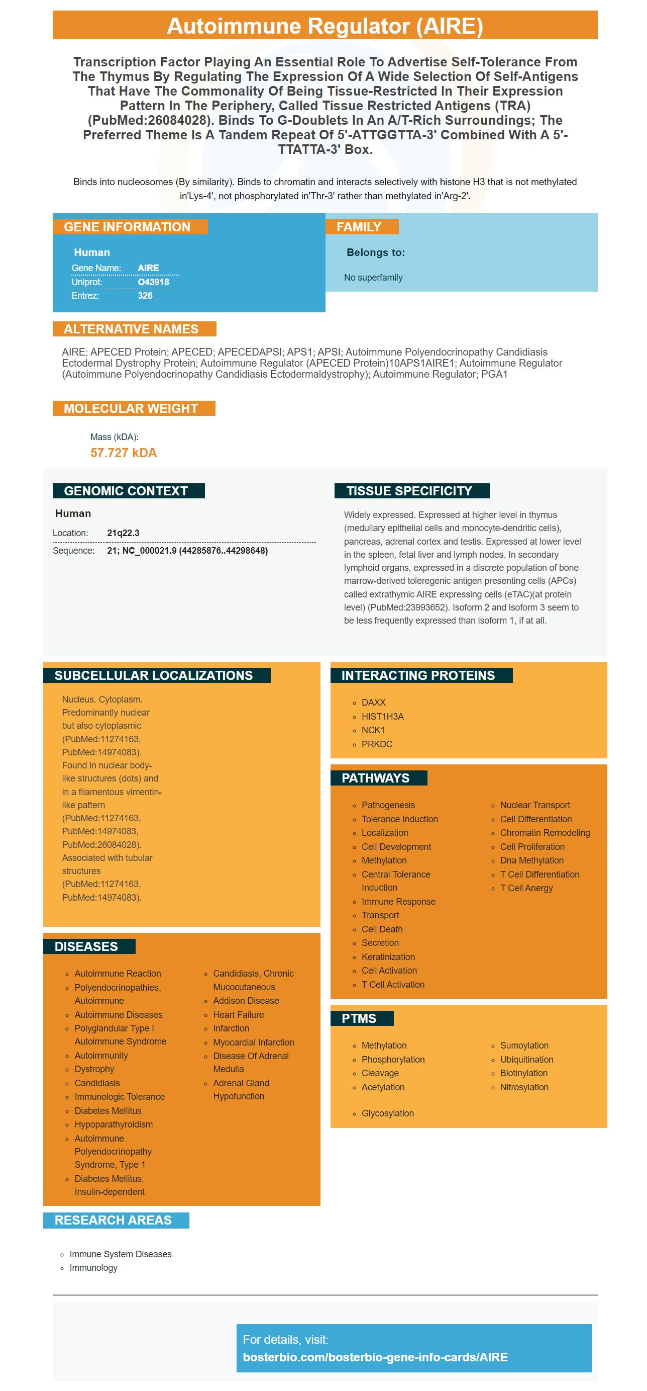
Binds into nucleosomes (By similarity). Binds to chromatin and interacts selectively with histone H3 that is not methylated in'Lys-4', not phosphorylated in'Thr-3' rather than methylated in'Arg-2'.
| Human | |
|---|---|
| Gene Name: | AIRE |
| Uniprot: | O43918 |
| Entrez: | 326 |

| Belongs to: |
|---|
| No superfamily |

AIRE; APECED protein; APECED; APECEDAPSI; APS1; APSI; Autoimmune polyendocrinopathy candidiasis ectodermal dystrophy protein; autoimmune regulator (APECED protein)10APS1AIRE1; autoimmune regulator (autoimmune polyendocrinopathy candidiasis ectodermaldystrophy); Autoimmune Regulator; PGA1
Mass (kDA):
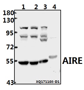
57.727 kDA

| Human | |
|---|---|
| Location: | 21q22.3 |
| Sequence: | 21; NC_000021.9 (44285876..44298648) |
Widely expressed. Expressed at higher level in thymus (medullary epithelial cells and monocyte-dendritic cells), pancreas, adrenal cortex and testis. Expressed at lower level in the spleen, fetal liver and lymph nodes. In secondary lymphoid organs, expressed in a discrete population of bone marrow-derived toleregenic antigen presenting cells (APCs) called extrathymic AIRE expressing cells (eTAC)(at protein level) (PubMed:23993652). Isoform 2 and isoform 3 seem to be less frequently expressed than isoform 1, if at all.
Nucleus. Cytoplasm. Predominantly nuclear but also cytoplasmic (PubMed:11274163, PubMed:14974083). Found in nuclear body-like structures (dots) and in a filamentous vimentin-like pattern (PubMed:11274163, PubMed:14974083, PubMed:26084028). Associated with tubular structures (PubMed:11274163, PubMed:14974083).


PMID: 9398839 by Nagamine K., et al. Positional cloning of the APECED gene.
PMID: 9398840 by Aaltonen J., et al. An autoimmune disease, APECED, caused by mutations in a novel gene featuring two PHD-type zinc-finger domains.