This website uses cookies to ensure you get the best experience on our website.


- Table of Contents
Plan ALDH3A2 staining in paraffin sections using the observed cytoplasmic tissue pattern and the catalog antibody's IHC conditions (HPA tissue IHC; datasheet PB9982). Choose cell-specific controls and account for variation in glandular staining across tissues (HPA tissue IHC).

Expected localisation, validated protocols, controls and antibodies — the at-a-glance facts below, then the full design guide.
| Expected localisation | Cytoplasmic tissue staining; ER and microsome membrane protein (HPA tissue IHC; UniProt) | |
| Staining pattern | Cytoplasmic staining in glandular, epithelial and neuronal cells (HPA tissue IHC) | |
| Antigen retrieval | EDTA pH 8.0 HIER, heat-mediated (datasheet PB9982) | |
| Positive control | Adrenal gland+4 more · see all | |
| Negative control | Appendix+4 more · see all |
| Fixation | Keep fixation consistent across sections (standard IHC practice; not target-specific) | |
| Caveat | Glandular staining varies by tissue (HPA tissue IHC) | |
| Regulation | Liver-enhanced RNA expression (HPA tissue RNA) | |
| Isoform / epitope | 2 isoforms; map the epitope to the cytoplasmic region (UniProt) |
The catalog antibody protocol uses EDTA pH 8.0 retrieval (datasheet PB9982). The published protocols below cover ovarian, esophageal, and breast tissues (PMC3458927; PMC7935981; PMC12715930).
| Sample | Paraffin-embedded human liver cancer tissue; fixative not specified (datasheet PB9982) |
| Fixation | Image fixative and duration unreported (datasheet PB9982); verify before use. |
| Sectioning | 4–5 µm sections on charged slides (standard) |
| Deparaffinisation | Xylene, graded ethanol series to water (standard) |
| Antigen retrieval | Heat retrieval: EDTA pH 8.0 (datasheet PB9982); 20 min, 95–100 °C (standard) |
| Peroxidase block | 3% H2O2, 10 min, room temperature (standard) |
| Blocking | 10% goat serum (datasheet PB9982) |
| Primary antibody | Rabbit anti-ALDH3A2, 0.5-1μg/ml (datasheet PB9982) |
| Primary incubation | Overnight at 4 °C (datasheet PB9982) |
| Detection | Streptavidin-biotin complex (SABC), DAB chromogen (datasheet PB9982) |
| Counterstain | Hematoxylin, blue, dehydrate and mount (standard) |
| Expected result | ALDH3A2-positive staining in glandular cells of adrenal gland (HPA tissue IHC: High). HPA tissue profile: Cytoplasmic expression in several tissues. No signal in the no-primary control. |
ALDH3A2 is an endoplasmic reticulum and microsome membrane protein with a cytoplasmic region spanning residues 1–463 and one transmembrane segment at 464–484 (UniProt P51648 topology). In paraffin section IHC, expect cytoplasmic staining in selected glandular and epithelial cells; adrenal glandular and bronchial respiratory epithelial cells are reported as High (HPA tissue IHC). HPA rates the tissue profile Enhanced, with medium consistency between staining and RNA expression (HPA tissue IHC).
| Distinct cytoplasmic staining in adrenal glandular or bronchial respiratory epithelial cells. | These are the supplied High IHC examples (HPA tissue IHC). Cytoplasmic staining is consistent with the reported tissue profile and with an ER or microsome membrane protein whose large region faces the cytoplasm (HPA tissue IHC; UniProt P51648 topology). Chromogenic IHC alone does not resolve the ER. |
| Cytoplasmic staining in colon, duodenum or endometrial glandular cells, or in the listed neuronal cells. | A Medium result can fit the HPA observations: glandular cells in those three tissues, and neuronal cells in caudate or cerebral cortex and Purkinje cells in cerebellum (HPA tissue IHC). Judge the identified cell population rather than treating every cell in a section as a positive control. |
| Predominantly nuclear, sharply surface restricted, or extracellular deposits. | These do not match HPA's cytoplasmic tissue profile or its approved ER and cytosol ICC-IF locations (HPA tissue IHC; HPA subcellular). Check morphology and detection controls before calling them ALDH3A2; misplaced signal can reflect staining artefact (general IHC practice). |
| Strong signal in cells listed as Not detected, such as appendix glandular cells or bone marrow hematopoietic cells. | That conflicts with the supplied cell specific IHC observations (HPA tissue IHC). Cross-reactivity or endogenous detection activity is possible, but the image alone cannot identify the cause (general IHC practice). Confirm the cell identity and compare a detection control before assigning target expression. |
| Diffuse color across cells and tissue spaces, or no staining in a known positive section. | Neither gives a readable cell specific pattern (general IHC practice). Diffuse color warrants background and detection controls; a blank adrenal or bronchus section warrants checking section quality and the IHC run before interpreting a sample as negative (HPA tissue IHC; general IHC practice). |
| Cell and tissue choice | Adrenal glandular and bronchial respiratory epithelial cells provide High reference patterns; colon, duodenum and endometrial glandular cells are Medium (HPA tissue IHC). Appendix glandular and bone marrow hematopoietic cells are listed as Not detected; use the specified cell type when comparing sections (HPA tissue IHC). |
| Antibody validation | HPA lists HPA014769 as IHC Enhanced and CAB020692 as IHC Approved (HPA antibodies). These labels describe antibody validation; the tissue profile separately has Enhanced reliability and medium staining to RNA consistency (HPA tissue IHC). Compare patterns with that distinction in mind. |
| Topology and epitope interpretation | Residues 1–463 are cytoplasmic and 464–484 form the transmembrane segment (UniProt P51648 topology). No antibody epitope is supplied, so topology cannot establish epitope exposure or an ALDH3A2 specific antigen retrieval requirement. |
| Isoforms and liver evidence | UniProt lists 2 isoforms and reports protein detection in liver (UniProt P51648). HPA reports liver enhanced RNA specificity (HPA tissue IHC). Neither item supplies an isoform specific IHC pattern or a liver IHC intensity in this payload; do not assign either. |
| IF/ICC: What localization is expected? | HPA approves endoplasmic reticulum and cytosol localization in ICC-IF and lists images from OE19 and SiHa (HPA subcellular). This is a localization comparison only; it does not turn an IF/ICC method into an IHC-P protocol. |
| Situation | Likely cause | Next action |
|---|---|---|
| Adrenal glandular or bronchial respiratory epithelial cells show no clear signal. | A blank expected positive may reflect a failed IHC run or unsuitable section; it does not by itself establish absence of ALDH3A2 (HPA tissue IHC; general IHC practice). | Review tissue morphology, run controls and the catalog antibody's IHC-P instructions. Check the prescribed retrieval, primary antibody dilution and detection steps as general IHC practice; no ALDH3A2 specific retrieval effect is supplied. |
| Color is widespread with little cell definition. | Background from nonspecific binding or the detection system can obscure the cytoplasmic pattern (general IHC practice). | Compare an omission control, inspect blocking and washes, and review primary antibody concentration and detection exposure (general IHC practice). Reassess only cells whose boundaries remain interpretable. |
| Signal is strong in an HPA Not detected cell population. | Cross-reactivity or endogenous detection activity is possible; HPA's Not detected label is an observation for that named cell population, not proof that the whole organ must be blank (HPA tissue IHC; general IHC practice). | Verify cell identity and compare omission and detection controls. If chromogenic enzyme detection is used, assess its endogenous activity control as appropriate (general IHC practice). |
| Most color appears nuclear or extracellular. | The dominant compartment disagrees with HPA's cytoplasmic tissue profile and approved ER and cytosol ICC-IF locations (HPA tissue IHC; HPA subcellular). | Inspect morphology and background controls, then compare a section containing an HPA High cell population. Do not score compartment mismatches as a confirmed ALDH3A2 positive (general IHC practice). |
| A liver section stains, but its intensity seems inconsistent with other tissues. | UniProt reports liver protein and HPA reports liver enhanced RNA specificity, but the supplied HPA IHC payload gives no liver staining level (UniProt P51648; HPA tissue IHC). | Record the observed liver cell type, compartment and intensity without assigning an expected IHC grade. Use the listed High IHC cell populations for an intensity comparison (HPA tissue IHC). |
| Two antibodies give different staining patterns. | HPA records different IHC validation statuses for HPA014769 and CAB020692; status alone does not explain a particular disagreement (HPA antibodies). | Compare the same named cell populations, compartment and run controls under each antibody's validated IHC conditions (HPA tissue IHC; general IHC practice). Treat discordant staining as unresolved until the pattern is supported. |

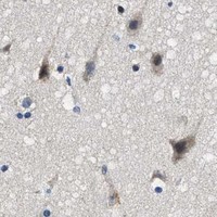


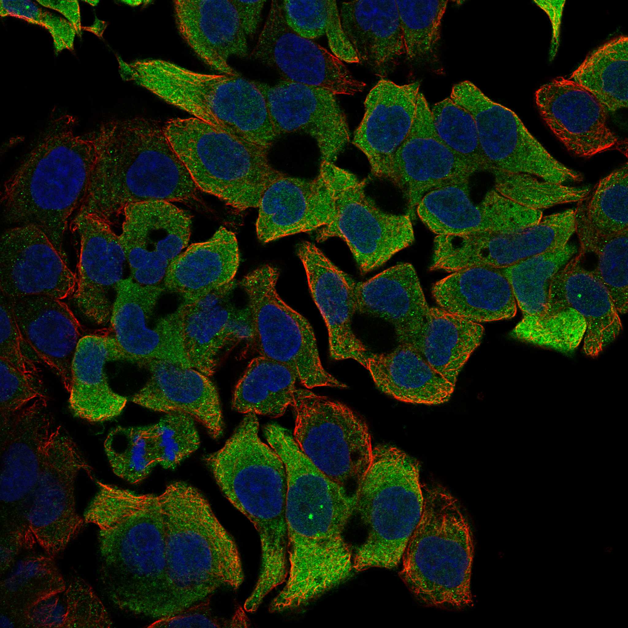
Comprehensive Human Protein Atlas IHC scoring per tissue (reliability: Enhanced — Medium consistency between antibody staining and RNA expression data.). Rows are taken directly from the HPA tissue chart — click any row's HPA link to view the source.
| Tissue | Cell type | Level | Evidence | Source |
|---|---|---|---|---|
| Adrenal gland | Glandular cells | High | Protein (IHC) | HPA → |
| Bronchus | Respiratory epithelial cells | High | Protein (IHC) | HPA → |
| Caudate | Neuronal cells | Medium | Protein (IHC) | HPA → |
| Cerebellum | Purkinje cells | Medium | Protein (IHC) | HPA → |
| Cerebral cortex | Neuronal cells | Medium | Protein (IHC) | HPA → |
| Tissue | Cell type | Level | Evidence | Source |
|---|---|---|---|---|
| Appendix | Glandular cells | Not detected | Protein (IHC) | HPA → |
| Bone marrow | Hematopoietic cells | Not detected | Protein (IHC) | HPA → |
| Breast | Adipocytes | Not detected | Protein (IHC) | HPA → |
| Cervix | Glandular cells | Not detected | Protein (IHC) | HPA → |
| Fallopian tube | Glandular cells | Not detected | Protein (IHC) | HPA → |
Troubleshoot ALDH3A2 staining in paraffin sections by checking retrieval, cellular distribution, controls and scoring before interpreting chromogenic signal.
PB9982 has real IHC data from a human liver cancer paraffin section (PB9982 image caption); the catalog lists human and rat reactivity, with no IF data (PB9982 catalog).
PB9982 is the sole rendered SKU and is listed for IHC and WB with human and rat reactivity (PB9982 catalog). Its IHC image shows staining in a human liver cancer paraffin section (PB9982 image caption).
Which to pick: Choose PB9982 for human tissue IHC in paraffin sections (PB9982 image caption); the fixative is unreported (PB9982 image caption). No listed SKU has IF/ICC validation or an IF image (PB9982 catalog). PB9982 lists rat reactivity, but the supplied IHC image documents human tissue only (PB9982 catalog; PB9982 image caption).