This website uses cookies to ensure you get the best experience on our website.


- Table of Contents
Plan ALKBH2 IHC-P using the catalog antibody at a starting concentration of 10 μg/mL (datasheet: A06325). Expect general nuclear staining and compare high-staining testis preleptotene spermatocytes with appendix glandular cells where staining was not detected (HPA tissue IHC).

Expected localisation, validated protocols, controls and antibodies — the at-a-glance facts below, then the full design guide.
| Expected localisation | Nuclear staining in tissue (HPA tissue IHC) | |
| Staining pattern | General nuclear staining across tissue cell types (HPA tissue IHC) | |
| Antigen retrieval | Tris-EDTA pH 9.0 HIER, 95–98 °C, 20 min (rule: nuclear antigen) | |
| Positive control | Adipose tissue+4 more · see all | |
| Negative control | Appendix+3 more · see all |
| Fixation | Keep fixation consistent across sections (standard IHC practice; not target-specific) | |
| Caveat | Appendix glandular cells may lack staining (HPA tissue IHC) | |
| Regulation | No expression regulation specified (UniProt) | |
| Isoform / epitope | 2 isoforms; epitope differences unspecified (UniProt) |
Compare the catalog antibody’s IHC-P protocol (datasheet) with a published colorectal tissue protocol (PMC7731553).
| Sample | Tissue sections; selected-image fixative not specified (standard IHC workflow) |
| Fixation | Image fixative and duration unreported (datasheet A06325); verify before use. |
| Sectioning | 4–5 µm sections on charged slides (standard) |
| Deparaffinisation | Xylene, graded ethanol series to water (standard) |
| Antigen retrieval | Heat-induced epitope retrieval in Tris-EDTA buffer, pH 9.0, 20 min at 95–98 °C (standard rule: nuclear antigen) |
| Peroxidase block | 3% H2O2, 10 min, room temperature (standard) |
| Blocking | 10% normal serum of the secondary host, 30 min, room temperature (standard) |
| Primary antibody | Rabbit anti-ALKBH2, 10 μg/mL (datasheet A06325) |
| Primary incubation | Overnight at 4 °C (standard) |
| Detection | HRP-polymer secondary, DAB chromogen 5–10 min (standard) |
| Counterstain | Hematoxylin, blue, dehydrate and mount (standard) |
| Expected result | ALKBH2-positive staining in adipocytes of adipose tissue (HPA tissue IHC: High). HPA tissue profile: General nuclear expression. No signal in the no-primary control. |
ALKBH2 is a nuclear protein found in the nucleoplasm and nucleolus, with relocation to replication foci during S phase; it has no transmembrane segment (UniProt Q6NS38). In paraffin sections, expect nuclear staining in selected cell populations, including adipocytes, bone marrow hematopoietic cells, melanocytes and preleptotene spermatocytes (HPA: High in each). The HPA tissue profile is Approved, with medium consistency between staining and RNA expression (HPA tissue IHC).
| Distinct nuclear staining in adipocytes, marrow hematopoietic cells, melanocytes or preleptotene spermatocytes (HPA: High in each). | This fits the reported general nuclear tissue pattern (HPA tissue IHC) and ALKBH2 localization to nucleoplasm and nucleolus (UniProt Q6NS38). Compare intensity within the identified cell population: HPA levels describe observed staining, not a required intensity for every cell or section. |
| Predominantly cytoplasmic or membrane-like staining, with little discernible nuclear signal. | Review as a possible staining artefact because UniProt places ALKBH2 in the nucleus and reports no transmembrane segment (UniProt Q6NS38). HPA reports cytosol and primary cilium as uncertain additional ICC-IF locations (HPA subcellular), so those observations alone do not establish an expected paraffin IHC pattern. |
| Strong signal in appendix glandular cells or pancreatic exocrine glandular cells, especially without nuclear contrast (HPA: Not detected in those cells). | Investigate possible antibody cross-reactivity or endogenous chromogenic activity (general IHC practice). HPA's result applies to the named cells in its sampled tissues; it is not proof that every cell in either organ must be negative (HPA tissue IHC). |
| Diffuse brown deposit across cells, connective tissue or empty areas, obscuring nuclear boundaries. | The distribution is difficult to score against ALKBH2's nuclear localization (UniProt Q6NS38; HPA tissue IHC). Uneven chromogen deposition, inadequate blocking or insufficient washing can produce background (general chromogenic IHC practice); inspect controls before assigning it to ALKBH2. |
| No nuclear signal in a technically evaluable section containing an HPA High cell population (HPA tissue IHC). | Check assay performance before calling ALKBH2 absent: HPA reports high staining in specific cell populations, not uniformly across their entire tissues (HPA tissue IHC). Confirm those cells are present, then compare a positive control processed in the same run (general IHC practice). |
| Tissue and cell selection (HPA tissue IHC) | HPA reports High staining in adipocytes, bone marrow hematopoietic cells, melanocytes and preleptotene spermatocytes, but Not detected in appendix glandular cells, lymph node germinal center cells, pancreatic exocrine glandular cells and spleen red-pulp cells (HPA tissue IHC). Select and score the named cell population, not the organ as a whole. |
| Nuclear localization and cell cycle (UniProt Q6NS38) | UniProt places ALKBH2 in nucleoplasm and nucleolus and reports relocation to replication foci during S phase (UniProt Q6NS38). Nuclear distribution can therefore vary among cells; a visible focus is not required to score an otherwise credible nuclear IHC signal. |
| Evidence and antibody validation (HPA tissue IHC; HPA antibodies) | The HPA tissue profile is Approved with medium staining-to-RNA consistency (HPA tissue IHC). CAB011198 is listed as IHC Approved, while the two listed rabbit antibodies have no IHC status in this payload (HPA antibodies). Interpret a discordant result alongside the antibody and tissue controls actually used. |
| IF/ICC Q&A: where should fluorescence localize? (HPA subcellular) | Mainly to the nucleoplasm: HPA calls that location supported (HPA subcellular). Its additional cytosol and primary-cilium assignments are uncertain, while UniProt also lists nucleolar localization (HPA subcellular; UniProt Q6NS38). This IF/ICC evidence informs compartment interpretation; it supplies no paraffin IHC protocol. |
| Situation | Likely cause | Next action |
|---|---|---|
| No signal in the intended positive cells (HPA: High in the named populations). | The expected cells may be absent from the section, or the staining run may have failed (HPA tissue IHC; general IHC practice). | Verify cell identity on the counterstained section and inspect a same-run positive control; then check primary antibody application, detection reagents and assay-validated retrieval steps (general IHC practice). |
| Nuclear signal is weak despite visible, intact target cells. | Low contrast can make a true nuclear pattern hard to separate from counterstain or background (general chromogenic IHC practice). HPA's High level does not guarantee that intensity in every preparation (HPA tissue IHC). | Compare positive and omission controls from the same run; review the chosen antibody dilution, detection development and counterstain against the validated assay settings (general IHC practice). |
| Brown staining dominates cytoplasm or cell edges. | This conflicts with the main nuclear assignment and absence of a transmembrane segment (UniProt Q6NS38). Background or cross-reactivity is possible (general IHC practice). | Check whether nuclei also show discrete signal, examine the primary-omission control, and reassess blocking and washes before scoring the extranuclear colour (general IHC practice). |
| HPA Not detected cell populations show strong chromogen. | The result is discordant for those specific cells and may reflect cross-reactivity or endogenous detection activity (HPA tissue IHC; general chromogenic IHC practice). | Confirm the cell type, review a primary-omission control and the endogenous-enzyme blocking step, then compare an expected positive cell population in the same run (general IHC practice). |
| Background covers the section and obscures individual nuclei. | Nonspecific reagent binding, residual detection activity or uneven chromogen development can reduce contrast (general chromogenic IHC practice). | Inspect the omission control and background distribution; review blocking, wash stringency and chromogen development within the validated workflow before interpreting ALKBH2 intensity (general IHC practice). |
| An IF/ICC image shows cytosol or primary-cilium fluorescence. | HPA lists those additional locations as uncertain; its supported main location is nucleoplasm (HPA subcellular). | Treat the extranuclear fluorescence as provisional and assess nuclear colocalization and controls within the separate IF/ICC assay; do not transfer its distribution directly to chromogenic paraffin IHC (HPA subcellular; general IF/IHC practice). |


Comprehensive Human Protein Atlas IHC scoring per tissue (reliability: Approved — Medium consistency between antibody staining and RNA expression data.). Rows are taken directly from the HPA tissue chart — click any row's HPA link to view the source.
| Tissue | Cell type | Level | Evidence | Source |
|---|---|---|---|---|
| Adipose tissue | Adipocytes | High | Protein (IHC) | HPA → |
| Bone marrow | Hematopoietic cells | High | Protein (IHC) | HPA → |
| Skin | Melanocytes | High | Protein (IHC) | HPA → |
| Soft tissue | Fibroblasts | Medium | Protein (IHC) | HPA → |
| Testis | Preleptotene spermatocytes | High | Protein (IHC) | HPA → |
| Tissue | Cell type | Level | Evidence | Source |
|---|---|---|---|---|
| Appendix | Glandular cells | Not detected | Protein (IHC) | HPA → |
| Lymph node | Germinal center cells | Not detected | Protein (IHC) | HPA → |
| Pancreas | Exocrine glandular cells | Not detected | Protein (IHC) | HPA → |
| Spleen | Cells in red pulp | Not detected | Protein (IHC) | HPA → |
Troubleshoot ALKBH2 chromogenic IHC in paraffin sections by checking nuclear staining, tissue controls, detection background and scoring before interpreting biological differences (UniProt Q6NS38; HPA tissue IHC).
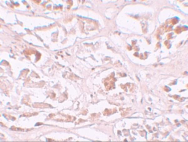
The IHC-validated ALKBH2 antibody has human kidney tissue data (catalog image: human kidney) and lists human, mouse, and rat reactivity (catalog: reactivity). IF/ICC data are absent (catalog: applications and images).
A06325 will render with an IHC image of human kidney tissue at 10 μg/mL (catalog image caption). Its listed applications include IHC-P, and its listed reactivity covers human, mouse, and rat (catalog: applications and reactivity).
Which to pick: Choose A06325 for paraffin-section IHC because IHC-P is listed and its own image shows human kidney tissue staining at 10 μg/mL (catalog: applications and image caption); the fixative is unreported (catalog image caption). No IF/ICC option is supported by the listed applications or images (catalog: applications and images). A06325 lists mouse and rat reactivity, but the supplied IHC image documents human tissue only (catalog: reactivity and image caption).