ALPK1 / Alpha-protein kinase 1 · IHC design guide

Design Immunohistochemistry for ALPK1

Plan ALPK1 staining in paraffin sections using the catalog antibody’s IHC dilution range of 1:100–1:300 (datasheet A08171). Compare cytoplasmic staining in adrenal glandular cells with HPA’s reported absence of staining in fallopian tube glandular cells, while accounting for medium consistency between staining and RNA data (HPA tissue IHC).

Evidence assembled Oct 2026 · For research use; verify linked source records and product datasheet before use
Immunohistochemistry protocol sheet for ALPK1 (IHC for ALPK1): expected localisation General cytoplasmic staining in tissue (HPA tissue IHC), antibody A08171, validated IHC image, and IHC protocol steps
Printable ALPK1 IHC protocol sheet — expected localisation General cytoplasmic staining in tissue (HPA tissue IHC), antibody A08171, controls and protocol steps. Open the full ALPK1 IHC guide →

ALPK1 Immunohistochemistry Experimental Design Guide

Expected localisation, validated protocols, controls and antibodies — the at-a-glance facts below, then the full design guide.

Must know before staining
Expected localisation General cytoplasmic staining in tissue (HPA tissue IHC)
Staining pattern Cytoplasmic staining in glandular and neuronal cells (HPA tissue IHC)
Antigen retrieval Tris-EDTA pH 9.0 HIER, heat-mediated (datasheet A08171)
Positive control ⓘ Adrenal gland+4 more · see all
Negative control ⓘ Fallopian tube+4 more · see all
Important caveats
Reasons your staining may differ from the expected pattern.
Fixation Keep fixation consistent between sections. (standard IHC practice; not target-specific)
Caveat Staining and RNA show medium consistency (HPA tissue IHC)
Regulation Expression regulation unreported (UniProt)
Isoform / epitope 2 isoforms; epitope coverage is unknown (UniProt)
Section 1

Recommended ALPK1 IHC & IF Protocols

The catalog antibody’s IHC-P protocol uses heat-mediated retrieval (datasheet A08171). One published renal IHC protocol provides additional ALPK1 staining parameters (PMC9893546).

Recommended immunohistochemistry (IHC-P) protocol parameters
SampleParaffin-embedded human Gastric adenocarcinoma tissue; fixative not specified (datasheet A08171)
FixationImage fixative and duration unreported (datasheet A08171); verify before use.
Sectioning4–5 µm sections on charged slides (standard)
DeparaffinisationXylene, graded ethanol series to water (standard)
Antigen retrievalHeat retrieval: Tris-EDTA pH 9.0 (datasheet A08171); 20 min, 95–100 °C (standard)
Peroxidase block3% H2O2, 10 min, room temperature (standard)
Blocking10% normal serum of the secondary host, 30 min, room temperature (standard)
Primary antibodyRabbit anti-ALPK1, 1:100-1:300 (datasheet A08171)
Primary incubationOvernight at 4 °C (standard)
DetectionHRP-polymer secondary, DAB chromogen 5–10 min (standard)
CounterstainHematoxylin, blue, dehydrate and mount (standard)
Expected resultALPK1-positive staining in glandular cells of adrenal gland (HPA tissue IHC: Medium). HPA tissue profile: General cytoplasmic expression. No signal in the no-primary control.
💡Decision noteStart with Tris-EDTA pH 9.0 heat-mediated retrieval (datasheet A08171); the published protocol uses a microwave but does not specify the retrieval buffer (PMC9893546).
Section 2

What Is the Expected ALPK1 Staining Pattern?

ALPK1 is a cytosolic protein with reported centrosome and ciliary base localization and no transmembrane segment (UniProt Q96QP1). In paraffin IHC, expect mainly cytoplasmic staining in selected glandular, respiratory epithelial, and neuronal cells (HPA: general cytoplasmic expression; selected cells Medium). HPA rates the tissue IHC evidence Approved, with medium consistency between staining and RNA expression; external verification is pending (HPA: reliability description).

What am I looking at on my slide?
Moderate cytoplasmic staining appears in adrenal or breast glandular cells, bronchial respiratory epithelium, or the listed neuronal cells (HPA: Medium).This fits the reported IHC pattern (HPA: general cytoplasmic expression). Judge the named cell population, not the whole section: other cells in the same tissue are not established positives by these examples (HPA: tissue IHC).
Staining is predominantly nuclear or outlines the plasma membrane, with little cytoplasmic signal.This departs from the reported cytoplasmic IHC profile and the absence of a transmembrane segment (HPA: tissue IHC; UniProt Q96QP1 topology). Review staining distribution and controls before calling such signal ALPK1; compartment alone cannot identify its cause (general IHC practice).
Strong staining appears in pancreatic exocrine glandular cells or lymph-node germinal-center cells (HPA: Not detected).Those named cells are useful checks for unexpected signal, though they are not universal ALPK1-negative tissues (HPA: tissue IHC). Consider antibody cross-reactivity or endogenous detection activity, particularly if control sections also develop color (general IHC practice).
Color is spread across stroma, empty spaces, or many unrelated cell types without clear cytoplasmic boundaries.That distribution is difficult to score as the reported cell-associated cytoplasmic pattern (HPA: tissue IHC). Diffuse background can arise from nonspecific binding, detection chemistry, or inadequate washing; assess the run controls (general IHC practice).
A selected positive cell population has no detectable staining, including bronchial respiratory epithelium or cerebellar Purkinje cells (HPA: Medium).A negative result in a reported moderate-staining population warrants a technical check, but HPA's Approved rating has medium RNA–staining consistency and awaits external verification (HPA: reliability description). Do not infer absence of ALPK1 from one failed section.
💡Expected ALPK1 appearanceCall a section positive when the listed glandular, respiratory epithelial, or neuronal cells show discernible, chiefly cytoplasmic, approximately moderate staining (HPA: Medium; general cytoplasmic expression); isolated nuclear, membrane-rim, or diffuse noncellular color is an unexpected pattern (HPA: tissue IHC; UniProt Q96QP1 topology).
How each factor affects the staining
Choice of comparison cellsUse a listed Medium cell population as a positive reference and a listed Not detected population as a check for unexpected staining (HPA: tissue IHC). These labels apply to the specified cells, not every cell in either tissue.
Strength of the IHC evidenceHPA calls tissue staining Approved but reports only medium consistency with RNA and pending external verification (HPA: reliability description). Treat a matching pattern as supportive rather than conclusive identification of ALPK1.
Compartment and protein architectureA cytoplasmic IHC pattern accords with cytosolic localization and no transmembrane segment (HPA: tissue IHC; UniProt Q96QP1). Centrosome and ciliary-base locations describe finer structures and need not resolve as puncta in chromogenic sections (UniProt Q96QP1; general microscopy practice).
Isoforms and processingUniProt lists two isoforms, one chain spanning residues 1–1244, and no signal peptide, propeptide, or annotated glycosylation sites (UniProt Q96QP1). The payload gives no antibody epitope or isoform-specific IHC result, so the stain cannot be assigned to an isoform.
IF/ICC Q: Should ALPK1 appear punctate?In IF/ICC, centrosome and basal-body localization is supported, and HPA027435 has Supported ICC validation (HPA: subcellular; HPA: antibodies). That higher-resolution observation does not require puncta in paraffin chromogenic IHC (general microscopy practice); IF/ICC methods belong in its separate guide.
Why is my staining missing, weak or wrong?
SituationLikely causeNext action
No signal in a reported Medium cell population (HPA: tissue IHC).The section or IHC run may have failed, or this specimen may differ from the HPA example (general IHC practice; HPA: medium consistency).Check tissue preservation, retrieval and detection controls, then compare another listed positive cell population before interpreting a negative result (general IHC practice; HPA: tissue IHC).
Color develops in cells reported Not detected, such as pancreatic exocrine glandular cells (HPA: tissue IHC).Cross-reactivity or endogenous enzyme activity may contribute, depending on the detection system (general IHC practice).Examine the no-primary control and detection block; reassess specificity in the named cell population rather than treating the entire tissue as a negative control (general IHC practice; HPA: tissue IHC).
The dominant signal is nuclear or forms a crisp cell-surface rim.The pattern conflicts with reported cytoplasmic staining and ALPK1's lack of a transmembrane segment (HPA: tissue IHC; UniProt Q96QP1 topology).Compare a known positive cell population and no-primary control; review antibody concentration and detection conditions before scoring the unexpected compartment (general IHC practice; HPA: tissue IHC).
A diffuse brown haze obscures cell boundaries.Nonspecific binding, residual detection activity, or excessive chromogen development can obscure localization (general IHC practice).Inspect no-primary controls, blocking, washes, and development time; score only cellular signal that remains distinguishable from background (general IHC practice).
Cytoplasmic staining is visible, but no centrosomal dot can be identified in paraffin IHC.The general cytoplasmic IHC profile and the supported centrosome or basal-body IF localization describe observations at different resolution (HPA: tissue IHC; HPA: subcellular).Score the IHC result against the reported cytoplasmic pattern; consult the separate IF/ICC guide if the question requires subcellular puncta (HPA: tissue IHC; HPA: subcellular).

Sample controls for ALPK1 IHC & IF

🧪Run adrenal gland first: glandular cells should stain at the HPA Medium level (HPA: adrenal gland, glandular cells, Medium). Use parathyroid glandular cells as a tissue negative (HPA: parathyroid gland, glandular cells, Not detected); on the adrenal slide, use neighboring cells without specific staining as an internal background comparator rather than assuming they are ALPK1-negative.
Positive control tissue: Adrenal gland (Glandular cells, HPA Medium)
Negative control tissue: Fallopian tube (HPA Not detected)
ICC-IF cell lines (HPA subcellular resource): HPA ICC-IF images show ALPK1 in A-431, U-251MG, U2OS, ASC52telo, RPTEC/TERT1, hTERT-RPE1 (serum starved), with annotated localisation: Centrosome (supported), Basal body (supported) (HPA subcellular).
Technical controls: Include no-primary (secondary-only) and host- and clonality-matched isotype controls, plus an ALPK1 knockout sample or a peptide-block control if the immunizing peptide is available (standard IHC practice). Check endogenous peroxidase background in the adrenal section before interpreting chromogenic signal (standard chromogenic IHC practice).
⚠️Feasibility: A target-specific fixation window or fixation effect is unreported in the supplied evidence; the selected A08171 paraffin-section caption does not state a fixative (A08171 tissue-IHC caption: fixative not stated). That caption uses Tris-EDTA at pH 9.0 for retrieval and a 1:200 primary dilution, providing a starting condition without establishing retrieval dependence (A08171 tissue-IHC caption). ICC-IF images support centrosome and basal-body localization, but the evidence does not establish that IF or frozen sections are easier than paraffin IHC; assess adrenal background staining alongside the controls (HPA: centrosome and basal body supported; standard IHC practice).

HPA tissue IHC evidence for ALPK1

Comprehensive Human Protein Atlas IHC scoring per tissue (reliability: Approved — Medium consistency between antibody staining and RNA expression data. Pending external verification.). Rows are taken directly from the HPA tissue chart — click any row's HPA link to view the source.

Positive expression · recommended positive controls

TissueCell typeLevelEvidenceSource
Adrenal gland Glandular cells Medium Protein (IHC) HPA →
Breast Glandular cells Medium Protein (IHC) HPA →
Bronchus Respiratory epithelial cells Medium Protein (IHC) HPA →
Caudate Neuronal cells Medium Protein (IHC) HPA →
Cerebellum Purkinje cells Medium Protein (IHC) HPA →

Undetected expression · recommended negative controls

TissueCell typeLevelEvidenceSource
Fallopian tube Glandular cells Not detected Protein (IHC) HPA →
Lymph node Germinal center cells Not detected Protein (IHC) HPA →
Pancreas Exocrine glandular cells Not detected Protein (IHC) HPA →
Parathyroid gland Glandular cells Not detected Protein (IHC) HPA →
Tonsil Germinal center cells Not detected Protein (IHC) HPA →
Section 3

Advanced ALPK1 IHC Tips

Troubleshoot ALPK1 staining in paraffin sections using the catalog antibody’s tissue image, HPA staining patterns and known intracellular localisation.

What retrieval should I try first when ALPK1 staining is weak?
Start with heat-mediated Tris-EDTA retrieval at pH 9.0 (datasheet A08171). The catalog image used this retrieval for paraffin-embedded human gastric adenocarcinoma, with primary antibody at 1:200 overnight at 4°C (datasheet A08171). If staining remains weak, compare a milder retrieval condition on adjacent sections while keeping antibody dilution and detection constant (standard IHC practice). Record retrieval temperature and duration for each run because those details are absent from the caption (datasheet A08171). Judge improvement by cleaner cellular staining and preserved morphology, rather than by stronger signal alone (standard IHC practice).
Could fixation explain weak or uneven ALPK1 staining?
ALPK1-specific sensitivity to fixation is unknown: the selected paraffin-section caption does not state a fixative (datasheet A08171). Record each specimen’s fixative and fixation duration before comparing staining across blocks (standard IHC practice). Use similarly processed sections when assessing the documented pH 9.0 retrieval and 1:200 primary dilution (datasheet A08171; standard IHC practice). If staining varies across a section, inspect preservation and compare adjacent sections under matched retrieval and detection conditions (standard IHC practice). Neither HPA tissue patterns nor ALPK1’s intracellular topology establishes a target-specific fixation effect (HPA tissue IHC; UniProt Q96QP1 topology).
Which staining compartments are plausible for ALPK1 in tissue sections?
Expect predominantly cytoplasmic staining in tissue IHC (HPA tissue IHC: general cytoplasmic expression). ALPK1 is annotated in the cytosol, at spindle poles and centrosomes, and at the base of primary cilia (UniProt Q96QP1 subcellular location). A small punctum can therefore merit closer inspection, but routine chromogenic sections may not resolve a centrosome from nearby cytoplasmic stain (UniProt Q96QP1 subcellular location; standard IHC practice). Compare any punctate pattern with the broader cellular distribution in the same section and with controls processed together (standard IHC practice). Predominantly nuclear staining should prompt reassessment of specificity and detection artefacts (UniProt Q96QP1 subcellular location; standard IHC practice).
How should I troubleshoot discordant staining between ALPK1 antibodies?
ALPK1 has 2 annotated isoforms, so first check which sequence each antibody recognises (UniProt Q96QP1 isoforms; standard IHC practice). The provided catalog caption establishes staining conditions but does not identify the antibody’s epitope (datasheet A08171). ALPK1 has no annotated transmembrane segment, and its alpha-type protein kinase domain spans residues 1017–1237 (UniProt Q96QP1 topology and domains). Do not assign discordant staining to an isoform without epitope mapping and an independently validated comparison (standard IHC practice). Run the antibodies on adjacent sections with matched scoring, then investigate differences in retrieval or background before making a biological claim (standard IHC practice).
How can IF help assess an uncertain ALPK1 IHC pattern?
Use IF as an orthogonal localisation check while interpreting chromogenic IHC; HPA reports supported centrosome and basal-body localisation in ICC/IF (HPA subcellular). Multiplex ALPK1 with a validated marker for the cell population being scored, then compare overlap and cellular boundaries (standard IF practice). Choose fluorophores and imaging channels after checking tissue autofluorescence, with particular care for weak puncta (standard IF practice). ALPK1 lacks a transmembrane segment, so permeabilise cells when testing an intracellular epitope; confirm the antibody’s epitope before setting permeabilisation conditions (UniProt Q96QP1 topology; standard IF practice). The pH 9.0 retrieval and 1:200 dilution document paraffin-section IHC, not an IF protocol (datasheet A08171).
What should I change when diffuse brown signal obscures ALPK1?
Check a no-primary control for detection background and review tissue edges, folds and necrotic areas separately from intact cells (standard IHC practice). Quench endogenous peroxidase before horseradish-peroxidase detection and develop DAB under matched conditions across sections (standard chromogenic IHC practice). If the control is clean, titrate around the documented 1:200 primary dilution and review wash stringency while preserving the pH 9.0 retrieval baseline (datasheet A08171; standard IHC practice). ALPK1 is broadly cytoplasmic in HPA tissue IHC, so diffuse cellular staining alone does not prove background (HPA tissue IHC). Compare staining intensity with cellular outlines and control tissue rather than relying on overall brown area (standard IHC practice).
How should I score ALPK1 across paraffin sections? ⚠ ANSWER MARKED FOR VERIFICATION
Define the scored cell population and region before reviewing stain intensity, then apply the same rule to every section (standard IHC practice). For cellular cytoplasmic staining, report an H-score from 0–300 or the percentage of positive cells at prespecified thresholds (HPA tissue IHC: general cytoplasmic expression; standard IHC practice). Normalise positive-cell counts to the number of eligible cells in the same region; use cells per mm² when reporting spatial density (standard IHC practice). Exclude folds, necrosis and poorly preserved areas using a recorded rule (standard IHC practice). Keep retrieval, detection and image-analysis thresholds consistent across the comparison (standard IHC practice).
How can I distinguish credible ALPK1 staining from artefact?
Look for staining within intact cell boundaries, mainly in cytoplasm, before calling a section positive (HPA tissue IHC: general cytoplasmic expression; standard IHC practice). HPA reports medium staining in breast glandular cells and no detection in lymph-node germinal-center cells; these are reference patterns, not universal controls for every specimen (HPA tissue IHC). Reassess isolated nuclear signal against ALPK1’s cytosolic and centrosomal annotations (UniProt Q96QP1 subcellular location). Discount staining confined to edges, necrosis or damaged areas, and check a no-primary control for endogenous enzyme signal (standard IHC practice). HPA rates tissue staining Approved but notes medium RNA agreement and pending external verification, so confirm consequential findings independently (HPA tissue IHC reliability).
Boster reagents

Best ALPK1 / Alpha-protein kinase 1 IHC Antibodies

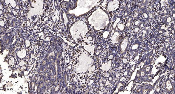
A08171 has real ALPK1 IHC data from paraffin-embedded human gastric adenocarcinoma (IHC image caption); IF is listed for the antibody, with human and mouse reactivity (catalog: applications/reactivity).

Real IHC data Immunohistochemical analysis of paraffin-embedded human Gastric adenocarcinoma. 1, Antibody was diluted at 1:200 (4° overnight). 2, Tris-EDTA, pH9.0 was used for antigen retrieval. 3, Secondary antibody was diluted at 1:200 (room temperature, 45min).
Anti-alpha-protein Kinase 1 ALPK1 Antibody
Cat # A08171

A08171 is pictured in IHC on paraffin-embedded human gastric adenocarcinoma (IHC image caption). A08171 also lists IF, but provides no IF image; human and mouse reactivity is catalog-listed (catalog: applications/reactivity/IF image alts).

Which to pick: For tissue IHC, choose A08171: its paraffin-section image documents 1:200 primary antibody and Tris-EDTA retrieval at pH 9.0; the fixative is unreported (IHC image caption). For IF, A08171 is catalog-listed at 1:200–1:1000, but no IF image or ICC application is provided (catalog: applications/IF dilution/IF image alts). For human or mouse work, A08171 is the listed rabbit polyclonal option, although its pictured IHC evidence is human only (catalog: host/clonality/reactivity; IHC image caption).

Each figure is that product's own IHC / IF validation image from its datasheet.

References

  1. UniProt Consortium. UniProt entry Q96QP1 (ALPK1_HUMAN, Alpha-protein kinase 1).
  2. Human Protein Atlas. ALPK1 tissue IHC expression (reliability: Approved).
  3. Human Protein Atlas. ALPK1 subcellular location (ICC-IF): Localized to the centrosome and basal body..
  4. Human Protein Atlas. ALPK1 antibody validation summary (2 antibodies).
  5. ALPK1 regulates streptozotocin-induced nephropathy through CCL2 and CCL5 expressions. Journal of cellular and molecular medicine 2019 — PMC6815771.
  6. Alpha-kinase1 promotes tubular injury and interstitial inflammation in diabetic nephropathy by canonical pyroptosis pathway. Biological research 2023 — PMC9893546.
  7. Investigating the role of the ALPK1 signaling pathway in the pathogenesis of diabetic retinopathy. Scientific reports 2025 — PMC12475159.
  8. Fusobacterium nucleatum promotes colorectal cancer cells adhesion to endothelial cells and facilitates extravasation and metastasis by inducing ALPK1/NF-κB/ICAM1 axis. Gut microbes 2022 — PMC8890384.
  9. PubMed PMID:10021370 — UniProt-cited evidence.
  10. PubMed PMID:14702039 — UniProt-cited evidence.
  11. PubMed PMID:15815621 — UniProt-cited evidence.