This website uses cookies to ensure you get the best experience on our website.


- Table of Contents
Plan chromogenic ATXN2 IHC on paraffin sections using cytoplasmic CNS staining as a reference (HPA tissue IHC). This guide pairs high-staining neuronal examples with cell types where staining was undetected (HPA tissue IHC) and flags the 5 documented isoforms for epitope review (UniProt).

Expected localisation, validated protocols, controls and antibodies — the at-a-glance facts below, then the full design guide.
| Expected localisation | Cytoplasmic; most abundant in CNS (HPA tissue IHC) | |
| Staining pattern | Neuronal cytoplasm and cerebellar granular-layer processes (HPA tissue IHC) | |
| Antigen retrieval | EDTA pH 8.0 HIER, heat-mediated (datasheet PA2098) | |
| Positive control | Caudate+4 more · see all | |
| Negative control | Adipose tissue+4 more · see all |
| Fixation | Keep fixation consistent across sections. (standard IHC practice; not target-specific) | |
| Caveat | Staining shows medium consistency with RNA expression (HPA tissue IHC) | |
| Regulation | Isoform abundance varies in the CNS (UniProt) | |
| Isoform / epitope | 5 isoforms; check antibody epitope coverage (UniProt) |
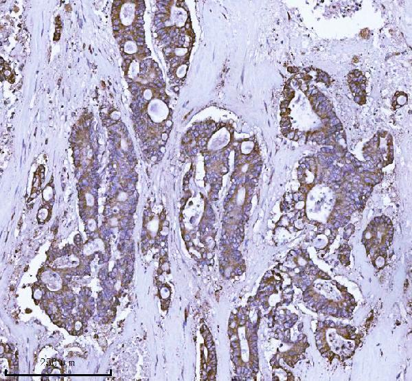
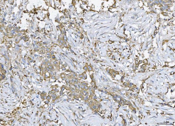
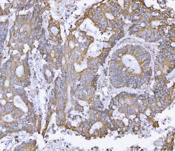
The catalog antibody uses EDTA pH 8.0 heat retrieval (datasheet PA2098). The published protocols below describe ATXN2 staining in brain sections (PMC7594343; PMC13076406).
| Sample | Paraffin-embedded human breast cancer tissue; fixative not specified (datasheet PA2098) |
| Fixation | Image fixative and duration unreported (datasheet PA2098); verify before use. |
| Sectioning | 4–5 µm sections on charged slides (standard) |
| Deparaffinisation | Xylene, graded ethanol series to water (standard) |
| Antigen retrieval | Heat retrieval: EDTA pH 8.0 (datasheet PA2098); 20 min, 95–100 °C (standard) |
| Peroxidase block | 3% H2O2, 10 min, room temperature (standard) |
| Blocking | 10% goat serum (datasheet PA2098) |
| Primary antibody | Rabbit anti-ATXN2, 2-5 μg/ml (datasheet PA2098) |
| Primary incubation | Overnight at 4 °C (datasheet PA2098) |
| Detection | HRP-conjugated secondary, DAB chromogen (datasheet PA2098) |
| Counterstain | Hematoxylin, blue, dehydrate and mount (standard) |
| Expected result | ATXN2-positive staining in neuronal cells of caudate (HPA tissue IHC: High). HPA tissue profile: Cytoplasmic expression in several tissues, most abundant in CNS. No signal in the no-primary control. |
ATXN2 should appear mainly in the cytoplasm of stained cells (UniProt Q99700: cytoplasm; HPA: cytoplasmic tissue expression). Strong IHC staining is reported in neuronal cells of the caudate and cerebral cortex, cerebellar granular-layer processes, and selected non-neural cells (HPA: High in each). ATXN2 has no transmembrane segment, so a membrane-only pattern would be unexpected (UniProt Q99700: topology). HPA rates the tissue profile Enhanced, with medium consistency between staining and RNA data (HPA: reliability).
| Clear cytoplasmic staining in caudate or cerebral-cortex neurons, with recognizable tissue structure. | This matches ATXN2 localisation and reported High neuronal staining (UniProt Q99700: cytoplasm; HPA: High in caudate and cerebral-cortex neuronal cells). Compare cells within the section before assigning an overall tissue score (general IHC practice). |
| Predominantly nuclear or sharply membrane-only staining, without a convincing cytoplasmic component. | That compartment is inconsistent with the supplied localisation evidence (UniProt Q99700: cytoplasm and no transmembrane segment; HPA: cytoplasmic expression). Treat it as suspect and check staining controls and morphology before scoring (general IHC practice). |
| Strong staining in a cell population reported as not detected, while expected positive cells are weak. | For example, strong adipocyte or skeletal-myocyte staining conflicts with those cell-specific observations (HPA: Not detected in adipocytes and skeletal myocytes). Check cell identification, antibody specificity, and endogenous chromogen activity (general IHC practice). |
| Diffuse color across stroma, empty spaces, and multiple cell compartments, obscuring cell borders. | This is poor evidence for the reported cell-associated cytoplasmic pattern (HPA: cytoplasmic expression). Review no-primary and detection controls, blocking, washing, and chromogen development (general IHC practice); do not score background as ATXN2. |
| No detectable staining in caudate neurons or another documented High population. | A blank known-positive control makes a negative study section hard to interpret (HPA: High in caudate neuronal cells; general IHC practice). Verify tissue identity and assay performance before calling the study section negative; HPA tissue levels are observations, not guarantees for every specimen. |
| Compartment and topology | Cytoplasmic staining is expected, and ATXN2 lacks a transmembrane segment (UniProt Q99700: subcellular location and topology). The protein's EGFR-trafficking function does not by itself establish a membrane IHC pattern (UniProt Q99700: function). |
| Cell-specific tissue expression | HPA reports High staining in lung macrophages, gallbladder glandular cells, and nasopharyngeal basal cells, alongside CNS populations (HPA: tissue IHC). Low or Not detected calls apply to the named cells, not automatically to every cell in that tissue (HPA: tissue IHC). |
| Isoforms and tissue selection | UniProt lists 5 isoforms; isoform 1 predominates in brain and spinal cord, while isoform 4 is more abundant in cerebellum (UniProt Q99700: isoforms and tissue specificity). The supplied evidence does not map this assay's epitope to an isoform. |
| Strength of reference pattern | HPA calls the tissue profile Enhanced but reports medium consistency between antibody staining and RNA expression (HPA: reliability). Its listed IHC antibodies HPA018295, HPA020339, and HPA021146 each have Enhanced validation (HPA: antibodies); this does not validate every antibody or specimen. |
| IF/ICC cross-check: where should signal appear? | Expect cytosolic signal in an appropriately validated IF/ICC assay (HPA: Cytosol, approved). HPA018295 has Enhanced ICC validation (HPA: antibodies). That subcellular observation supports compartment interpretation; it is not an IHC intensity standard. |
| Situation | Likely cause | Next action |
|---|---|---|
| Positive-control neurons are blank. | The expected population may be absent from the section, or the staining workflow may have failed (HPA: High in caudate neuronal cells; general IHC practice). | Confirm the cell population on the counterstained section; check the antibody's IHC instructions, reagent integrity, retrieval, and detection controls (general IHC practice). Target-specific fixation sensitivity is unreported in the supplied evidence. |
| Every cell looks strongly positive. | Widespread color can reflect background or overdeveloped chromogen rather than the cell-specific HPA pattern (HPA: tissue IHC; general IHC practice). | Compare a no-primary control, inspect unstained spaces, and review blocking, washing, antibody concentration, and development time (general IHC practice). Score only interpretable cell-associated signal. |
| Staining appears mainly nuclear. | A nuclear-dominant result conflicts with the reported cytoplasmic localisation (UniProt Q99700: cytoplasm; HPA: cytoplasmic expression). | Recheck cellular boundaries and counterstain, then compare positive and no-primary controls; repeat with an IHC-validated antibody if the pattern persists (general IHC practice; HPA: IHC antibody validation). |
| Adipocytes or skeletal myocytes stain strongly. | Those populations are reported Not detected by HPA; misidentified cells, cross-reactivity, or endogenous detection activity are possible explanations (HPA: adipocytes and skeletal myocytes; general IHC practice). | Verify the stained cell type, inspect detection-only controls, and compare a documented positive population on the same run (general IHC practice; HPA: High in caudate neuronal cells). |
| Skeletal muscle looks negative despite a UniProt expression entry. | UniProt reports expression in skeletal muscle, whereas HPA reports myocytes Not detected by tissue IHC (UniProt Q99700: tissue specificity; HPA: skeletal-muscle myocytes). These statements describe different measurements. | Do not use skeletal myocytes alone as the positive control; include a documented High IHC population and report the cell type and assay context (HPA: tissue IHC; general IHC practice). |
| Only some cells in a mixed tissue stain. | The HPA calls are cell-specific: lung macrophages are High, while other lung cell populations are not characterized by this payload (HPA: High in lung macrophages). | Identify cells by morphology and score each relevant population separately; avoid extending a macrophage result to the whole tissue (HPA: lung macrophages; general IHC practice). |
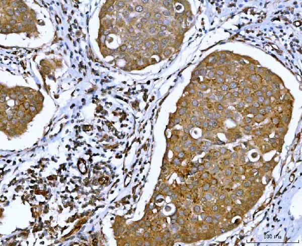
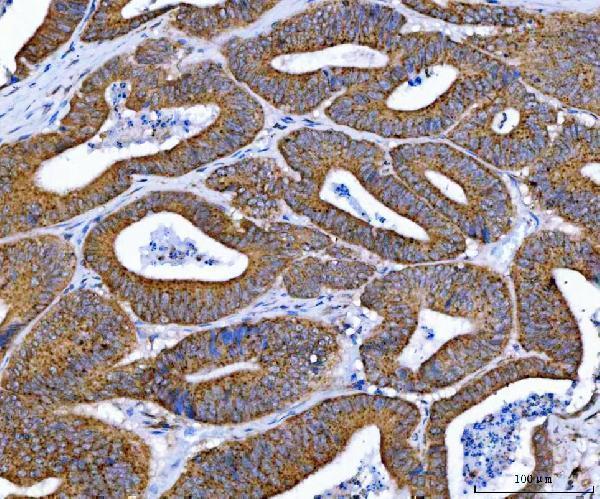
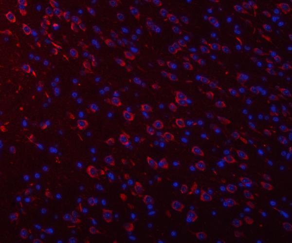
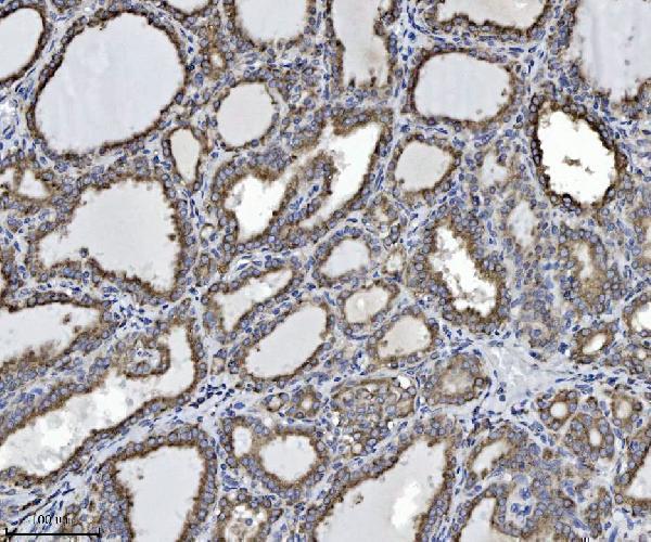


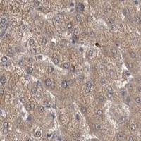
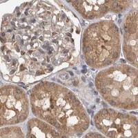


Comprehensive Human Protein Atlas IHC scoring per tissue (reliability: Enhanced — Medium consistency between antibody staining and RNA expression data.). Rows are taken directly from the HPA tissue chart — click any row's HPA link to view the source.
| Tissue | Cell type | Level | Evidence | Source |
|---|---|---|---|---|
| Caudate | Neuronal cells | High | Protein (IHC) | HPA → |
| Cerebellum | Processes in granular layer | High | Protein (IHC) | HPA → |
| Cerebral cortex | Neuronal cells | High | Protein (IHC) | HPA → |
| Gallbladder | Glandular cells | High | Protein (IHC) | HPA → |
| Lung | Macrophages | High | Protein (IHC) | HPA → |
| Tissue | Cell type | Level | Evidence | Source |
|---|---|---|---|---|
| Adipose tissue | Adipocytes | Not detected | Protein (IHC) | HPA → |
| Cervix | Squamous epithelial cells | Not detected | Protein (IHC) | HPA → |
| Ovary | Follicle cells | Not detected | Protein (IHC) | HPA → |
| Skeletal muscle | Myocytes | Not detected | Protein (IHC) | HPA → |
| Spleen | Cells in red pulp | Not detected | Protein (IHC) | HPA → |
Use compartment, cell type and matched controls to assess ATXN2 staining in paraffin sections; interpret weak signals alongside retrieval and detection conditions.
Three anti-ATXN2 antibodies have IHC images from human paraffin sections; PA2098 also has mouse and rat brain IHC and IF images (catalog image captions).
A01915 has human thyroiditis, colorectal adenocarcinoma, tonsil and breast cancer IHC images; PB9483 has human breast cancer, colonic adenoma cancer, glioma and placenta IHC images (A01915 and PB9483 image captions). PA2098 has human breast and rectal cancer plus mouse and rat brain IHC images, and mouse and rat brain IF images (PA2098 image captions).
Which to pick: Choose PA2098 for cross-species tissue IHC or mouse and rat IF; ICC is listed as an application, but the supplied ICC evidence contains no image (PA2098 applications, reactivity and image captions). For human tissue IHC, A01915 is a polyclonal option with human IHC images, while PB9483 has human IHC images; its listed mouse and rat reactivity is not demonstrated by the supplied IHC images (A01915 dilution data and image captions; PB9483 reactivity and image captions). Each SKU’s own IHC caption describes paraffin sections, EDTA retrieval at pH 8.0 and primary antibody at 2 μg/ml; the fixative is unreported (PA2098, A01915 and PB9483 IHC image captions).