This website uses cookies to ensure you get the best experience on our website.


- Table of Contents
Plan B4GALT1 IHC in paraffin sections using the granular staining reported in most cells as a reference (HPA tissue IHC). This guide covers fixation, staining controls and interpretation alongside the expected Golgi localization (UniProt).

Expected localisation, validated protocols, controls and antibodies — the at-a-glance facts below, then the full design guide.
| Expected localisation | Granular tissue staining (HPA tissue IHC); Golgi expected (UniProt) | |
| Staining pattern | Granular staining in most cells (HPA tissue IHC) | |
| Antigen retrieval | Tris-EDTA pH 9.0 HIER, heat-mediated (datasheet A03993T1) | |
| Positive control | Adrenal gland+4 more · see all | |
| Negative control | Adipose tissue+4 more · see all |
| Fixation | Matched tissue-IHC evidence does not establish the fixation claim. Validate the specimen-specific method before use. (selected-SKU IHC image A03993T1) | |
| Caveat | A secreted form may complicate cell-associated scoring (UniProt) | |
| Regulation | No specific expression regulator reported (UniProt) | |
| Isoform / epitope | 2 isoforms; map epitopes to cytoplasmic or lumenal regions (UniProt) |
The catalog antibody’s IHC-P protocol is accompanied by three published B4GALT1 IHC protocols (PMC5968541; PMC5078046; PMC10618527).
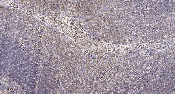
| Sample | Paraffin-embedded human tonsil tissue; fixative not specified (datasheet A03993T1) |
| Fixation | Image fixative and duration unreported (datasheet A03993T1); verify before use. |
| Sectioning | 4–5 µm sections on charged slides (standard) |
| Deparaffinisation | Xylene, graded ethanol series to water (standard) |
| Antigen retrieval | Heat retrieval: Tris-EDTA pH 9.0 (datasheet A03993T1); 20 min, 95–100 °C (standard) |
| Peroxidase block | 3% H2O2, 10 min, room temperature (standard) |
| Blocking | 10% normal serum of the secondary host, 30 min, room temperature (standard) |
| Primary antibody | Rabbit anti-B4GALT1, 1:100-1:300 (datasheet A03993T1) |
| Primary incubation | Overnight at 4 °C (standard) |
| Detection | HRP-polymer secondary, DAB chromogen 5–10 min (standard) |
| Counterstain | Hematoxylin, blue, dehydrate and mount (standard) |
| Expected result | B4GALT1-positive staining in glandular cells of adrenal gland (HPA tissue IHC: High). HPA tissue profile: Distinct expression with a granular pattern in most cells. No signal in the no-primary control. |
B4GALT1 is a Golgi stack membrane protein with a luminal catalytic region; UniProt also lists cell surface and secreted locations (UniProt P15291 topology and subcellular location). In tissue IHC, expect granular staining in many cells, including strong staining in several glandular epithelia and bronchial basal cells (HPA tissue IHC). HPA rates the tissue staining reliability Enhanced, while noting medium consistency with RNA expression (HPA tissue IHC).
| Discrete granular staining in colon or duodenal glandular cells, with a concentrated intracellular pattern (HPA: High in these cells; HPA: granular pattern in most cells). | This fits the observed tissue pattern and the expected Golgi enrichment (HPA tissue IHC; UniProt P15291 subcellular location). Judge the stained cell population and pattern together; intensity alone cannot establish the compartment in a chromogenic section (standard IHC interpretation). |
| Predominantly nuclear or uniformly diffuse cytoplasmic staining, without a discernible granular component. | This conflicts with the reported Golgi and granular pattern (UniProt P15291 subcellular location; HPA tissue IHC). Consider background or an antibody-specific artefact; inspect cellular detail and controls before calling the section positive (standard IHC practice). |
| Strong staining of adipocytes, cardiomyocytes, or skeletal myocytes. | HPA reports these specific cell populations as Not detected (HPA tissue IHC). Treat conspicuous staining there as a possible cross-reactive signal or endogenous detection activity, and compare it with a negative reagent control; the HPA result does not rule out every cell in those tissues (standard IHC interpretation). |
| Broad, hazy color across cells and extracellular areas, with little difference between positive and control sections. | A field-wide signal lacks the distinct cellular granularity reported for B4GALT1 (HPA tissue IHC). Background from detection chemistry or insufficient blocking is plausible; assess the control section before assigning staining to the target (standard IHC practice). |
| No staining in glandular cells of an otherwise interpretable colon or duodenum section. | These are HPA High cell populations, so an absent signal warrants checking assay performance (HPA tissue IHC). A single negative section does not establish target absence; review tissue preservation, retrieval, antibody application, detection, and a concurrently stained positive control (standard IHC practice). |
| Cell type and tissue context | HPA records High staining in glandular cells of adrenal gland, appendix, cervix, colon, duodenum, endometrium, and epididymis, and in bronchial basal cells (HPA tissue IHC). Its Not detected calls apply to the named cell populations, rather than every cell in an entire organ (HPA tissue IHC). |
| Golgi topology and compartment | B4GALT1 has a transmembrane segment at residues 25–44, a cytoplasmic segment at 1–24, and a luminal segment at 45–398 (UniProt P15291 topology). These annotations support an intracellular Golgi interpretation; they do not identify the epitope recognized by an unspecified antibody (UniProt P15291 topology). |
| Additional protein locations | UniProt also annotates cell membrane, cell surface, filopodium, and secreted locations (UniProt P15291 subcellular location). Limited peripheral staining may therefore merit review alongside the granular signal; a widespread surface-only pattern would not match HPA’s dominant tissue description (HPA tissue IHC). |
| Isoforms and processing | UniProt lists Long and Short isoforms and a processed B4GALT1 form (UniProt P15291 isoforms and processing). Without an antibody epitope or isoform-specific validation in this payload, their relative contributions to an IHC signal cannot be assigned; avoid reading staining differences as isoform changes. |
| Tissue evidence and antibody validation | HPA calls tissue staining Enhanced but reports only medium consistency with RNA expression; its RNA classification is low tissue specificity (HPA tissue IHC). Two listed antibodies, HPA010806 and HPA010807, each have Enhanced IHC status (HPA antibodies). These summaries support the reported pattern, not a claim that every specimen will stain equally. |
| IF/ICC: what pattern should appear? | A Golgi-localized signal is expected in ICC-IF (HPA subcellular: Golgi apparatus, Enhanced). This is a localization cross-check for interpreting IHC granularity, not an IHC protocol option; the HPA ICC-IF finding does not specify an IHC dilution or retrieval condition (HPA subcellular). |
| Situation | Likely cause | Next action |
|---|---|---|
| High-background section obscures cell boundaries or granules. | Detection reagent activity, inadequate blocking, or excessive antibody exposure can produce nonspecific color (standard IHC practice). | Compare a negative reagent control and reduce the source of background according to the assay controls; score B4GALT1 only where a distinct cellular granular pattern remains (HPA tissue IHC; standard IHC practice). |
| Expected glandular cells are unstained in colon or duodenum. | These populations are reported High, so a failed staining run or an unsuitable section is possible (HPA tissue IHC; standard IHC practice). | Check a concurrent known-positive section, tissue integrity, retrieval execution, antibody application, and detection reagents (standard IHC practice). No target-specific fixation sensitivity is established by the supplied HPA or UniProt records. |
| Nuclei stain strongly while granular cytoplasmic signal is absent. | A nuclear-dominant pattern conflicts with the reported Golgi location (HPA subcellular; UniProt P15291 subcellular location). | Inspect the counterstain and negative reagent control, then assess whether the chromogen can be distinguished from nuclear color; do not score nuclear color alone as B4GALT1 (standard IHC practice). |
| Adipocytes, cardiomyocytes, or myocytes are strongly positive. | HPA calls these specific cells Not detected; cross-reactivity or endogenous detection activity is possible (HPA tissue IHC; standard IHC practice). | Compare the same cell type in a negative reagent control and check whether the stain has the expected granular distribution (HPA tissue IHC; standard IHC practice). If discordance persists, verify with an independently validated antibody. |
| A weak or negative brain section is being used to reject the assay. | UniProt reports very low expression in fetal and adult brain; HPA reports low neuronal staining in cerebral cortex and Not detected staining in several listed brain cell populations (UniProt P15291 tissue specificity; HPA tissue IHC). | Use a reported High glandular population as the positive performance check, and interpret the sampled brain cell type against its own HPA entry (HPA tissue IHC; standard IHC practice). |
| Staining is mainly peripheral, with little granular intracellular color. | Surface localization is annotated, but a predominantly peripheral pattern differs from HPA’s granular tissue profile (UniProt P15291 subcellular location; HPA tissue IHC). | Review morphology, background controls, and a reported High glandular population before scoring; record peripheral staining separately if reproducible (standard IHC practice). |


Comprehensive Human Protein Atlas IHC scoring per tissue (reliability: Enhanced — Medium consistency between antibody staining and RNA expression data.). Rows are taken directly from the HPA tissue chart — click any row's HPA link to view the source.
| Tissue | Cell type | Level | Evidence | Source |
|---|---|---|---|---|
| Adrenal gland | Glandular cells | High | Protein (IHC) | HPA → |
| Appendix | Glandular cells | High | Protein (IHC) | HPA → |
| Bronchus | Basal cells | High | Protein (IHC) | HPA → |
| Cervix | Glandular cells | High | Protein (IHC) | HPA → |
| Colon | Glandular cells | High | Protein (IHC) | HPA → |
| Tissue | Cell type | Level | Evidence | Source |
|---|---|---|---|---|
| Adipose tissue | Adipocytes | Not detected | Protein (IHC) | HPA → |
| Caudate | Glial cells | Not detected | Protein (IHC) | HPA → |
| Cerebellum | Cells in granular layer | Not detected | Protein (IHC) | HPA → |
| Heart muscle | Cardiomyocytes | Not detected | Protein (IHC) | HPA → |
| Hippocampus | Glial cells | Not detected | Protein (IHC) | HPA → |
Troubleshoot B4GALT1 staining by checking retrieval, compartment pattern, controls and scoring before interpreting changes in chromogenic signal.
A03993T1 has an IHC image from paraffin-embedded human tonsil (IHC image caption); IF is listed without an IF image (catalog applications; IF image alts). Human and mouse reactivity are listed (catalog reactivity).
A03993T1 will render with its own IHC image of paraffin-embedded human tonsil (IHC image caption). A03993T1 is listed for IHC and IF in human and mouse, but no IF image or mouse sample is provided (catalog applications; catalog reactivity; IF image alts; IHC image caption).
Which to pick: Choose A03993T1 for paraffin-section IHC: its human tonsil image used 1:200 primary antibody overnight at 4°C with Tris-EDTA retrieval at pH 9.0 (IHC image caption). For IF/ICC, A03993T1 lists IF at 1:50, but provides no IF image or ICC validation (catalog dilution; IF image alts; catalog applications). For human or mouse samples, A03993T1 lists both species and has a rabbit host with no clone specified; the IHC image documents human tissue only, and its fixative is unreported (catalog reactivity; catalog host; catalog clone; IHC image caption).