This website uses cookies to ensure you get the best experience on our website.


- Table of Contents
Plan B4GALT2 paraffin-section IHC around granular tissue staining (HPA tissue IHC) and an expected Golgi membrane location (UniProt). Start with the catalog antibody’s 1:100–1:300 IHC dilution range (datasheet A10774T2), using glandular cells in colon as a positive reference (HPA tissue IHC).

Expected localisation, validated protocols, controls and antibodies — the at-a-glance facts below, then the full design guide.
| Expected localisation | Golgi stack membrane expected; tissue compartment unverified (UniProt) | |
| Staining pattern | Granular staining in several tissues, including glandular cells (HPA tissue IHC) | |
| Antigen retrieval | Tris-EDTA pH 9.0 HIER, heat-mediated (datasheet A10774T2) | |
| Positive control | Adrenal gland+4 more · see all | |
| Negative control | Adipose tissue+4 more · see all |
| Fixation | Keep fixation consistent across sections. (standard IHC practice; not target-specific) | |
| Caveat | Presumed off-target staining may confound interpretation (HPA tissue IHC) | |
| Regulation | Expression varies by tissue (UniProt) | |
| Isoform / epitope | 3 isoforms; epitope maps unknown; cytosolic versus lumenal location matters (UniProt) |
The catalog antibody’s IHC-P protocol (datasheet A10774T2) is accompanied by one published lung adenocarcinoma IHC protocol (PMC11865799).
| Sample | Paraffin-embedded human cervical carcinoma tissue; fixative not specified (datasheet A10774T2) |
| Fixation | Image fixative and duration unreported (datasheet A10774T2); verify before use. |
| Sectioning | 4–5 µm sections on charged slides (standard) |
| Deparaffinisation | Xylene, graded ethanol series to water (standard) |
| Antigen retrieval | Heat retrieval: Tris-EDTA pH 9.0 (datasheet A10774T2); 20 min, 95–100 °C (standard) |
| Peroxidase block | 3% H2O2, 10 min, room temperature (standard) |
| Blocking | 10% normal serum of the secondary host, 30 min, room temperature (standard) |
| Primary antibody | Rabbit anti-B4GALT2, 1:100-1:300 (datasheet A10774T2) |
| Primary incubation | Overnight at 4 °C (standard) |
| Detection | HRP-polymer secondary, DAB chromogen 5–10 min (standard) |
| Counterstain | Hematoxylin, blue, dehydrate and mount (standard) |
| Expected result | B4GALT2-positive staining in glandular cells of adrenal gland (HPA tissue IHC: High). HPA tissue profile: Granular expression in several tissues. No signal in the no-primary control. |
B4GALT2 is a Golgi stack membrane protein with a cytoplasmic N terminus and lumenal catalytic region (UniProt O60909 topology). Expect granular cytoplasmic staining in the appropriate cells, including colon and duodenal glandular cells (HPA: High in both). HPA rates tissue IHC reliability Supported, with medium agreement between staining and RNA data; presumed off-target staining was observed and disregarded (HPA: tissue IHC reliability).
| Granular cytoplasmic staining in colon or duodenal glandular cells (HPA: High in both cell types). | Consistent with the reported tissue pattern and Golgi localization (HPA: tissue IHC; UniProt O60909 subcellular location). Score the relevant cells and their staining intensity; a positive region does not imply that every cell in the section is positive. |
| Predominantly nuclear or continuous plasma membrane staining, with little granular cytoplasmic signal. | This differs from the Golgi pattern expected from UniProt and HPA (UniProt O60909 subcellular location; HPA: subcellular location). Treat it as suspect and compare a known-positive tissue section and detection controls before assigning B4GALT2 expression. |
| Strong staining in adipocytes or esophageal squamous epithelial cells (HPA: Not detected in these cells). | These are unexpected cell-specific results, even if another cell population in the tissue stains. Consider antibody cross-reactivity or chromogenic detection activity; HPA reports presumed off-target staining in its tissue assessment (HPA: tissue IHC reliability). |
| Uniform haze across cells, extracellular spaces, or the whole section. | A broad background pattern obscures the cell-restricted, granular pattern reported by HPA (HPA: tissue IHC profile). Compare a no-primary control, review blocking and washes, and avoid scoring haze as B4GALT2 staining. |
| No signal in colon glandular cells on a section expected to be positive (HPA: High in colon glandular cells). | The result is inconclusive until the staining run is checked. Review tissue integrity, antigen retrieval, primary antibody use and chromogenic detection; the HPA tissue record does not identify a B4GALT2-specific cause of signal loss. |
| Golgi membrane topology (UniProt O60909 topology) | B4GALT2 has one membrane segment at residues 16–36; residues 1–15 face the cytoplasm and 37–372 face the lumen (UniProt O60909 topology). This supports an intracellular Golgi pattern, but does not predict how an unspecified antibody epitope responds to antigen retrieval. |
| Cell type within a tissue (HPA: tissue IHC) | Interpret staining at the cell level. HPA reports High staining in fallopian tube ciliated cell bodies and Not detected in ovarian stroma cells (HPA: tissue IHC). The latter does not make the entire ovary a negative control; UniProt reports relatively high expression in ovary (UniProt O60909 tissue specificity). |
| Antibody evidence (HPA: antibody validation) | The listed IHC antibody HPA047739 has Supported IHC validation; HPA007620 has Supported ICC validation, with no IHC status listed (HPA: antibody validation). HPA also notes presumed off-target tissue staining, so evaluate unexpected patterns against cell-specific references (HPA: tissue IHC reliability). |
| IF/ICC: what localization should be expected? | HPA reports mainly Golgi apparatus and vesicle localization in ICC-IF, with Golgi supported and vesicles approved (HPA: subcellular). This informs image interpretation; the ICC-IF record does not establish an IHC-P staining protocol or a dilution. |
| Isoforms and glycosylation (UniProt O60909) | UniProt lists three isoforms and glycosylation sites at residues 66, 71 and 357 (UniProt O60909). Without an antibody epitope or isoform-specific staining data, these annotations cannot establish which isoforms an IHC signal detects or explain an unexpected tissue result. |
| Situation | Likely cause | Next action |
|---|---|---|
| Known-positive glandular cells have no visible signal (HPA: High in colon glandular cells). | A staining-run failure is possible; the supplied sources do not establish B4GALT2-specific fixation sensitivity. | Check a positive control section, reagent application, retrieval settings and detection steps. Repeat only after identifying a plausible run-level issue; do not attribute the loss to fixation from these records. |
| Staining is broad and diffuse instead of granular (HPA: granular tissue expression). | Nonspecific antibody binding or detection background may obscure the expected pattern. | Inspect the no-primary control, blocking, washes and primary concentration. Compare cell boundaries and unstained regions before assigning a positive score. |
| Adipocytes stain strongly (HPA: Not detected in adipocytes). | Cross-reactivity or endogenous detection activity is possible; HPA reports presumed off-target tissue staining (HPA: tissue IHC reliability). | Compare no-primary and known-positive sections, then reassess whether the signal is cell-specific and granular. Do not treat adipocyte staining alone as confirmation. |
| Staining appears mainly nuclear or along the cell surface. | The compartment conflicts with the Golgi location reported by UniProt and HPA (UniProt O60909; HPA: subcellular). | Review morphology, counterstain and detection controls. Confirm a granular intracellular pattern in a known-positive cell type before interpreting the unusual compartment. |
| Ovarian stroma is unstained, but other ovarian cells stain. | HPA reports Not detected specifically in ovarian stroma cells; that observation does not describe every ovarian cell (HPA: tissue IHC). | Score the cell populations separately. Compare the stained population with tissue morphology and avoid calling the entire section either positive or negative from stromal staining alone. |
| IF/ICC shows vesicle signal in addition to Golgi signal. | HPA lists vesicles as an approved location and Golgi as supported (HPA: subcellular). | Describe each compartment separately and use the IF/ICC guide for that application. Do not transfer ICC-IF validation or settings to paraffin-section IHC (HPA: antibody validation). |


Comprehensive Human Protein Atlas IHC scoring per tissue (reliability: Supported — Medium consistency between antibody staining and RNA expression data. Presumed off target binding observed and disregarded.). Rows are taken directly from the HPA tissue chart — click any row's HPA link to view the source.
| Tissue | Cell type | Level | Evidence | Source |
|---|---|---|---|---|
| Adrenal gland | Glandular cells | High | Protein (IHC) | HPA → |
| Appendix | Lymphoid tissue | High | Protein (IHC) | HPA → |
| Colon | Glandular cells | High | Protein (IHC) | HPA → |
| Duodenum | Glandular cells | High | Protein (IHC) | HPA → |
| Endometrium | Glandular cells | High | Protein (IHC) | HPA → |
| Tissue | Cell type | Level | Evidence | Source |
|---|---|---|---|---|
| Adipose tissue | Adipocytes | Not detected | Protein (IHC) | HPA → |
| Caudate | Glial cells | Not detected | Protein (IHC) | HPA → |
| Cerebellum | Cells in granular layer | Not detected | Protein (IHC) | HPA → |
| Cerebral cortex | Endothelial cells | Not detected | Protein (IHC) | HPA → |
| Esophagus | Squamous epithelial cells | Not detected | Protein (IHC) | HPA → |
Troubleshoot B4GALT2 staining in paraffin sections by checking retrieval, cellular pattern, controls, and cell type before scoring chromogenic signal.
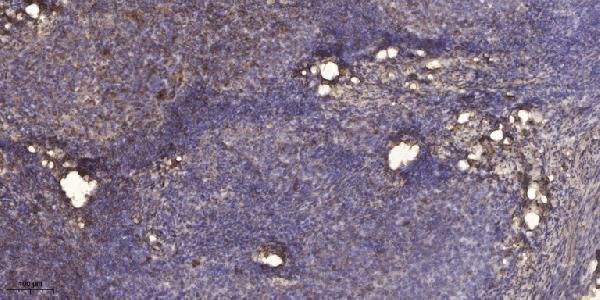
A10774T2 has an IHC image of human paraffin-embedded cervical carcinoma (catalog IHC image caption); IF and human/mouse reactivity are listed, but no IF image is supplied (catalog applications/reactivity; IF image alts).
A10774T2 is shown staining paraffin-embedded human cervical carcinoma by IHC (catalog IHC image caption). It lists IHC and IF applications and human/mouse reactivity; no IF image is supplied (catalog applications/reactivity; IF image alts).
Which to pick: For tissue IHC, choose A10774T2: its image caption documents paraffin-embedded human cervical carcinoma, 1:200 primary antibody dilution and Tris-EDTA retrieval at pH 9.0; the fixative is unreported (catalog IHC image caption). For IF, A10774T2 lists 1:50, but supplies no IF image or ICC application entry (catalog IF dilution/applications; IF image alts). For cross-species work, A10774T2 lists human and mouse reactivity, although its IHC image shows human tissue only; rabbit host is listed and clonality is unreported (catalog reactivity/host/clone; IHC image caption).