This website uses cookies to ensure you get the best experience on our website.


- Table of Contents
Plan chromogenic IHC for BAIAP2L2 in paraffin sections using a primary antibody dilution of 1:100–1:300 (datasheet A09681-1). Assess cytoplasmic staining in gastrointestinal glandular cells while accounting for presumed off-target binding and low RNA–IHC consistency (HPA tissue IHC).

Expected localisation, validated protocols, controls and antibodies — the at-a-glance facts below, then the full design guide.
| Expected localisation | Cytoplasmic staining (HPA tissue IHC); junction and vesicle membrane location annotated (UniProt) | |
| Staining pattern | Gastrointestinal glandular cells show high cytoplasmic staining (HPA tissue IHC) | |
| Antigen retrieval | Tris-EDTA pH 9.0 HIER, heat-mediated (datasheet A09681-1) | |
| Positive control | Colon+4 more · see all | |
| Negative control | Bone marrow+4 more · see all |
| Fixation | Keep fixation consistent across paraffin sections. (standard IHC practice; not target-specific) | |
| Caveat | Presumed off-target staining and low RNA–IHC consistency (HPA tissue IHC) | |
| Regulation | Intestine/kidney group-enriched RNA (HPA RNA) | |
| Isoform / epitope | 2 isoforms; no transmembrane segment; epitope effects unknown (UniProt) |
The catalog antibody's IHC-P protocol (datasheet A09681-1) is accompanied by one published osteosarcoma tissue protocol (PMC8577457).
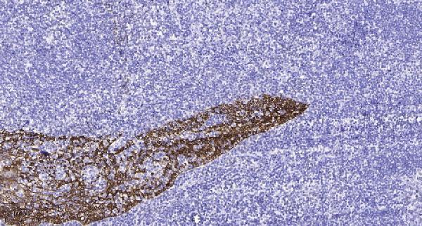
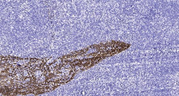
| Sample | Paraffin-embedded human tonsil tissue; fixative not specified (datasheet A09681-1) |
| Fixation | Image fixative and duration unreported (datasheet A09681-1); verify before use. |
| Sectioning | 4–5 µm sections on charged slides (standard) |
| Deparaffinisation | Xylene, graded ethanol series to water (standard) |
| Antigen retrieval | Heat retrieval: Tris-EDTA pH 9.0 (datasheet A09681-1); 20 min, 95–100 °C (standard) |
| Peroxidase block | 3% H2O2, 10 min, room temperature (standard) |
| Blocking | 10% normal serum of the secondary host, 30 min, room temperature (standard) |
| Primary antibody | Rabbit anti-BAIAP2L2, 1:100-1:300 (datasheet A09681-1) |
| Primary incubation | Overnight at 4 °C (standard) |
| Detection | HRP-polymer secondary, DAB chromogen 5–10 min (standard) |
| Counterstain | Hematoxylin, blue, dehydrate and mount (standard) |
| Expected result | BAIAP2L2-positive staining in glandular cells of colon (HPA tissue IHC: High). HPA tissue profile: Cytoplasmic expression in several different tissue types, most abundant in gastrointestinal tract. No signal in the no-primary control. |
BAIAP2L2 is observed as cytoplasmic staining, strongest in gastrointestinal glandular cells (HPA: tissue IHC). UniProt places the protein at cell membranes, cell junctions, and cytoplasmic vesicle membranes, including RAB13-positive vesicles and intercellular contacts; it has no transmembrane segment (UniProt Q6UXY1). Interpret the pattern cautiously: HPA rates the IHC evidence Approved but reports low agreement with RNA data and disregarded presumed off-target staining (HPA: reliability description).
| Strong cytoplasmic stain in colon, duodenal, small-intestinal, stomach, or gallbladder glandular cells. | This matches the reported High glandular-cell staining in those tissues (HPA: tissue IHC). Membrane-adjacent detail at cell contacts is plausible from UniProt localisation, but HPA describes the tissue pattern chiefly as cytoplasmic (UniProt Q6UXY1; HPA: tissue IHC). |
| Predominantly nuclear stain, with little cytoplasmic signal in gastrointestinal glandular cells. | A nuclear-dominant result conflicts with the reported cytoplasmic IHC pattern and membrane, junction, and vesicle localisation (HPA: tissue IHC; UniProt Q6UXY1). Treat it as an unconfirmed pattern; review controls and antibody specificity before scoring it as BAIAP2L2 (general IHC practice). |
| Strong stain in cells HPA lists as undetected, such as bone-marrow hematopoietic cells. | That result conflicts with HPA's cell-specific observation (HPA: bone marrow, hematopoietic cells, Not detected). Consider cross-reactivity or endogenous detection activity, then compare a reagent control and an independent antibody before assigning target expression (general IHC practice; HPA: reliability description). |
| Diffuse stain covers glandular cells and surrounding tissue without a clear cellular boundary. | A broad haze cannot establish the reported glandular-cell pattern (HPA: tissue IHC). Nonspecific antibody binding or chromogen background may obscure localisation; examine a control lacking primary antibody and compare staining with tissue morphology (general IHC practice). |
| No stain in a gastrointestinal glandular-cell positive reference section. | Absence of signal conflicts with HPA's High observations in the listed gastrointestinal glandular cells (HPA: tissue IHC). Check section integrity, detection reagents, and the antibody's validated IHC conditions before interpreting other sections as negative (general IHC practice). |
| Observed tissue and cell distribution | Colon, duodenum, gallbladder, small intestine, and stomach glandular cells are High; appendix glandular cells are Medium (HPA: tissue IHC). Choose the exact cell population when comparing slides, since whole-tissue positivity does not describe every cell (general IHC practice). |
| Compartment and topology | HPA reports cytoplasmic staining, while UniProt locates BAIAP2L2 at the cell membrane, junctions, and cytoplasmic vesicle membranes; there is no transmembrane segment (HPA: tissue IHC; UniProt Q6UXY1). Do not require an exclusively surface-membrane outline to call a glandular-cell result concordant. |
| IHC evidence and specificity | HPA calls the tissue IHC result Approved, while noting low consistency with RNA expression and presumed off-target binding that was disregarded (HPA: reliability description). HPA lists HPA003043 as IHC Approved, not Enhanced (HPA: antibodies). Use tissue and cell context when interpreting unexpected staining. |
| RNA versus protein readout | HPA reports RNA group enrichment in intestine and kidney, but the supplied IHC observations identify gastrointestinal glandular-cell staining and give no kidney IHC level (HPA: RNA specificity; HPA: tissue IHC). RNA enrichment alone cannot assign a kidney protein-staining intensity. |
| Situation | Likely cause | Next action |
|---|---|---|
| Positive reference section is blank. | A detection or staining-step failure is possible when a cell population reported as High is unstained (HPA: tissue IHC; general IHC practice). | Confirm that the reference section contains the relevant glandular cells, then check primary antibody, detection reagent, and counterstain performance against the catalog antibody's IHC-P instructions (general IHC practice). |
| All tissue regions stain evenly. | Diffuse background can arise from nonspecific binding or the detection system and prevents assessment of HPA's cell-specific pattern (general IHC practice; HPA: tissue IHC). | Inspect a section processed without primary antibody; review blocking, washes, and detection development under the assay's established conditions (general IHC practice). |
| Unexpectedly strong nuclear signal appears. | Nuclear dominance is unsupported by the supplied tissue pattern or UniProt localisation (HPA: tissue IHC; UniProt Q6UXY1). | Check cell boundaries and counterstain, compare a positive reference and reagent control, and seek independent antibody agreement before assigning nuclear localisation (general IHC practice). |
| Strong staining appears in an HPA-undetected cell population. | Cross-reactivity or endogenous detection activity is possible; HPA also reports presumed off-target staining in its reliability assessment (general IHC practice; HPA: reliability description). | Compare the same cell population with a no-primary control and, where available, an independently validated antibody; record the discrepancy instead of calling it confirmed expression (general IHC practice). |
| A kidney section is expected to be strongly positive from RNA data. | The payload reports kidney RNA enrichment but supplies no kidney tissue-IHC staining level (HPA: RNA specificity; HPA: tissue IHC). | Use a listed gastrointestinal glandular-cell positive section to assess IHC performance; treat any kidney result as a separate observation requiring validation (HPA: tissue IHC; general IHC practice). |
| Can IF/ICC images establish the expected subcellular pattern? | The supplied HPA subcellular record has no main location or ICC-IF image-bearing cell lines, although UniProt annotates membrane, junction, and vesicle localisation (HPA: subcellular; UniProt Q6UXY1). | Do not infer an observed IF/ICC pattern from the IHC slide. Interpret IF/ICC with its own controls and guide; use the UniProt locations as hypotheses, not an image-validated result (HPA: subcellular; general IF practice). |


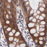

Comprehensive Human Protein Atlas IHC scoring per tissue (reliability: Approved — Low consistency between antibody staining and RNA expression data. Presumed off target binding observed and disregarded.). Rows are taken directly from the HPA tissue chart — click any row's HPA link to view the source.
| Tissue | Cell type | Level | Evidence | Source |
|---|---|---|---|---|
| Colon | Glandular cells | High | Protein (IHC) | HPA → |
| Duodenum | Glandular cells | High | Protein (IHC) | HPA → |
| Gallbladder | Glandular cells | High | Protein (IHC) | HPA → |
| Small intestine | Glandular cells | High | Protein (IHC) | HPA → |
| Stomach | Glandular cells | High | Protein (IHC) | HPA → |
| Tissue | Cell type | Level | Evidence | Source |
|---|---|---|---|---|
| Bone marrow | Hematopoietic cells | Not detected | Protein (IHC) | HPA → |
| Breast | Adipocytes | Not detected | Protein (IHC) | HPA → |
| Cervix | Glandular cells | Not detected | Protein (IHC) | HPA → |
| Epididymis | Glandular cells | Not detected | Protein (IHC) | HPA → |
| Fallopian tube | Glandular cells | Not detected | Protein (IHC) | HPA → |
Troubleshoot BAIAP2L2 staining in paraffin sections by checking retrieval, tissue pattern, compartment, and controls before interpreting chromogenic signal.
Both antibodies have human tonsil paraffin-section IHC images and list IF and human/mouse reactivity (catalog applications, reactivity and IHC captions); no IF image is supplied (catalog image records).
A09681-1 lists IHC and IF for human and mouse; its IHC image shows paraffin-embedded human tonsil (catalog applications and reactivity; A09681-1 IHC caption). A30580 lists IHC, IF and ICC for human and mouse; its IHC image also shows paraffin-embedded human tonsil (catalog applications and reactivity; A30580 IHC caption).
Which to pick: For tissue IHC, either SKU has its own paraffin-section example; each caption reports 1:200 primary antibody overnight at 4°C with Tris-EDTA retrieval at pH 9.0 (A09681-1 IHC caption; A30580 IHC caption). For IF, either lists the application; choose A30580 if ICC is needed, since only its application list includes ICC (catalog applications). Both are rabbit polyclonal antibodies listing human and mouse reactivity, but their pictured IHC samples are human tonsil and neither caption reports the fixative (catalog host, dilution records and reactivity; A09681-1 IHC caption; A30580 IHC caption).