BTK / Tyrosine-protein kinase BTK · IHC design guide

Design Immunohistochemistry for BTK

Plan BTK IHC around cytoplasmic staining in lymphoid and infiltrating immune cells (HPA tissue IHC). Tonsil germinal center cells provide a positive reference (HPA tissue IHC); catalog antibody A00245 has an IHC starting concentration of 5 μg/mL (datasheet: A00245).

Evidence assembled Oct 2026 · For research use; verify linked source records and product datasheet before use
Immunohistochemistry protocol sheet for BTK (IHC for BTK): expected localisation Cytoplasmic in lymphoid and infiltrating immune cells (HPA tissue IHC), antibody A00245, validated IHC image, and IHC protocol steps
Printable BTK IHC protocol sheet — expected localisation Cytoplasmic in lymphoid and infiltrating immune cells (HPA tissue IHC), antibody A00245, controls and protocol steps. Open the full BTK IHC guide →

BTK Immunohistochemistry Experimental Design Guide

Expected localisation, validated protocols, controls and antibodies — the at-a-glance facts below, then the full design guide.

Must know before staining
Expected localisation Cytoplasmic in lymphoid and infiltrating immune cells (HPA tissue IHC)
Staining pattern Cytoplasmic staining in germinal centers and immune cells (HPA tissue IHC)
Antigen retrieval Tris-EDTA pH 9.0 HIER, 95–98 °C, 20 min (rule: nuclear antigen)
Positive control ⓘ Appendix+4 more · see all
Negative control ⓘ Adipose tissue+4 more · see all
Important caveats
Reasons your staining may differ from the expected pattern.
Fixation Keep fixation consistent across sections. (standard IHC practice; not target-specific)
Caveat Infiltrating immune cells can complicate tissue scoring (HPA tissue IHC)
Regulation BCR engagement recruits BTK to the membrane (UniProt)
Isoform / epitope 2 isoforms (BTK-A/C); epitope coverage unknown (UniProt)
Section 1

Recommended BTK IHC & IF Protocols

The catalog antibody’s IHC-P protocol is accompanied by published BTK protocols for liver tumor and ovarian cancer tissues (PMC7007841; PMC4537012).

Recommended immunohistochemistry (IHC-P) protocol parameters
SampleTissue sections; selected-image fixative not specified (standard IHC workflow)
FixationImage fixative and duration unreported (datasheet A00245); verify before use.
Sectioning4–5 µm sections on charged slides (standard)
DeparaffinisationXylene, graded ethanol series to water (standard)
Antigen retrievalHeat-induced epitope retrieval in Tris-EDTA buffer, pH 9.0, 20 min at 95–98 °C (standard rule: nuclear antigen)
Peroxidase block3% H2O2, 10 min, room temperature (standard)
Blocking10% normal serum of the secondary host, 30 min, room temperature (standard)
Primary antibodyRabbit anti-BTK, 5 μg/mL (datasheet A00245)
Primary incubationOvernight at 4 °C (standard)
DetectionHRP-polymer secondary, DAB chromogen 5–10 min (standard)
CounterstainHematoxylin, blue, dehydrate and mount (standard)
Expected resultBTK-positive staining in germinal center cells of appendix (HPA tissue IHC: High). HPA tissue profile: Cytoplasmic expression in lymphoid tissues and infiltrating immune cells in several different tissue types. No signal in the no-primary control.
💡Decision noteStart with this page’s Tris-EDTA pH 9.0 retrieval at 95–98 °C for 20 min; compare the published citrate method if needed (page retrieval; PMC7007841).
Section 2

What Is the Expected BTK Staining Pattern?

BTK is predominantly cytosolic, with plasma membrane recruitment after B-cell receptor engagement and a smaller nuclear pool; it has no transmembrane segment (UniProt Q06187 localization and topology). In IHC, expect cytoplasmic staining in lymphoid tissues and infiltrating immune cells (HPA: Enhanced tissue IHC; high consistency with RNA data). Judge staining by cell type as well as compartment.

What am I looking at on my slide?
Cytoplasmic staining in tonsil or lymph node germinal center cells; immune cells stand out from surrounding tissue.This matches the reported IHC pattern (HPA: High in tonsil and lymph node germinal center cells; cytoplasmic tissue profile). Compare cells within the same section before assigning a score.
Strong, uniform nuclear staining replaces cytoplasmic staining across most cells.Treat this as a possible artifact and check the staining run. A BTK fraction can enter the nucleus, so nuclear signal alone is not proof of error (UniProt Q06187 localization).
Respiratory epithelial cells in bronchus stain strongly while expected immune cells do not.Question antibody cross-reactivity or endogenous chromogenic activity before calling epithelial BTK expression (HPA: bronchial respiratory epithelial cells Not detected; immune-cell cytoplasmic tissue profile).
Diffuse color covers cells and extracellular areas without a clear cytoplasmic pattern.The result cannot be scored confidently against the reported cell-specific pattern (HPA: cytoplasmic expression in lymphoid tissues and infiltrating immune cells). Check background and detection controls (general IHC practice).
No staining appears in germinal center cells of a tonsil section used as a positive control.The run needs review before interpreting an unknown as negative (HPA: High in tonsil germinal center cells). Check tissue integrity, antibody application and detection controls (general IHC practice).
💡Expected BTK appearanceCall a result positive when distinct cytoplasmic color marks expected immune cells, with strong germinal center staining in tonsil or lymph node (HPA: High); widespread epithelial or extracellular color is suspect against the HPA tissue profile.
How each factor affects the staining
Cell lineage and tissue contextBTK is predominantly expressed in B lymphocytes (UniProt Q06187 tissue specificity). HPA also reports High staining in placental Hofbauer cells and splenic red-pulp cells, and Medium staining in lung macrophages and bone-marrow hematopoietic cells (HPA: tissue IHC).
Cellular redistributionPredominantly cytosolic BTK can move to the plasma membrane after B-cell receptor engagement (UniProt Q06187 localization). Membrane accentuation can fit BTK biology, but a single static section does not establish receptor activation.
Antibody and assay evidenceFour listed antibodies have Enhanced IHC status: HPA001198, HPA002028, CAB016689 and CAB080300 (HPA: antibody validation). That supports the reported tissue pattern; it does not establish performance for an unspecified catalog antibody or staining protocol.
IF/ICC Q&A: what localization should be checked?Look for cytosol and plasma membrane signal (HPA: supported ICC-IF locations). HPA lists HEL and REH images and Supported ICC status for HPA002028 (HPA: subcellular and antibody records). This localization cross-check is not an IF protocol.
Why is my staining missing, weak or wrong?
SituationLikely causeNext action
Tonsil control has no germinal center signal.A technical failure is possible; expected staining is High there (HPA: tonsil germinal center cells).Review section quality, antibody application and detection controls before scoring unknown sections (general IHC practice).
All nuclei stain strongly, including cells without clear cytoplasmic signal.The distribution conflicts with predominant cytosolic BTK, although a nuclear fraction exists (UniProt Q06187 localization).Compare with a tissue control and a detection-only control; reassess whether signal follows expected immune cells (general IHC practice; HPA: tissue profile).
Bronchial respiratory epithelium stains prominently.Cross-reactivity or endogenous detection activity is possible (HPA: respiratory epithelial cells Not detected).Check a detection-only control and evaluate nearby immune cells separately before assigning epithelial positivity (general IHC practice).
Broad brown haze obscures cell boundaries.Background from the staining workflow may hide the reported cytoplasmic immune-cell pattern (HPA: tissue IHC profile).Check blocking, washes and detection-only controls; adjust the workflow using its validated instructions (general IHC practice).
A membrane rim appears in some otherwise expected immune cells.BTK can translocate to the plasma membrane after B-cell receptor engagement (UniProt Q06187 localization).Retain cell-type and cytoplasmic context in the score; do not infer activation from morphology alone (UniProt Q06187 localization).
An unexpected tissue is called positive based on scattered colored cells.Infiltrating immune cells can account for focal staining (HPA: cytoplasmic expression in infiltrating immune cells across tissue types).Identify the stained cell population before assigning positivity to resident tissue cells; compare with the specified HPA cell-level pattern (HPA: tissue IHC).

Sample controls for BTK IHC & IF

🧪Run tonsil first and look for staining in germinal center cells (HPA: High in tonsil germinal center cells). Run adipose tissue as the negative tissue, focusing on adipocytes (HPA: Not detected in adipocytes); on the tonsil slide, treat neighboring cells without specific staining as internal background comparators, without assuming every neighboring cell is BTK-negative (UniProt Q06187: predominantly expressed in B-lymphocytes).
Positive control tissue: Appendix (Germinal center cells, HPA High)
Negative control tissue: Adipose tissue (HPA Not detected)
ICC-IF cell lines (HPA subcellular resource): HPA ICC-IF images show BTK in HEL, REH, with annotated localisation: Plasma membrane (supported), Cytosol (supported) (HPA subcellular).
Technical controls: Include a no-primary, secondary-only control; a nonimmune control matched to the primary antibody’s host species, Ig class and clonality; and BTK-knockout tissue or a validated peptide-block control (standard IHC practice). On tonsil, check for endogenous peroxidase signal before interpreting chromogenic staining (standard IHC practice).
⚠️Feasibility: A target-specific fixation window or fixation effect is unreported, and the selected A00245 mouse-brain IHC caption does not state its fixative (selected-SKU caption: fixative not stated). Retrieval dependency is unreported, so optimize antigen retrieval for paraffin sections empirically (standard IHC practice). The supplied evidence does not establish whether frozen sections or IF are easier; for IF/ICC, expect cytosolic and plasma-membrane localization and check tissue autofluorescence with an unstained section (HPA subcellular: cytosol and plasma membrane; standard IF practice).

HPA tissue IHC evidence for BTK

Comprehensive Human Protein Atlas IHC scoring per tissue (reliability: Enhanced — High consistency between antibody staining and RNA expression data.). Rows are taken directly from the HPA tissue chart — click any row's HPA link to view the source.

Positive expression · recommended positive controls

TissueCell typeLevelEvidenceSource
Appendix Germinal center cells High Protein (IHC) HPA →
Lymph node Germinal center cells High Protein (IHC) HPA →
Placenta Hofbauer cells High Protein (IHC) HPA →
Spleen Cells in red pulp High Protein (IHC) HPA →
Tonsil Germinal center cells High Protein (IHC) HPA →

Undetected expression · recommended negative controls

TissueCell typeLevelEvidenceSource
Adipose tissue Adipocytes Not detected Protein (IHC) HPA →
Adrenal gland Glandular cells Not detected Protein (IHC) HPA →
Breast Adipocytes Not detected Protein (IHC) HPA →
Bronchus Respiratory epithelial cells Not detected Protein (IHC) HPA →
Caudate Glial cells Not detected Protein (IHC) HPA →
Section 3

Advanced BTK IHC Tips

Troubleshoot BTK staining in paraffin sections by checking retrieval, cell identity, subcellular pattern and detection controls before comparing signal intensity.

How should I adjust retrieval when BTK staining is weak in paraffin sections?
Start with Tris-EDTA at pH 9.0, heated to 95–98 °C for 20 min (page retrieval setting). Cool sections consistently before blocking, and compare a known positive lymphoid section alongside the test section (standard IHC practice; HPA: high staining in tonsil germinal center cells). If staining remains weak, test a milder retrieval condition on adjacent sections while holding antibody concentration and detection time constant (standard IHC practice). Assess whether stronger retrieval increases specific cytoplasmic staining without lifting tissue or increasing diffuse background (HPA: cytoplasmic expression in lymphoid tissues; standard IHC practice). Record the selected condition for every staining run (standard IHC practice).
Can fixation account for inconsistent BTK staining across paraffin blocks?
Target-specific BTK sensitivity to fixation is unknown from the supplied evidence; the selected tissue caption states a staining concentration of 5 µg/mL but gives no fixative (A00245 tissue-IHC caption). Compare sections with documented processing histories, using identical retrieval, antibody incubation and detection conditions (standard IHC practice). Inspect tissue preservation and staining of a positive control processed with the same batch before attributing weak signal to fixation (standard IHC practice; HPA: high staining in appendix germinal center cells). If processing histories differ, optimise on matched sections and report the difference as a possible preanalytical variable, without assigning BTK a particular fixation sensitivity (standard IHC practice).
Where should BTK appear in a convincing chromogenic IHC section?
Expect predominantly cytoplasmic signal in appropriate immune cells at baseline (UniProt Q06187: predominantly cytosolic at steady state; HPA: cytoplasmic expression in lymphoid tissues and infiltrating immune cells). BTK can move to the plasma membrane after B-cell receptor engagement through its PH domain, so a peripheral pattern can be biologically plausible (UniProt Q06187: BCR-dependent translocation). A nuclear fraction is also reported, but nuclear staining alone should prompt a control review (UniProt Q06187: nuclear–cytoplasmic shuttling; standard IHC practice). Compare cell morphology and location with a positive lymphoid control, and check whether an apparent membrane rim persists across adjacent cells and sections (HPA: high staining in lymph node germinal center cells; standard IHC practice).
Could isoforms or epitope placement change what my BTK IHC antibody detects?
BTK has 2 reported isoforms, BTK-A and BTK-C, so verify the antibody's stated immunogen or epitope before interpreting discordant staining (UniProt Q06187: isoforms; standard IHC practice). The PH domain spans residues 3–133, SH3 214–274, SH2 281–377 and kinase domain 402–655 (UniProt Q06187: domains). If epitope mapping is unavailable, staining cannot establish which isoform the catalog antibody recognizes (standard IHC interpretation). Compare independently mapped antibodies, where available, on adjacent positive and negative control sections using the same retrieval and scoring scheme (standard IHC practice). Document epitope uncertainty alongside any compartment-specific result (standard IHC reporting practice).
How can IF help check BTK cell identity and subcellular staining?
On the separate IF/ICC guide, multiplex BTK with an independently validated B-cell marker to test cell identity while keeping the chromogenic section as the primary readout (UniProt Q06187: predominant B-lymphocyte expression; standard IF practice). Choose spectrally separated fluorophores after checking tissue autofluorescence with an unstained control, and include single-label controls for bleed-through (standard IF practice). For a cytosolic or nuclear epitope, use controlled permeabilisation; for a membrane-associated pattern, compare permeabilised and minimally permeabilised preparations according to epitope accessibility (UniProt Q06187: cytosol, nucleus and plasma membrane; standard IF practice). Interpret rim staining in light of B-cell receptor activation rather than requiring it in every cell (UniProt Q06187: activation-dependent membrane translocation).
What causes diffuse brown staining that obscures BTK-positive immune cells?
Check a no-primary control for endogenous peroxidase or nonspecific detection signal before changing the BTK antibody concentration (standard chromogenic IHC practice). Apply a peroxidase block, appropriate protein block and thorough washes, then keep DAB development consistent across comparison sections (standard chromogenic IHC practice). Titrate the primary antibody on adjacent sections; 5 µg/mL is reported for a mouse brain tissue image, but its fixative is unstated and that caption does not establish an optimum for other samples (A00245 tissue-IHC caption). Judge improvements by whether cytoplasmic immune-cell signal remains distinguishable from surrounding tissue (HPA: cytoplasmic staining in lymphoid tissues and infiltrating immune cells; standard IHC practice).
How should I score BTK staining when immune-cell abundance varies? ⚠ ANSWER MARKED FOR VERIFICATION
Define the scoring compartment and eligible cell population before reviewing the chromogenic slides (standard IHC quantification practice; HPA: cytoplasmic expression in lymphoid tissues and infiltrating immune cells). Within each region, record percent BTK-positive eligible cells and intensity, then calculate an H-score if intensity is reproducibly graded (standard IHC quantification practice). Where infiltration varies, also report BTK-positive cell density per mm² and normalise positive counts to the total eligible immune-cell count, rather than total tissue area alone (standard IHC quantification practice). Use one threshold, counterstain and exposure or scan setting across sections, with a positive control on each run (standard IHC quantification practice).
How do I distinguish true BTK signal from staining artefact?
A convincing result places cytoplasmic signal in morphologically appropriate immune cells, with possible membrane enrichment in activated cells (HPA: cytoplasmic staining in lymphoid tissues and infiltrating immune cells; UniProt Q06187: BCR-dependent membrane translocation). Check a known positive lymphoid section and a no-primary control before treating isolated nuclear or epithelial staining as BTK (HPA: high staining in tonsil germinal center cells; standard IHC practice). Discount staining restricted to section edges, necrotic areas or visibly damaged tissue, and investigate granular brown signal reproduced by the no-primary control as a detection artefact (standard chromogenic IHC practice). Interpret nuclear staining cautiously because a BTK fraction shuttles through the nucleus, while steady-state BTK is predominantly cytosolic (UniProt Q06187: subcellular localisation).
Boster reagents

Best BTK / Tyrosine-protein kinase BTK IHC Antibodies

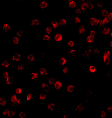
The catalog antibodies cover human tonsil and mouse brain IHC, plus Daudi cell and mouse brain IF (IHC and IF image captions).

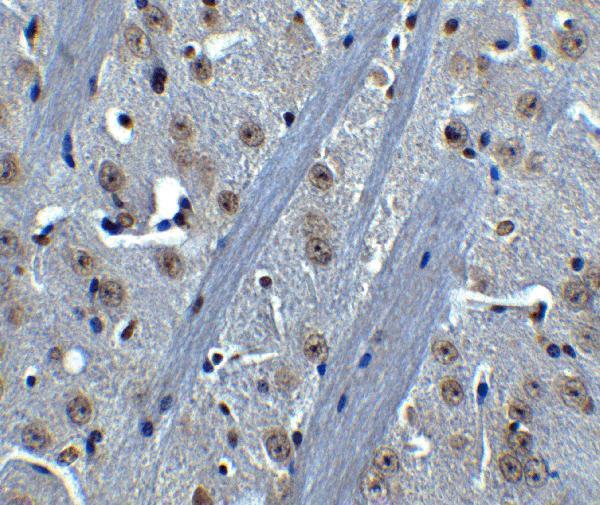
Real IHC data Immunohistochemistry of BTK in mouse brain tissue with BTK antibody at 5 μg/mL.
Anti-Tyrosine-protein kinase BTK BTK Antibody
Cat # A00245
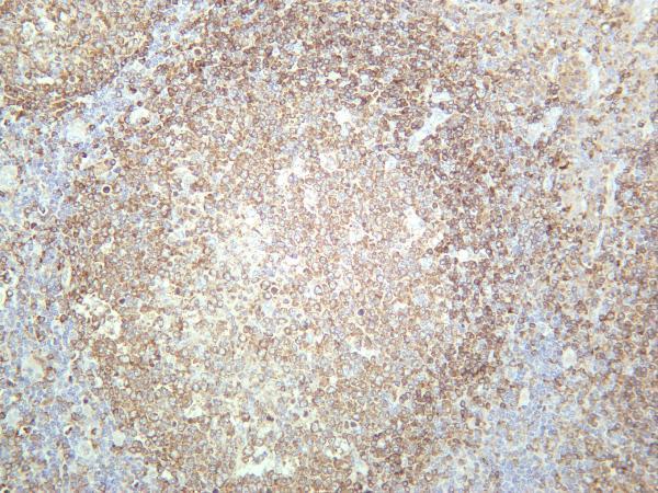
Real IHC data Human tonsil was stained with anti-BTK rabbit antibody
Anti-BTK Rabbit Monoclonal Antibody
Cat # M00245-4

A00245 has an IHC-P mouse brain image and IF images of Daudi cells and mouse brain (catalog applications; image captions). M00245-4 has a human tonsil IHC image and lists IF for human samples (catalog applications, reactivity, and IHC image caption).

Which to pick: For paraffin-section IHC or human and mouse reactivity, choose A00245; its mouse brain IHC image uses 5 μg/mL, while the fixative is unreported (A00245: IHC-P application, reactivity, IHC image caption). For human tonsil IHC, M00245-4 is a rabbit monoclonal with a 1:1000–1:4000 IHC range; its image caption does not report section processing or fixative (M00245-4: catalog description, dilution, IHC image caption). For IF/ICC, A00245 has Daudi cell IF and ICC images; M00245-4 lists IF for human samples, while P00245 is specific to phospho-BTK Y551 and lists ICC/IF without an IHC application (A00245: IF image captions; M00245-4 and P00245: catalog applications and reactivity).

Each figure is that product's own IHC / IF validation image from its datasheet.

References

  1. UniProt Consortium. UniProt entry Q06187 (BTK_HUMAN, Tyrosine-protein kinase BTK).
  2. Human Protein Atlas. BTK tissue IHC expression (reliability: Enhanced).
  3. Human Protein Atlas. BTK subcellular location (ICC-IF): Localized to the plasma membrane and cytosol..
  4. Human Protein Atlas. BTK antibody validation summary (4 antibodies).
  5. Combination of acalabrutinib with lenalidomide and rituximab in relapsed/refractory aggressive B-cell non-Hodgkin lymphoma: a single-arm phase II trial. Nature communications 2024 — PMC10981676.
  6. Ibrutinib Potentiates Antihepatocarcinogenic Efficacy of Sorafenib by Targeting EGFR in Tumor Cells and BTK in Immune Cells in the Stroma. Molecular cancer therapeutics 2020 — PMC7007841.
  7. Bruton's tyrosine kinase (Btk) inhibitor ibrutinib suppresses stem-like traits in ovarian cancer. Oncotarget 2015 — PMC4537012.
  8. Bortezomib enhances the anti-cancer effect of the novel Bruton's tyrosine kinase inhibitor (BGB-3111) in mantle cell lymphoma expressing BTK. Aging 2021 — PMC8457562.
  9. PubMed PMID:8380905 — UniProt-cited evidence.
  10. PubMed PMID:8090769 — UniProt-cited evidence.
  11. PubMed PMID:7927535 — UniProt-cited evidence.