This website uses cookies to ensure you get the best experience on our website.


- Table of Contents
Plan chromogenic BUB3 IHC on paraffin sections using 0.5–1 μg/mL catalog antibody (datasheet: PA1645 IHC-P). Expect widespread nuclear staining, with high signal in colon glandular cells and no detection in liver cholangiocytes (HPA tissue IHC).

Expected localisation, validated protocols, controls and antibodies — the at-a-glance facts below, then the full design guide.
| Expected localisation | Nuclear staining in tissue (HPA tissue IHC) | |
| Staining pattern | Ubiquitous nuclear staining across cell types (HPA tissue IHC) | |
| Antigen retrieval | Tris-EDTA pH 9.0 HIER, 95–98 °C, 20 min (rule: nuclear antigen) | |
| Positive control | Adrenal gland+4 more · see all | |
| Negative control | Liver |
| Fixation | Keep fixation consistent (standard IHC practice; not target-specific) | |
| Caveat | Kinetochore localisation varies with meiotic stage (UniProt) | |
| Regulation | Expression regulation unreported (UniProt) | |
| Isoform / epitope | 2 isoforms; epitope differences unreported (UniProt) |
Compare the catalog antibody’s IHC-P protocol with two published BUB3 IHC workflows (PMC7190565; PMC8601638).
| Sample | Tissue sections; selected-image fixative not specified (standard IHC workflow) |
| Fixation | Image fixative and duration unreported (datasheet PA1645); verify before use. |
| Sectioning | 4–5 µm sections on charged slides (standard) |
| Deparaffinisation | Xylene, graded ethanol series to water (standard) |
| Antigen retrieval | Heat-induced epitope retrieval in Tris-EDTA buffer, pH 9.0, 20 min at 95–98 °C (standard rule: nuclear antigen) |
| Peroxidase block | 3% H2O2, 10 min, room temperature (standard) |
| Blocking | 10% normal serum of the secondary host, 30 min, room temperature (standard) |
| Primary antibody | Rabbit anti-BUB3, 0.5-1μg/ml (datasheet PA1645) |
| Primary incubation | Overnight at 4 °C (standard) |
| Detection | HRP-polymer secondary, DAB chromogen 5–10 min (standard) |
| Counterstain | Hematoxylin, blue, dehydrate and mount (standard) |
| Expected result | BUB3-positive staining in glandular cells of adrenal gland (HPA tissue IHC: High). HPA tissue profile: Ubiquitous nuclear expression. No signal in the no-primary control. |
BUB3 is primarily nuclear, with centromere and kinetochore localization at defined cell-cycle stages (UniProt O43684: localization). Expect nuclear staining across many tissue cell types, including strong staining in the listed glandular and epithelial cells (HPA: ubiquitous nuclear expression; High in listed cells). HPA rates the tissue IHC pattern Supported, with medium consistency between staining and RNA data (HPA: reliability). BUB3 has no transmembrane segment (UniProt O43684: topology).
| Nuclear chromogen in adrenal, colon, or gallbladder glandular cells. | This fits the reported high staining in those cells and the broad nuclear profile (HPA: High in listed glandular cells; ubiquitous nuclear expression). Compare nuclei with neighboring cells and a matched negative control before scoring intensity; the HPA rating is Supported, with medium staining–RNA consistency (HPA: reliability). |
| Discrete staining associated with chromosomes in a cell undergoing division. | A cell-cycle-specific pattern can fit BUB3 localization to centromeres and kinetochores (UniProt O43684: localization). Evaluate the cell's morphology and counterstain before calling such a focus artefactual. UniProt describes timed kinetochore localization during oocyte meiosis; it does not make every nuclear punctum in a routine tissue section diagnostic of BUB3 (UniProt O43684: localization). |
| Predominantly membranous or extracellular staining, with little nuclear signal. | This conflicts with the reported nuclear distribution and absence of a transmembrane segment (HPA: ubiquitous nuclear expression; UniProt O43684: topology). Treat the pattern as suspect, then compare with a no-primary control and an independently validated antibody where available (general IHC practice). |
| Strong staining confined to cholangiocytes, smooth muscle cells, or adipocytes. | Check cell identification and controls before assigning the stain to BUB3: cholangiocytes are Not detected, while smooth muscle cells and adipocytes are Low in the cited HPA tissue profile (HPA: listed cell levels). Cross-reactivity or endogenous detection activity is possible; these HPA observations do not rule out staining in other cells of the same tissue. |
| Even chromogen across nuclei, cytoplasm, and empty spaces. | This lacks the compartment contrast expected from BUB3's nuclear profile (HPA: ubiquitous nuclear expression). Examine the no-primary control and tissue morphology. Widespread haze may reflect nonspecific binding, incomplete blocking, detection background, or excessive chromogen development (general IHC practice); appearance alone cannot identify which cause applies. |
| Tissue and cell selection | Use a reported High population to judge whether the assay can reveal nuclear BUB3; colon glandular cells and cervix squamous epithelial cells are examples (HPA: High). Cholangiocytes are a useful low-signal comparator only in their identified compartment: HPA reports Not detected there, not in every liver cell (HPA: liver cholangiocytes). |
| Antibody validation | Match the antibody identifier to its application. HPA003601 and CAB037075 have Supported IHC status; HPA071982 has no IHC status in the supplied list (HPA: antibody validation). An Enhanced ICC rating for an antibody should not be substituted for IHC validation (HPA: antibody validation). |
| Antigen retrieval | No BUB3-specific retrieval condition or fixation-sensitivity result is supplied. For paraffin-section IHC, follow the selected antibody's validated IHC instructions and assess retrieval with positive tissue and a no-primary control (general IHC practice). Changes in signal after retrieval are assay observations, not evidence here of a defined BUB3 fixation effect. |
| Isoforms and epitope coverage | UniProt lists two BUB3 isoforms and a 1–328 main chain, but the supplied evidence gives no antibody epitope or isoform-specific staining data (UniProt O43684: isoforms and processing). Do not explain a missing stain by isoform exclusion without antibody-specific evidence. |
| IF/ICC Q: What localization should appear? | A: HPA reports enhanced nucleoplasmic localization in ICC-IF and lists images from A-431, U-251MG, U2OS, and REH (HPA: subcellular). This supports a localization expectation for IF/ICC; it supplies no IF protocol here and does not replace tissue IHC validation (HPA: subcellular; antibody validation). |
| Detection background | Endogenous enzyme activity and nonspecific secondary-antibody binding can produce chromogenic color independently of the primary antibody (general IHC practice). Use appropriate detection controls and inspect whether color tracks the expected nuclei (HPA: ubiquitous nuclear expression); a colored compartment alone does not prove antigen recognition. |
| Situation | Likely cause | Next action |
|---|---|---|
| No nuclear signal in a reported High population. | The assay may lack usable signal, or the sampled cells may have been misidentified; HPA reports High staining in colon glandular cells, for example (HPA: colon). The supplied sources do not identify a BUB3-specific fixation cause. | Verify tissue and cell identity, review the antibody's IHC validation and instructions, and compare the run with a known-positive section processed alongside it (HPA: antibody validation; general IHC practice). |
| Cytoplasmic or membrane color dominates the section. | That distribution conflicts with nuclear BUB3 and its lack of a transmembrane segment (HPA: ubiquitous nuclear expression; UniProt O43684: topology). Nonspecific binding or detection background may contribute (general IHC practice). | Inspect the no-primary control and counterstained nuclei, then reassess antibody concentration and detection controls according to the assay instructions (general IHC practice). Do not score dominant membrane color as the expected pattern. |
| Cholangiocytes appear strongly positive. | HPA reports BUB3 as Not detected in cholangiocytes; misidentified cells, cross-reactivity, or endogenous chromogen are possible explanations (HPA: liver cholangiocytes; general IHC practice). | Confirm the cell compartment on the counterstained section and compare no-primary and independent-antibody controls where available (general IHC practice). Avoid extending this cholangiocyte observation to all liver cells. |
| Every cell and empty area has similar color. | Uniform field-wide color obscures the nuclear contrast expected for BUB3 (HPA: ubiquitous nuclear expression). Background from detection chemistry or excessive development is possible (general IHC practice). | Check the no-primary section, reagent blocking, washes, and chromogen development against the assay instructions (general IHC practice). Score BUB3 only where identifiable nuclei show signal above the local background. |
| Different antibodies give conflicting nuclear patterns. | Validation is antibody- and application-specific: two listed antibodies are Supported for IHC, while one has no supplied IHC status (HPA: antibody validation). Epitope and isoform coverage cannot be inferred from this payload (UniProt O43684: isoforms). | Record each antibody identifier and its IHC status, compare matched sections and controls, and interpret only reproducible cell-specific nuclear staining (HPA: antibody validation and tissue profile; general IHC practice). |
| A focal chromosomal signal is called background. | BUB3 can occupy centromeres and kinetochores at defined stages, including the stated interval in oocyte meiosis (UniProt O43684: localization). A focal mark is still insufficient to identify the antigen by itself. | Review cell-cycle morphology, nuclear counterstain, and control sections before classifying the focus (general IHC practice). Report it separately from the broad nuclear tissue pattern described by HPA (HPA: ubiquitous nuclear expression). |


Comprehensive Human Protein Atlas IHC scoring per tissue (reliability: Supported — Medium consistency between antibody staining and RNA expression data.). Rows are taken directly from the HPA tissue chart — click any row's HPA link to view the source.
| Tissue | Cell type | Level | Evidence | Source |
|---|---|---|---|---|
| Adrenal gland | Glandular cells | High | Protein (IHC) | HPA → |
| Cervix | Squamous epithelial cells | High | Protein (IHC) | HPA → |
| Colon | Glandular cells | High | Protein (IHC) | HPA → |
| Fallopian tube | Glandular cells | High | Protein (IHC) | HPA → |
| Gallbladder | Glandular cells | High | Protein (IHC) | HPA → |
| Tissue | Cell type | Level | Evidence | Source |
|---|---|---|---|---|
| Liver | Cholangiocytes | Not detected | Protein (IHC) | HPA → |
Troubleshoot BUB3 chromogenic IHC in paraffin sections by checking nuclear staining, retrieval, controls and cell level scoring (UniProt O43684 localisation; HPA tissue IHC).
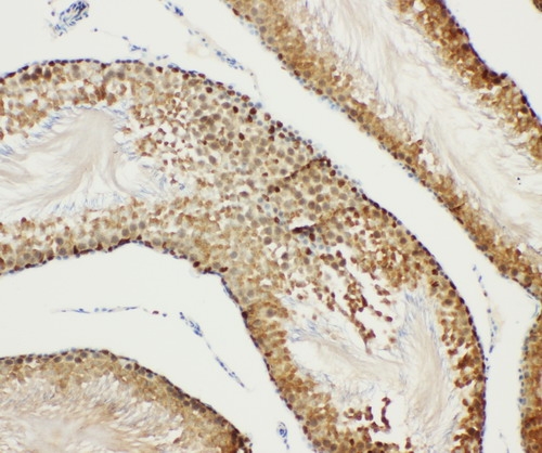
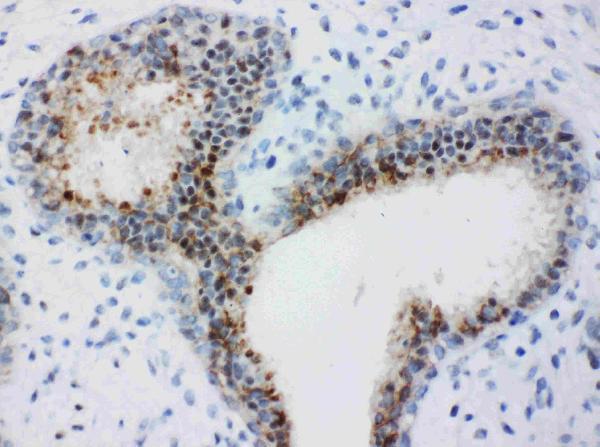
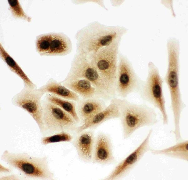
PA1645 has IHC images of rat testis and human mammary cancer paraffin sections, plus ICC images of A549 and HeLa cells (catalog image captions).
PA1645 is the only SKU with a rendered card; its IHC figure shows rat testis in a paraffin section (card figure caption). Its other captions show human mammary cancer paraffin sections and A549 and HeLa ICC samples (catalog image captions).
Which to pick: Choose PA1645 for tissue IHC because its captions document rat testis and human mammary cancer paraffin sections; the fixative is unreported (PA1645 IHC image captions). For IF/ICC, PA1645 has A549 and HeLa ICC images, while M03118-1 lists ICC/IF without an image in the payload (PA1645 ICC image captions; M03118-1 catalog applications and image alts). Both list human, mouse and rat reactivity; M03118-1 is a rabbit monoclonal, while PA1645 has no clone listed (catalog reactivity and clone fields).