This website uses cookies to ensure you get the best experience on our website.


- Table of Contents
Plan chromogenic CALCA IHC in paraffin sections using thyroid and bronchial epithelial subsets as positive reference tissue (HPA tissue IHC). This guide covers fixation, controls, and interpretation of a secreted target (UniProt).

Expected localisation, validated protocols, controls and antibodies — the at-a-glance facts below, then the full design guide.
| Expected localisation | Secreted (UniProt); tissue compartment unreported (HPA tissue IHC) | |
| Staining pattern | Thyroid and bronchial epithelial subsets; compartment unclear (HPA tissue IHC) | |
| Antigen retrieval | EDTA pH 8.0 HIER, heat-mediated (datasheet PB9936) | |
| Positive control | Bronchus+1 more · see all | |
| Negative control | Adipose tissue+4 more · see all |
| Fixation | Keep fixation consistent across paraffin sections. (standard IHC practice; not target-specific) | |
| Caveat | Secreted protein: RNA and protein locations may differ (HPA tissue IHC) | |
| Regulation | No expression regulator specified (UniProt) | |
| Isoform / epitope | 3 isoforms; check epitopes across processing regions (UniProt) |
The catalog antibody protocol uses EDTA pH 8.0 retrieval (datasheet PB9936). The published CALCA IHC protocol below uses citrate retrieval (PMC5716875).
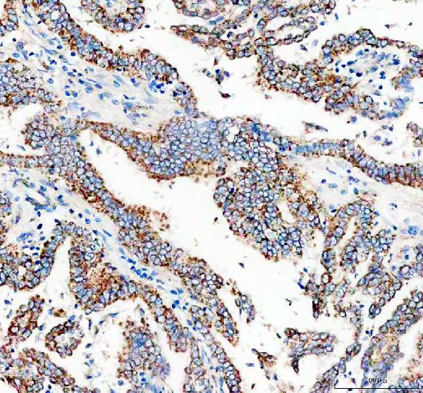
| Sample | Paraffin-embedded human lung cancer tissue; fixative not specified (datasheet PB9936) |
| Fixation | Image fixative and duration unreported (datasheet PB9936); verify before use. |
| Sectioning | 4–5 µm sections on charged slides (standard) |
| Deparaffinisation | Xylene, graded ethanol series to water (standard) |
| Antigen retrieval | Heat retrieval: EDTA pH 8.0 (datasheet PB9936); 20 min, 95–100 °C (standard) |
| Peroxidase block | 3% H2O2, 10 min, room temperature (standard) |
| Blocking | 10% goat serum (datasheet PB9936) |
| Primary antibody | Rabbit anti-CALCA, 0.5-1μg/ml recommended; image 2 μg/ml (datasheet PB9936) |
| Primary incubation | Overnight at 4 °C (datasheet PB9936) |
| Detection | HRP-conjugated secondary, DAB chromogen (datasheet PB9936) |
| Counterstain | Hematoxylin, blue, dehydrate and mount (standard) |
| Expected result | CALCA-positive staining in respiratory epithelial cells of bronchus (HPA tissue IHC: Medium). HPA tissue profile: Expressed in a subset of thyroid epithelial cells and bronchial epithelial neuroendocrine cells. No signal in the no-primary control. |
CALCA is a secreted peptide precursor with no transmembrane segment (UniProt P01258: secreted; topology). In paraffin-section IHC, expect selective staining in a subset of thyroid epithelial cells and bronchial epithelial neuroendocrine cells; HPA reports medium staining in thyroid glandular and bronchial respiratory epithelial cells (HPA tissue IHC: Enhanced). HPA does not assign CALCA a precise intracellular compartment (HPA subcellular: main location unavailable).
| A subset of thyroid glandular cells stains at moderate intensity while neighbouring cells remain unstained. | This fits the reported thyroid pattern (HPA tissue IHC: thyroid glandular cells, Medium; subset of thyroid epithelial cells). Score the fraction of stained cells as well as intensity: uniform staining of every glandular cell would be less consistent with the reported subset pattern. HPA does not define a required nuclear, cytoplasmic or granular distribution (HPA subcellular: main location unavailable). |
| Scattered bronchial epithelial cells stain at moderate intensity within a mostly unstained lining. | This fits the reported subset of bronchial epithelial neuroendocrine cells (HPA tissue IHC: bronchus respiratory epithelial cells, Medium; profile). Use cell identity and the selective distribution together when reading the slide. A positive bronchial section need not show continuous staining along the epithelium (HPA tissue IHC: subset pattern). |
| Staining appears mainly as a sharp membrane rim or isolated nuclear signal. | Review it as a compartment mismatch: CALCA is secreted and has no transmembrane segment (UniProt P01258: secreted; topology). Neither source specifies a precise intracellular staining pattern (HPA subcellular: main location unavailable). A membrane rim or nuclear-only result therefore needs further validation rather than a confident CALCA localisation call. |
| Broad staining appears in cells outside the reported positive subsets. | This is less consistent with the selective tissue pattern (HPA tissue IHC: thyroid and bronchus profiles). In chromogenic IHC, cross-reactivity or endogenous detection activity can produce apparent positives (general IHC practice). Check the cell type, negative controls and detection controls before attributing widespread colour to CALCA. |
| The slide shows diffuse colour, or a known-positive thyroid or bronchus section has no signal. | Diffuse colour that does not resolve into the reported cell subsets is background rather than a readable positive pattern (HPA tissue IHC: subset pattern; general IHC practice). An unstained section is inconclusive until a suitable positive control works. HPA reports medium staining, not universal staining of every cell in these tissues (HPA tissue IHC: positive entries). |
| Secreted protein and tissue interpretation (UniProt P01258: secreted). | RNA and protein locations may differ, so do not use tissue RNA enrichment alone to accept or reject a stained section (HPA tissue IHC: reliability description; RNA group enriched in parathyroid and thyroid gland). HPA provides no internal RNA correlation for this staining assessment (HPA tissue IHC: reliability description). |
| Antibody validation (HPA antibodies: HPA064453 IHC Enhanced; HPA059886 IHC Supported). | The validation labels differ by antibody. Record which antibody produced the image when comparing results; the overall tissue reliability is Enhanced, while the two listed antibodies do not carry identical IHC validation labels (HPA tissue IHC: reliability; HPA antibodies). |
| Precursor processing and alternative isoforms (UniProt P01258: signal 1–25, propeptide 26–82; isoforms 1, 2 and 3). | The record does not locate the antibody epitope or show which processed form or isoform it detects (UniProt P01258: processing and isoforms; HPA antibodies: validation summary). Interpret staining as antibody reactivity to CALCA-related antigen without assigning a particular molecular form from the slide alone. |
| Subcellular resolution (HPA subcellular: Secreted; main location unavailable; no ICC-IF images). | A specific intracellular organelle or punctate pattern is not established by the supplied sources. Use the documented tissue and cell distribution as the primary IHC expectation (HPA tissue IHC: thyroid and bronchus profile), and avoid treating a proposed compartment as an independent pass criterion. |
| Situation | Likely cause | Next action |
|---|---|---|
| A thyroid section is entirely unstained, including the expected positive subset. | A failed IHC run or weak detection is possible (general IHC practice); the reported positive population is only a subset and has medium staining (HPA tissue IHC: thyroid). | Check that a separate known-positive control develops and that the counterstain does not obscure weak chromogen. Review antibody incubation, antigen retrieval and detection settings as general IHC variables; the sources provide no CALCA-specific retrieval condition or fixation-sensitivity claim. |
| Bronchial staining is continuous across most epithelial cells. | That distribution exceeds the reported neuroendocrine subset (HPA tissue IHC: bronchus profile). Cross-reactivity or excessive detection may broaden apparent staining (general IHC practice). | Compare the cell distribution with the positive control, reduce nonspecific detection through routine IHC optimisation, and inspect a negative control. Do not score a continuous epithelial sheet as matching the HPA subset solely because the tissue is bronchus. |
| Colour appears in expected-negative cells or across the entire section. | HPA reports several cell populations as not detected, including adipocytes in adipose tissue and glandular cells in adrenal gland (HPA tissue IHC: negative entries). Endogenous enzyme activity or nonspecific binding can add chromogenic colour (general IHC practice). | Inspect a no-primary detection control and use an appropriate endogenous-activity block for the detection chemistry (general IHC practice). Compare tissue and cell identity with the HPA negative entries before calling cross-reactivity; a negative entry applies to the listed cell population. |
| Only nuclear staining or a crisp cell-edge rim is visible. | Neither distribution is established as a CALCA pattern; the protein is secreted without a transmembrane segment (UniProt P01258: localisation and topology; HPA subcellular: main location unavailable). | Check morphology, background and detection controls, then repeat with an independently validated antibody if available (general IHC practice). Report the compartment mismatch rather than reclassifying the pattern as confirmed CALCA. |
| Diffuse haze prevents scoring the expected positive subset. | Background may arise from nonspecific antibody binding, incomplete blocking or detection chemistry (general IHC practice). The HPA pattern requires a discernible subset of cells (HPA tissue IHC: profile). | Compare no-primary and positive-control sections, then optimise blocking, washes and antibody concentration as general IHC steps. Reassess whether distinct thyroid or bronchial cells remain stained above the local background (HPA tissue IHC: positive entries). |
| What localisation should an IF/ICC image show? | A precise IF/ICC compartment cannot be inferred: HPA lists CALCA as secreted but provides no main location or cell-line ICC-IF images (HPA subcellular: summary and image list). | Use the separate IF/ICC guide for that application. Here, interpret the paraffin-section chromogenic IHC result by the documented thyroid and bronchial cell subsets (HPA tissue IHC: profile), without assigning an IF/ICC-specific pattern. |


Comprehensive Human Protein Atlas IHC scoring per tissue (reliability: Enhanced — Secreted protein, tissue location of RNA and protein is expected to differ. External characterization data supports antibody staining but no internal RNA data available for correlation.). Rows are taken directly from the HPA tissue chart — click any row's HPA link to view the source.
| Tissue | Cell type | Level | Evidence | Source |
|---|---|---|---|---|
| Bronchus | Respiratory epithelial cells | Medium | Protein (IHC) | HPA → |
| Thyroid gland | Glandular cells | Medium | Protein (IHC) | HPA → |
| Tissue | Cell type | Level | Evidence | Source |
|---|---|---|---|---|
| Adipose tissue | Adipocytes | Not detected | Protein (IHC) | HPA → |
| Adrenal gland | Glandular cells | Not detected | Protein (IHC) | HPA → |
| Appendix | Glandular cells | Not detected | Protein (IHC) | HPA → |
| Bone marrow | Hematopoietic cells | Not detected | Protein (IHC) | HPA → |
| Breast | Adipocytes | Not detected | Protein (IHC) | HPA → |
Troubleshooting CALCA staining in paraffin-section IHC, with one entry on multiplex immunofluorescence.
PB9936 has a human paraffin-section IHC image (PB9936 IHC image caption); M02352-6 lists IHC and IF/ICC for human, mouse and rat (catalog applications/reactivity).
PB9936 is the rendered card, with IHC imagery from a human lung cancer paraffin section (PB9936 IHC image caption). M02352-6 lists human, mouse and rat IHC/IF/ICC applications, but has no IHC or IF image in the payload (catalog applications/reactivity/image alts).
Which to pick: Choose PB9936 for human paraffin-section chromogenic IHC, supported by its DAB-stained image; the caption does not report the fixative (PB9936 IHC image caption). For IF/ICC or mouse and rat samples, consider rabbit monoclonal M02352-6 based on its listed applications and reactivity (catalog host/clone/applications/reactivity).